Reading...
![]()
Play button
![]()
Play button
![]()
Use LEFT and RIGHT arrow keys to navigate between flashcards;
Use UP and DOWN arrow keys to flip the card;
H to show hint;
A reads text to speech;
144 Cards in this Set
- Front
- Back
|
Nervous System Overview - The NERVOUS system is the body’s
|
principal control and integrating system.
|
|
|
Nervous System Overview - The nervous system performs 3 main functions
|
i.sensory, ii. Integrative function, iii. motorsystem…
|
|
|
Nervous System Overview - SENSORY function is
|
detection of changes inside and outside the body by specialized cells called sensory RECEPTORS/INPUT.
|
|
|
Nervous System Overview - Sensory information is carried
|
to brain and/or spinal cord along sensory or AFFERENT neurons.
|
|
|
Nervous System Overview - INTEGRATIVE FUNCTION is
|
(HAPPENS AT INTE CENTER) —interpretation of the changes detected by the sensory mechanisms.
|
|
|
Nervous System Overview - The conscious awareness of sensory stimuli within the brain is called
|
PERCEPTION (THINK POPCORN SMELL).
|
|
|
Nervous System Overview - The integration of sensory stimuli occurs along
|
the INTERNEURONS
|
|
|
Nervous System Overview - INTERNEURONS
|
connect neurons within the brain and spinal cord over short distances.
|
|
|
Nervous System Overview - MOTOR SYSTEM function is
|
reacting to changes through the action of the organ systems such as the glands and muscles.
|
|
|
Nervous System Overview - Motor information is carried away from the brain toward
|
the spinal cord to the EFFECTORS along the motor or EFFERENT neurons.
|
|
|
The nervous system has 2 major divisions
|

the CNS (central nervous system) and PNS (periphial nervous system)
|
|
|
The CENTRAL nervous system (CNS) consists of:
|

1. BRAIN, 2. SPINAL CORD (MUCH INVOLUNTARY REGULATION OCCURS)
|
|
|
The PERIPHIAL nervous system (PNS) consists of:
|
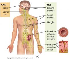
CRANIAL nerves which originate from the brain.
|
|
|
How many cranial nerves in the brain
|
12 NERVES
|
|
|
SPINAL nerves
|
originate from the spinal cord.
|
|
|
Spinal nerves EXTEND
|
THROUGHOUT THE BODY
|
|
|
The peripheral nervous system is further subdivided into:
|
i. sns - sematic nervous system ii. Ans – autonomic nervous system iii. ENS - Enteric nervous system
|
|
|
SEMATIC nervous system (SNS)
|
includes neurons that conduct impulses to SKELETAL MUSCLES
|
|
|
AUTONOMIC nervous system (ANS)
|
includes neurons that convey impulses from the brain to smooth muscles, cardiac muscle, and glands.
|
|
|
The autonomic nervous system is subdivided again into:
|
i. sympathetic division ii. Parasympathetic division
|
|
|
SYMPATHETIC division
|
generally increases the activity of an organ. (FIGHT OR FLIGHT)
|
|
|
PARASYMPATHETIC division
|
generally decreases the activity of an organ. (REST AND DIGEST)
|
|
|
ENTERIC nervous system (ENS) is part of the
|
autonomic nervous system
|
|
|
Enteric nervous system includes
|
involuntary sensory and motor neurons which control the gastrointestinal (GI) tract.
|
|
|
Enteric nervous system neurons function
|
independently of the ANS and CNS. (ANYTIME YOU SEE ENTERO THINK DIGESTION)
|
|
|
There are two types of nerve cells
|
i. neurons ii. neuroglia
|
|
|
NEURONS are
|
cells that conduct nerve impulses. 100 BILLION IN BRAIN 100 MILLION IN SPINAL CORD
|
|
|
NEUROGLIA are
|
cells that support and protect neurons.
|
|
|
Neuroglia (glial cells) are
|
smaller and more numerous than neurons.
|
|
|
There are 6 different types of neuroglia within
|
the CNS and PNS.
|
|
|
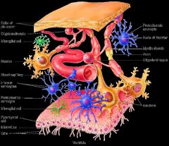
Neuroglia of the CNS:
|
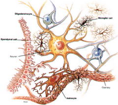
i. oligodendrocyte, ii. Astrocytes, iii. microglial cells, iiii. Ependymal cells
|
|
|
OLIGODENDROCYTE neuroglia
|

is the most common glial cell type.
|
|
|
oligodendrocytes Form
|

the myelin sheath around more than one axon in the CNS.
|
|
|
ASTROCYTES neurologia
|

attach to blood vessels forming a blood-brain barrier (BBB) that prevents harmful substances and organisms from entering the CNS.
|
|
|
MICROGLIAL CELL neurologia
|
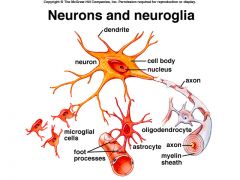
engage in phagocytosis of cellular debris and damaged nervous tissue.
|
|
|
PHAGOCYTOSIS is
|
the ingestion of a smaller cell or cell fragment, a microorganism, or foreign particles by means of the local infolding of a cell's membrane – BREAKDOWN of cells
|
|
|
EPENDYMAL CELLS neurolgia
|

produce the cerebrospinal fluid (CSF) of the CNS.
|
|
|
Neuroglia of the PNS:
|

i. schwann cells, ii. satellite
|
|
|
SCHWANN CELLS Neuroglia
|
produce the myelin sheath around single axons and participate in axon regeneration.
|
|
|
SATELLITE Neuroglia
|
are flat cells surrounding neuronal cell bodies in peripheral ganglia. They support neurons in the PNS ganglia.
|
|
|
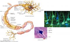
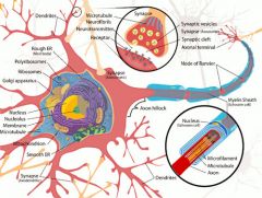
Most neurons consist of three distinct portions.
|
i. cell body, ii. Dendrites, iii. axon
|
|
|
CELL BODY (soma) contains
|
a single nucleus and granular cytoplasm.
|
|
|
DENDRITES receive
|
the stimuli and convey nerve impulses to the cell body.
|
|
|
AXON conducts
|
impulses from the neuron to the dendrites of another neuron or to an effector organ of the body.
|
|
|
AXOLEMA is
|
The cellular membrane of the axon
|
|
|
AXOPLASM is
|
the cytoplasm contained by the Axolema
|
|
|
axon COLLATERAL are
|
Side branches of the axon which occur at right angles.
|
|
|
TELODENDRIA aka
|
axon terminals
|
|
|
Telondendria
|
The fine filaments terminating the axon and axon collaterals also known as axon terminals
|
|
|
SYNAPTIC END BULBS are
|
The round nodules at the ends of telodendria
|
|
|
SYNAPTIC VESSICLES are
|
the membrane-enclosed sacs contained by the synaptic end bulbs NEUROTRANSMITTERS are chemical signals stored by the synaptic vessicles,
|
|
|
The SYNAPSE is
|
the site of communication between two neurons or between a neuron and an EFFECTOR cell.
|
|
|
Neurons are either
|
covered or uncovered
|
|
|
Covered or MYELINATED neurons possess
|
a white phospholipid, MYELIN (FATTY LAYER) , which covers the axons forming the myelin sheath.
|
|
|
MYELIN is produced by two different types of neuroglia
|
i. oligodendrocytes ii. Schwann cells
|
|
|
OLIGODENDROCYTES form
|
the myelin sheath of the axons within the CNS.
|
|
|
Myelinated neurons form
|
the WHITE matter of the CNS.
|
|
|
SCHWWAN cells form
|
the myelin sheath of the axons within the PNS.
|
|
|
NODES OF RANVIER or neurofibril nodes are
|
gaps within the myelin sheath that separate Individual Schwann cells
|
|
|
Uncovered or UNMYELINATED neurons form
|
the GRAY matter of the CNS
|
|
|
Unmyelinated neurons are
|
SLOWER than the myelinated neurons.
|
|
|
gray matter is on the outside within the
|
brain
|
|
|
gray matter is on the inside within the
|
spine (think butterfly)
|
|
|
Classification of neurons is based upon
|
structural and functional criteria.
|
|
|
Structural classification of neurons
|
i. multipolar, ii. Bipolar, iii. unipolar…
|
|
|
MULTIPOLAR neurons have
|
multiple dendrites and one axon.
|
|
|
BIPOLAR neurons have
|
one dendrite and one axon.
|
|
|
Bipolar Neurons are associated with
|
the SPECIAL SENSES (EYE, EAR, OLFACTORY)
|
|
|
UNIPOLAR neurons have
|
only one process extending from the cell body.
|
|
|
Unipolar neurons are found
|
in the dorsal (sensory) root ganglia of the spine.
|
|
|
The unipolar process (extension) divides into:
|
i. A central branch which acts as an AXON, ii.A peripheral branch which acts as a DENDRYTE
|
|
|
Functional classification of neurons
|
i. afferent ii. Efferent iii. interneurons…
|
|
|
AFFERENT neurons are
|
(sensory) nerve cells that conduct impulses from the SENSORY RECEPTORS towards the BRAIN (OR CENTRAL NERVOUS SYSTEM
|
|
|
Afferent neurons are CORRELATED WITH
|
CNS
|
|
|
EFFERENT neurons are
|
(motor) nerve cells that conduct impulses from the BRAIN towards the EFFECTORS
|
|
|
Efferent neurons are CORRELATED WITH
|
PNS
|
|
|
INTERNEURONS are
|
(association neurons) that connect sensory to motor neurons. INTERNEURONS are CORRELATED WITH CNS
|
|
|
The cell bodies and axons of neurons form
|
bundles of ganglions and nucleus
|
|
|
Cell bodies are arranged into bundles called:
|
i. ganglions ii. nucleus
|
|
|
GANGLIONS bundles are
|
in the PNS.
|
|
|
NUCLEUS bundles are
|
in the CNS.
|
|
|
The myelinated axons of afferent and efferent neurons are arranged into bundles called:
|
i. nerves, ii. fiber
|
|
|
Myelinated NERVES bundles are located
|
in the PNS.
|
|
|
Myelinated FIBER (nerve) tracts bundles are located
|
in the CNS.
|
|
|
The ability to produce nerve impulse (nerve action potential) is dependent on:
|
Membrane potentials.
|
|
|
The membrane of a RESTING (nonconducting) neuron is
|
POSITIVELY charged outside and NEGATIVELY charged inside.
|
|
|
The membrane of a RESTING (nonconducting) neuron is said to be
|
POLARIZED and has a resting membrane potential.
|
|
|
POSITIVE charge OUTSIDE the cell is accounted for
|
by The presence of positive SODIUM ions (cations) outside the membrane accounts.
|
|
|
NEGATIVE charge INSIDE the cell is accounted for
|
by Organic phosphate groups (PO43-) and protein anions
|
|
|
Ion channels Open or close in response to
|
specific STIMULI allowing for the flow of ions across the membrane.
|
|
|
Ion channels Maintain and restore
|
ELECTRO CHEMICAL gradient across the plasma membrane.
|
|
|
Neurons are capable of responding to
|
stimuli and converting them to impulses through EXCITABILITY
|
|
|
A nerve impulse (action potential) is generated over three steps:
|
i. depolarization ii. Repolarization iii. refractrion
|
|
|
DEPOLARIZATION occurs when
|
the inside of the nerve becomes POSITIVE and the outside becomes NEGATIVE
|
|
|
Depolarization - The inside of the nerve becomes positive when
|
ion channels (gates) open and allow SODIUM ions (Na+) to enter. THE SODIUM CAUSES ADDITIONAL CHANNELS TO OPEN.
|
|
|
Depolarization - Sodium channels are
|
closed quickly stopping the inflow of Na+.
|
|
|
REPOLARIZATION occurs when
|
the inside of the nerve becomes NEGATIVE and the outside becomes POSITIVE again.
|
|
|
REPOLARIZATION - POTASSIUM channels are
|
opened at the same time as the sodium channels are being closed.
|
|
|
REPOLARIZATION - Open channels results in
|
the outflow of POTASSIUM ions (K+) causing the membrane potential to become negative again. In addition, SODIUM ions are pumped back out of the nerve fiber.
|
|
|
REPOLARIZATION - HIGH AMOUNT OF POTASSIUM WILL CAUSE
|
PUMP OF POTASSIUM TO STOP
|
|
|
The REFRACTORY period is
|
a period of time during which the membrane recovers and cannot initiated another action potential.
|
|
|
During The REFRACTORY period
|
the sodium channels are inactivated.
|
|
|
There are two types of nerve conduction…
|
i. continuous ii. Salutatory
|
|
|
CONTINOUS conduction occurs only in
|
unmyelinated nerve fibers.
|
|
|
SALTATORY conduction occurs only in
|
myelinated nerve fibers.
|
|
|
Nerve Conduction - The MYELIN sheath acts as an electrical insulator and polarization can only occur at the
|
NODES OF RANVIER (UNMYELINATED SECTIONS)
|
|
|
Nerve Conduction - In salutatory conduction the impulse
|
appears to “leap” from node to node through the interstitial fluids covering the myelin segments.
|
|
|
The speed of impulse conduction is
|
greatly INCREASED because of the skipped distances (myelinated) and involves less activity by the sodium-potassium pumps. (ADDITIONAL BENEFIT ACTION REQUIRES LESS ATP)
|
|
|
NERVES ARE NOT MYELINATED AT BIRTH. MYELINATION DEVELOPS WITH AGE. AFFECTING GROSS AND THEN FINE MOTOR SKILLS Tor F
|
T
|
|
|
The speed of nerve impulses depends upon
|
i. diameter ii. Myelination iii. temperature
|
|
|
Speed of nerve impulse - larger diameters conduct
|
impulses FASTER than those with smaller diameters.
|
|
|
Speed of nerve impulse - Myelinated fibers conduct
|
impulses FASTER than unmyelinated fibers.
|
|
|
Speed of nerve impulse - COLD nerve fibers do not conduct
|
impulses efficiently.
|
|
|
the application of ICE reduces pain sensations from injuries. Tor F
|
T
|
|
|
A synapse is a region where communication occurs between
|
two NEURONS or between a neuron and an EFFECTOR.
|
|
|
EACH NEURON FORMS SYNAPSES WITH 1000 OTHER NEURONS – SO THE NUMBER OF SYNAPSES THAT OCCUR ARE
|
IN THE 100 TRILLIONS
|
|
|
Synapses may be either:
|
i. electrical ii. chemical
|
|
|
ELECTRICAL SYNAPSE is where
|
impulses pass directly from one cell to another through GAP JUNCTIONS.
|
|
|
Electrical synapses are
|
rare and FASTER than chemical synapses and can contract in unison.
|
|
|
Electrical synapse are
|
FOUND IN HEART AND GI TRACT
|
|
|
CHEMICAL SYNAPSE is where
|
a neuron secretes a chemical substance called a NEUROTRANSMITTER that acts upon receptor sites of the next neuron.
|
|
|
CHEMICAL SYNAPSE GOES FROM
|
ELECTRICAL IMPULSE TO CHEMICAL BACK TO ELECTRICAL
|
|
|
SYNAPTIC VESSICLES are
|
packages within the synaptic end bulbs of the telodendria which contain the NEUROTRANSMITTER which allows for the conduction of an impulse from one neuron to another or from a neuron to an end organ.
|
|
|
The SYNAPTIC CLEFT exists
|
at each synapse and is filled with extracellular fluid which separates the synaptic end bulb of one neuron (presynaptic) from the next (postsynaptic) or to the end organ.
|
|
|
the NEUROTRANSMITTER is released
|
When an impulse arrives at the end bulb and the synaptic vesicles open into the synaptic cleft.
|
|
|
Neurotransmitter RECEPTORS on the postsynaptic neuron
|
bind/RECEIVE to the neurotransmitter and the impulse passes from one neuron to the next.
|
|
|
There are about 100 known neurotransmitters broken down into 2 classes
|
i. small-molecule neurotransmitters ii. Neuropeptides
|
|
|
Small-molecule Neurotransmitters
|
i. amino acids ii. Acetylcholine iii. Biogenic amines
|
|
|
Small-molecule Neurotransmitters - Amino acids are
|
-aminobutyric acid (GABA)—inhibitory synapses of the BRAIN.
|
|
|
Small-molecule Neurotransmitters – Glycine is
|
inhibitory synapses of the SPINAL CORD.
|
|
|
Small-molecule Neurotransmitters - ACETYLCHOLINE (ACh) is
|
the main neurotransmitter at neuromuscular junctions.
|
|
|
Small-molecule Neurotransmitters - Biogenic Amines are
|
i. norepinephrine ii. Dopamine iii. Serotonin iv. Epinephrine v. histamine
|
|
|
NOREPINEPHRINE responsible for
|
arousal, dreaming, regulating mood
|
|
|
DOPAMINE responsible for
|
emotional responses, addictive behavior, pleasure (ADDICTIONS)
|
|
|
SEROTONIN responsible for
|
control of mood, appetite, induction of sleep (DEPRESSION)
|
|
|
EPINEPHRINE responsible for
|
fight or flight response (ADRENALINE)
|
|
|
HISTAMINE responsible for
|
triggers inflammatory response (Allergies)
|
|
|
Neuropeptides
|
ENDORPHINS (EXERCISE)
|
|
|
Homeostatic Imbalances - Multiple sclerosis (MS)
|
i. Possible VIRAL or autoimmune etiology. Ii. Demyelinated islands of neuronal sclerosing (scarring) develop in the BRAIN and SPINAL CORD iii. Characterized by REMISSIONS and exacerbations of sensory and motor symptoms—areas of numbness, impaired gait, visual problems, and loss of bladder control iv. Symptoms usually appear in 20-40’S (AGE) v. Course of treatment is varied, but there is no CURE vi. Affects females twice as often as males.
|
|
|
Homeostatic Imbalances - Different drugs affect neurotransmitters
|
i. CURARE competes with Ach for receptor sites and prevents muscular contractions, ii. BOTULISM toxin inhibits the release of Ach to prevent muscle contraction, iii. BARBITUATES inhibit synaptic conduction, iv. AMPHETAMINES increase synaptic conduction, v. Antidepressants alter SERATONIN , DOPAMINE , and NOREPINEPHRINE levels.
|
|
|
Homeostatic Imbalances – Antidepressants
|
i. Selective serotonin reuptake inhibitors (SSRIs), ii. Selective serotonin reuptake enhancers COAXAL, iii. Norepinephrine reuptake inhibitors (NRIs) (ADRINOKS SERATAX), iv. Serotonin-norepinephrine reuptake inhibitors (SNRIs) (PRESTIK CYMBALTA), v. Norepinephrine-dopamine reuptake inhibitors (WELBUTRIN ZYBAN), vi. Monoamine oxidase inhibitors (MAOIs) MARPLAN, ARNARDAL, EMARDIPRIL)
|
|
|
Homeostatic Imbalances - AXONAL TRANSPORT occurs where
|
substances are picked up peripherally and taken back to the nerve cell body.
|
|
|
Homeostatic Imbalances - AXONAL TRANSPORT This mode of infection has been exploited by certain microorganisms
|
herpes virus, polio virus, rabies virus, and tetanus bacillus.
|
|
|
Homeostatic Imbalances - The HERPESZOSTER (CHICKEN POX) virus becomes
|
sequestered in the nerve ganglions of the spine to return periodically to cause peripheral tissue inflammation or SHINGLES
|