Reading...
![]()
Play button
![]()
Play button
![]()
Use LEFT and RIGHT arrow keys to navigate between flashcards;
Use UP and DOWN arrow keys to flip the card;
H to show hint;
A reads text to speech;
136 Cards in this Set
- Front
- Back
|
Platysma: Origin, Insertion, Action, and Nerve
|
Origin: Fascia: deltoid / pectoralis major
Insertion: Mandible, skin Action: Depress jaw Nerve: Facial Nerve (CN7) |
|
|
Sternocleidomastoid: Origin, Insertion, Action, and Nerve
|
Origin: Manubrium and Clavicle
Insertion: Mastoid Process Action: Turns head to the opposite side Nerve: Spinal Acessory Nerve (CN 11) |
|
|
Digastric Muscle: Origin, Insertion, Action, and Nerve
|
Origin:
Anterior: mandible, Posterior: mastoid notch Insertion: Hyoid body Action: Elevate hyoid, depresses mandible Nerve: Anterior: Mylohyoid (CN V) Posterior: Facial (CN VII) |
|
|
Mylohyoid: Origin, Insertion, Action, and Nerve
|
Origin: Mandible
Insertion: Raphe on Hyoid Action: Elevates Hyoid, Depresses Mandible Nerve: Mylohyoid (CN V) |
|
|
Sylohyoid : Origin, Insertion, Action, and Nerve
|
Origin: Styloid Process
Insertion: Body of Hyoid Action: Elevates Hyoid Nerve: Facial N. (CN VII) |
|
|
Geniohyoid: Origin, Insertion, Action, and Nerve
|
Origin: Genial tubercle of madible
Insertion: Body of Hyoid Action: Elevates Hyoid Nerve: C1 via CN XII |
|
|
Sternohyoid: Origin, Insertion, Action, and Nerve
|
Origin: Manubrium and Clavicle
Insertion: Body of Hyoid Action: Depress Hyoid Nerve: Ansa cervicalis |
|
|
Omohyoid: Origin, Insertion, Action, and Nerve
|
Origin: Suprascapular notch
Insertion: Body of Hyoid Action: Depress Hyoid Nerve: Ansa cervicalis |
|
|
Thyrohyoid: Origin, Insertion, Action, and Nerve
|
Origin: Thyroid cartilage
Insertion: Greater horn of hyoid Action: Depresses hyoid / larynx Nerve: C1 via CN XII |
|
|
Sternothyroid: Origin, Insertion, Action, and Nerve
|
Origin: Manubrium
Insertion: Thyroid Cartilage Action: Depress/retract hyoid/larynx Nerve: Ansa cervicalis (C1-3) |
|
|
Anterior Scalene Muscle: Origin, Insertion, Action, and Nerve
|
Origin: C3-C6 transverse process
Insertion: 1st rib Action: Laterally flexes neck and elevates 1st rib Nerve: C5-C8 nerve roots |
|
|
Middle Scalene: Origin, Insertion, Action, and Nerve
|
Origin: C2-7 transverse processes
Insertion: 1st rib Action: Laterally flexes neck and elevates 1st rib Nerve: C5-C8 nerve roots |
|
|
Posterior Scalenes: Origin, Insertion, Action, and Nerve
|
Origin: C4-6 transverse proceses
Insertion: 2nd rib Action: Laterally flexes neck and elevates 2nd rib Nerve: C5-C8 nerve roots |
|
|
Rectus capitis posterior major: Origin, Insertion, Action, and Nerve
|
Origin: Spine of axis
Insertion: Inferior nuchal line Action: Extend, rotate, laterally flex head Nerve: Suboccipital nerve |
|
|
Rectus capitis posterior minor: Origin, Insertion, Action, and Nerve
|
Origin: Posterior tubercle of atlas
Insertion: Occipital bone Action: Extend, laterally flex head Nerve: Suboccippital nerve |
|
|
Obliquus capitis Superior: Origin, Insertion, Action, and Nerve
|
Origin: Atlas transverse process
Insertion: Occipital bone Action: Extend, rotate, laterally flex Nerve: Suboccipital nerve |
|
|
Obliquus capitis inferior: Origin, Insertion, Action, and Nerve
|
Origin: Spine of axis
Insertion: Atlas transverse process Action: Extend, laterally rotate head Nerve: Suboccipital nerve |
|
|
Trapezius: Origin, Insertion, Action, Nerve, and Artery
|

Origin: Spinous processes of C7-T12, External occipital protuberance, superior nuchal line, nuchal ligament
Insertion: Lateral third of clavicle, spine of scapula, acromion Action: Adducts scapula, tilts chin, draws back acromion, rotates scapula Nerve: Spinal accessory n (CN XI) Artery: Dorsal scapular (branches of the subclavian a) |
|
|
Latissimus Dorsi: Origin, Insertion, Action, Nerve, and Artery
|
Origin: Spinous processes of T6-S5
Insertion: Floor of bicipital groove of humerus Action: Adducts, extends, and medially rotates humerus Nerve: Thoracodorsal n (c7-c8) Artery: Dorsal scapular (branch of the subclavian a), subscapular (branch off the axillary a) |
|
|
Levator scapulae: Origin, Insertion, Action, Nerve, and Artery
|

Origin: Transverse processes of C1-C4
Insertion: Superior / Medial scapula Action: Raises scapula or inclines neck to corresponding side if scapula is fixed Nerve: Dorsal scapular (c5) Artery: Dorsal Scapular a (off of subclavian) |
|
|
Rhomboid minor: Origin, Insertion, Action, Nerve, and Artery
|

Origin: Spinous processes of C7-T1, Ligamentum nuchae
Insertion: Root of scapular spine Action: Adducts and rotates scapula Nerve: Dorsal scapular n (c5) Artery: Dorsal scapular (off of subclavian) |
|
|
Rhomboid major: Origin, Insertion, Action, Nerve, and Artery
|

Origin: Spinous processes of T2-T5
Insertion: Medial border of scapula, between spine and inferior angle Action: Adducts and rotates scapula Nerve: Dorsal scapular n (c5) Artery: Dorsal scapular (off of subclavian) |
|
|
Serratus Posterior superior: Origin, Insertion, Action, and Nerve
|
Origin: Spinous processes of C7-T3
Insertion: Upper border of Ribs 2-5 Action: Elevate ribs Nerve: Intercostal n (T1-T4) |
|
|
Serratus Posterior inferior: Origin, Insertion, Action, and Nerve
|
Origin: Spinous processes of T11-L3
Insertion: lower border of Ribs 9-12 Action: Depresses ribs Nerve: Intercostal nerves (T9-T12) |
|
|
Splenius capitis: Origin, Insertion, Action, Nerve, and Artery
|
Origin: Ligamentum nuchae
Insertion: Mastoid and nuchal line Action: Laterally flex and rotates neck to the same side Nerve: Dorsal rami of inferior cervical nerves |
|
|
Splenius cervicis: Origin, Insertion, Action, Nerve, and Artery
|
Origin: Spinous process of T1-T6
Insertion: Transverse processes of C1-C4 Action: Laterally flex and rotate neck to the same side Nerve: Dorsal rami of inferior cervical nerves |
|
|
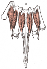
Iliocostalis: Origin, Insertion, Action, Nerve, and Artery
|
Origin: Sacrum, iliac crest, and lubmar spinous processes
Insertion: Ribs Action: Laterally flex, extend, and rotate head (to same side) and vertebral column Nerve: Dorsal rami of spinal nerves: |
|
|
Longissimus: Origin, Insertion, Action, and Nerve
|
Origin: Sacrum, Iliac crest, and lumbar spinous processes
Insertion: Thoracic and cervical spinous processes, & mastoid processe Action: Laterally flex, extend, and rotate head (to same side) and vertebral column Nerve: Dorsal rami of spinal nerves: |
|
|
Spinalis: Origin, Insertion, Action, and Nerve
|
Origin: Sacrum, Iliac crest, and lumbar spinous processes
Insertion: Spinous process of Thoracic spine Action: Laterally flex, extend, and rotate head (to same side) and vertebral column Nerve: Dorsal rami of spinal nerves: |
|
|
Semispinalis Capitus: Origin, Insertion, Action, and Nerve
|
Origin: Transverse process of T1-T6
Insertion: Nuchal ridge Action: Extends head Nerve: Dorsal primary rami |
|
|
Multifidus: Origin, Insertion, Action, and Nerve
|
Origin: Transverse procsseses of C2-S4 spine
Insertion: Spinous processess of C2-S4 spine (same number as origin) Action: Flex spine laterally, and rotates to the opposite side Nerve: Dorsal primary rami |
|
|
Rotatores: Origin, Insertion, Action, and Nerve
|
Origin: Transvese processes of spine
Insertion: Spinous processes (+1 from origin) Action: Rotates superior vertebra to the opposite direct Nerve: Dorsal primary rami |
|
|
Levator costarum: Origin, Insertion, Action, and Nerve,
|
Origin: Transverse process
Insertion: Brevis: rib-1, Longus: rib-2, Action: Elevates rib during inspiration Nerve: Dorsal primary rami |
|
|
Interspinales: Origin, Insertion, Action, and Nerve
|
Origin: Spinous process
Insertion: Spinous process + 1 Action: Extend column Nerve: Dorsal primary rami |
|
|
Intertransversarii: Origin, Insertion, Action, and Nerve
|
Origin: Transverse process
Insertion: Transverse process + 1 Action: Laterally flex spinal column Nerve: Dorsal primary rami |
|
|
Supraspinatus: Origin, Insertion, Action, Nerve, and Artery
|

Origin: Supraspinatus fossa of the scapula
Insertion: Superior facet of greater tuberosity of humerus and capsule of the shoulder joint Action: Assists deltoid in abducting arm, fixes head of humerus in glenoid cavity, rotates head of humerus laterally Nerve: Suprascapular n Artery: Suprascapular (transverse scapular) Trapped in impingement, #1 torn rotator cuff tendon |
|
|
Infraspinatus: Origin, Insertion, Action, Nerve, and Artery
|

Origin: Infraspinous fossa of scapula
Insertion: Middle facet of greater tuberosity of humerus Action: Externally rotates head of humerus with teres minor Nerve: Suprascapular Artery: Suprascapular (transverse scapular), scapular circumflex A weak external rotator, cuff tear or suscapular n. lesion in notch |
|
|
Teres minor: Origin, Insertion, Action, Nerve, and Artery
|

Origin: Dorsal surface of axillary border of scapula (inferior to origin of supraspinatus)
Insertion: Lower facet of greater tuberosity of humerus, capsle of shoulder joint, (inferior to the insertion of infraspinatus) Action: Adducts, externally rotates the head of humerus, and draws humerus towards glenoid fossa Nerve: Axillary n (specifically posterior branch of the axially) Artery: Scapular circumflex (a branch of the subscapular artery) Rarely torn rotator cuff tendon |
|
|
Subscapularis: Origin, Insertion, Action, Nerve, and Artery
|

Origin: Subscapular fossa of scapula
Insertion: Lesser tuberosity of humerus and capsule of the shoulder joint Action: Internally rotates and adducts humerus Nerve: Upper and lower subscapular Artery: Lateral thoracic (aka LIMA, branch of axillary artery), subscapular (branc of axillary artery) Test with lift off, lift off lag, and belly press tests (test internal rotation of humerus) at risk during anterior approach |
|
|
Deltoid: Origin, Insertion, Action, Nerve, and Artery
|

Origin: Lateral thrid of clavicle, upper surace of acromion, spine of scapula
Insertion: Deltoid tuberosity of humerus Action: Abducts arm Nerve: Axillary Artery: Posterior humeral circumflex (branch of axillary), deltoid branch of thoracoacromial artery |
|
|
Teres major: Origin, Insertion, Action, Nerve, and Artery
|
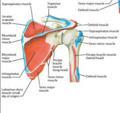
Origin: Dorsal surface of inferior angle of scapula
Insertion: Medial lip of bicipital groove of humerus Action: Adducts and medially rotates humerus and draws it back Nerve: Lower subscapular (C5-6) Artery: Scapular circumflex (branch of subscapular artery) |
|
|
Pectoralis major: Origin, Insertion, Action, Nerve, and Artery
|

Origin: Sternal half of clavicle, sternum to 7th rib, cartilages of true ribs, aponeurosis of external obliques
Insertion: Lateral lip of bicipital groove of humerus Action: Adducts arm, draws it forward, rotates it medially Nerve: Medial (C8-T1) and lateral (C5-C7) pectoral n Artery: Pectoral branch of thoracoacromial, perforating braches of internal thoracic Can rupture during weight lifting |
|
|
Pectoralis minor: Origin, Insertion, Action, Nerve, and Artery
|

Origin: Outer surface of upper margin of ribs 3-5
Insertion: Coracoid process of scapula Action: Lowers lateral angle of scapula, pulls shoulder forward, stablizes scapula Nerve: Medial pectoral n (C8-T1) Artery: Thoracoacromial and intercostal bracnes of internal thoracic, lateral thoracic (off axillary) |
|
|
Serratus Anterior: Origin, Insertion, Action, Nerve, and Artery
|
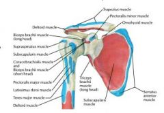
Origin: Lateral surace of Ribs 1-8
Insertion: Anteromedial border of scapula Action: Abducts scapula, raises ribs when scapula is fixed, (holds scapula to chest wall) Nerve: Long thoracic (C5-7) Artery: Lateral thoracic (off axillary artery) |
|
|
Subclavius: Origin, Insertion, Action, Nerve, and Artery
|
Origin: Upper border of 1st rib and its cartilage
Insertion: Groove on under surface of clavicle Action: Draws clavicle down and forward Nerve: Nerve to subclavius (c5 + 6) Artery: Clavicular branch of throracoacromial |
|
|
Corachobrachialis: Origin, Insertion, Action, Nerve, and Artery
|

Origin: Coracoid Process of Scapula
Insertion:Middle of medial border of humerus Action: Flexion and adduction of the arm Nerve: Musculocutaneous (C5-7, pierces coracobrachialis) Artery: Muscular branches of the brachial artery Part of the "conjoined tendon" |
|
|
Brachialis: Origin, Insertion, Action, Nerve, and Artery
|

Origin: Lower two thirds of anterior surface of the humerus
Insertion: Coronoid process and ulnar tuberosity (both on ulna) Action: Flexes forearm Nerve: Medial: Musculocutaneous (c5-c7), Lateral: Radial (C5-T1) Artery: Radial recurrent (branch of radial artery immediately below elbow), brachial (continuation of axillary after teres major) |
|
|
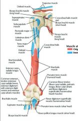
Biceps Brachii: Origin, Insertion, Action, Nerve, and Artery
|

Origin:
Short Head: coracoid process Long Head: supraglenoid tuberosity of scapula Insertion: Short head: Radial tuberosity Long head: Radial tuberosity Action: Supinate and flex forearm Nerve: Musculocutaneous (c5-7) Artery: Muscular branches of brachial |
|
|
Triceps Brachii: Origin, Insertion, Action, Nerve, and Artery
|
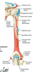
Origin:
Long Head: Infraglenoid tuberosity of scapula Lateral Head: Posteior and lateral surface of the proximal humerus Medial Head: Lower posterior surface of humerus Insertion: Olecranon Process Action: Extends forearmn, if arm is abducted, long head aids in abducting it Nerve: Radial nerve (c5-t1) Artery: Branch of profunda brachii Long head is the border of quadrangular and traiangular space and intreval Lateral head is a border in the lateral approach |
|
|
Conjoined tendon
|
Coracobrachialis and Short head of biceps brachii
|
|
|
Pronator Teres: Origin, Insertion, Action, Nerve, and Artery
|

Origin:
Humeral Head: from medial epicondylar ridge of humerus and common flexor tendon Ulnar (deep) Head: Medial side of coronoid process of ulna Insertion: Middle of lateral surface of radius Action:Pronates forearm, assists in flexing forearm Nerve: Median n (c5-t1) Artery: Anterior ulnar recurrent Can compress median nerve in pronator syndrome |
|
|
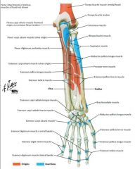
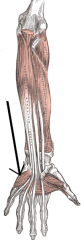
Flexor Carpi Radialis: Origin, Insertion, Action, Nerve, and Artery
|

Origin: Common flexor tendon from medial epicondyle of humerus, fascia of forearm
Insertion: base of 2nd and 3rd metacarpal bones Action: Flexes wrist, radial deviation (abducting) Nerve: Median Artery: Muscular branches of radial Radial artery is immediately lateral |
|
|
Palmaris Longus: Origin, Insertion, Action, Nerve, and Artery
|

Origin: Common flexor tendon from medial epicondyle of humerus
Insertion: Transverse carpal ligament and pamar aponeurosis Action: Flex wrist, assists in pronation and flexion of forearm Nerve: Median (c5-t1) Artery: Posterior ulnar recurrent |
|
|
Flexor Carpi Ulnaris: Origin, Insertion, Action, Nerve, and Artery
|

Origin:
Humeral Head: From common flexor tendon from medail epicondyle of humerus Ulnar Head: from olecranon and dorsal border of ulna Insertion: Pisiform, hook of hamate, 5th metacarpal bones Action: Flexes wrist and assists in adducting it, assists in flexing forearm Nerve: Ulnar (c8-t1) Artery: Posterior ulnar recurrent Most powerful wrist flexor. May compress ulnar nerve. |
|
|
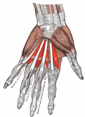
Flexor digitorum superficialis: Origin, Insertion, Action, Nerve, and Artery
|

Origin:
Humeral head: from common flexor tendon of medial epicondyle of humerus Ulnar head: from coronoid Process of ulna Radial Head: from oblique line of radius Insertion: Middle phalanges of medial 4 digits (no thumb!) Action: Flexes middle and proximal phalanx of medial 4 digits (flexes PIPJ) Nerve: Median (c5-t1) Artery: Muscular branches of ulnar, muscular branches of radial often considered a "middle flexor" because of its position between muscles |
|
|
Flexor digitorum profundus (FDP): Origin, Insertion, Action, Nerve, and Artery
|

Origin: Medial and anterior surface of ulna, interosseous membrane, deep fascia of forearm
Insertion: Distal phalanges of medial 4 digits Action: Flexes terminal phalanges of medial 4 digits, aids in flexing wrist Nerve: Anterior interosseus n ( a branch of the median nerve!) for index and middle finger, Ulnar n for ring and pinky Artery: Anterior interosseous of ulnar, muscular branches of ulnar use ok sign to test AIN (innervates the Flexor pollicis longus, Flexor digitorum profundus I & II, and the pronator quadratus) |
|
|
Flexor pollicis longus (FPL): Origin, Insertion, Action, Nerve, and Artery
|

Origin: Volar surface of radius, adjacent interosseous membrane, medial border of coronoid process of ulna
Insertion: Base of distal phalanx of thumb on palmar surface Action: Flexes thumb Nerve: Anterior interosseous n of median Artery: Anterior interosseous of ulnar |
|
|
Pronator Quadratus: Origin, Insertion, Action, Nerve, and Artery
|

Origin: Distal fourth of volar surface of ulna
Insertion: Distal fourth of lateral border on volar surface of radius Action: Pronates forearm Nerve: Anterior interosseous nerve (off of median) Artery: Volar interosseous of ulnar |
|
|
What innervates the three deep flexors of the hand and how is it tested
|

the Anterior Interosseous Nerve, tested by making the "OK" sign
|
|
|
Anconeous: Origin, Insertion, Action, Nerve, and Artery
|

Origin: Lateral epicondyle of humerus, posterior ligament of elbow joint
Insertion: Lateral side of olecranon and posterior surface of ulna Action: Assists tricep in extending forearm Nerve: Radial (c5-t1) Artery: Branch of profunda brachii |
|
|
Extensor digitorum communis (EDC): Origin, Insertion, Action, and Nerve
|

aka extensor digitorum
Origin: Common extensor origin on the Lateral epicondyle Insertion: MCP: Sag band, P2: Central slip, P3: terminal insert Action: Digit extension Nerve: Posterior interosseous nerve (of Radial) |
|
|
Extensor digit minimi: Origin, Insertion, Action, Nerve, and Artery
|

Origin: Common extensor tendon from lateral epicondyle of the humerus, intermuscular septa
Insertion: Dorsum of proximal phalanx of 5th digit Action:Extends 5th digit Nerve: Posterior interosseous of radial Artery: Posterior interosseous of ulnar |
|
|
Extensor Carpi Ulnaris: Origin, Insertion, Action, Nerve, and Artery
|
Origin: Freem common extensor tendon from lateral epicondyle of humerus and from posterior border of ulna
Insertion: Medial side of base of 5th metacarpal bone Action: Extends wrist, adducts hand Nerve: Posterior interosseus of radial Artery: Posterior interosseous of ulnar |
|
|
Brachioradialis: Origin, Insertion, Action, Nerve, and Artery
|

Origin: Proximal two thirds of lateral supracondylar ridge of humerus, lateral IM seputm
Insertion: Lateral side of base of styloid process of radius Action: Flexes forearm after flexion has been started by biceps and brachialis, may also act as a semipronator and semisupinator Nerve: Radial (C5-T1) Artery: Radial recurrent |
|
|
Extensor carpi radialis longus: Origin, Insertion, Action, Nerve, and Artery
|

Origin: Lateral third of lateral supracondylar ridge of humerus, lateral IM septum
Insertion: Dorsal surface of base of 2nd metacarpal bone Action: Extend wrist, adbucts hand Nerve: Radial (c5-t1) Artery: Muscular branches of radial, radial recurrent |
|
|
Extensor carpi radialis brevis: Origin, Insertion, Action, Nerve, and Artery
|

Origin: From common extensor from lateral condyle of humerus, radial collateral ligament of elbow joint, intermuscular septa
Insertion: Dorsal surface of base of 3rd metacarpal bone Action: Extends wrist, abducts hand Nerve: Posterior interosseous nerve of radial Artery: Muscular branches of radial, radial recurrent |
|
|
Supinator: Origin, Insertion, Action, Nerve, and Artery
|

Origin: Lateral epicondyle of humerus, lateral ligament of elbow joint and annular ligament of radius, supinator crest and fossa of ulna
Insertion: Latera and anterior surface of upper thrid of radius Action: supinates forearm Nerve:Deep branch of radial Artery: radial recurrent, and posterior interosseous of ulnar |
|
|
Abductor pollicis longus: Origin, Insertion, Action, Nerve, and Artery
|
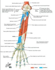
Origin: posterior surface of ulna, interossesous membrane, middle third of posterior surface of radius
Insertion: radial side of base of 1st metacarpal bone Action: Abducts and extends thumb and wrist (CMCJ) Nerve: Posterior interosseous of radial Artery: Posterior interosseous of ulnar |
|
|
Extensor pollicis longus: Origin, Insertion, Action, Nerve, and Artery
|

Origin: Middle third of posterior surface of ulna, interosseous membrane
Insertion: Base of distal phalanx of thumb Action: Extends terminal phalanx of thumb (IPJ) Nerve: Posterior interosseous of radial Artery: Posterior interosseous of ulnar tendon turns 45 degrees on Lister's tubercle |
|
|
Extensor polllicis brevis: Origin, Insertion, Action, Nerve, and Artery
|

Origin: Posterior surface of radius, interosseous membrane
Insertion: Base of proxima phalanx of thumb Action: Extends proximal phalanx of thumb (MCPJ) Nerve: Posterior interosseous of radial Artery: Posterior interosseous of ulnar radial border of snuffbox |
|
|
Extensor indicis proprius: Origin, Insertion, Action, Nerve, and Artery
|

Origin: Posterior surface of ulna, interosseous membrane
Insertion: Dorsum of proximal phalanx of index finger Action: Extends proximal phalanx of index finger Nerve: Posterior interosseous of radial Artery: Posterior interosseous of ulnar |
|
|
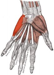
Abductor pollicis brevis: Origin, Insertion, Action, Nerve, and Artery
|

Origin: Transverse carpa ligament, scaphoid and trapezium
Insertion: Radial side of base of proximal phalanx of thumb Action: palmar pronation, abducts thumb, draws thumb at right anges to thumb. Primary muscle in opposition Nerve: Median (muscular branches) Artery: Superficial volar branch of radial |
|
|
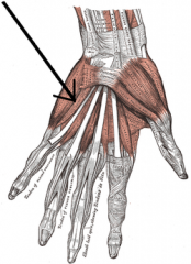
Flexor pollicis brevis: Origin, Insertion, Action, Nerve, and Artery
|
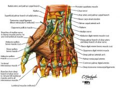
Origin:
1. Superficial head - transverse carpal ligament 2. Deep head - trapezium Insertion: 1. Superficial head - base of thumb 2. Proximal phalanx Action: Thumb MPC flexion Nerve: dual innervation 1. Superficial head: Median 2. Deep head: deep branch of ulnar nerve Artery: Superficial volar branch of radial |
|
|
Opponens pollicis: Origin, Insertion, Action, Nerve, and Artery
|

Origin: Transverse carpal ligament, trapezium
Insertion: Anterior surface of radial side of 1st metacarpal bone Action: Oppose (flex / abduct) thumb Nerve: Muscular branch of median Artery: Superficial volar branch of radial pronates / stabilizes thumb MC |
|
|
Adductor pollicis: Origin, Insertion, Action, Nerve, and Artery
|
Origin:
Oblique head: from trapezium, trapezoid, and capitate as well as base of 2nd and 3rd metacarpals Transverse head: from volar surface of 3rd metacarpal Insertion: Ulnar side of proximal phalanx of thumb Action: Adducts thumb, aids in opposition Nerve: Deep volar branch of ulnar Artery:Deep volar arch Test function with froment's test |
|
|
Palmaris brevis: Origin, Insertion, Action, Nerve, and Artery
|
Origin: Ulnar side of transverse carpal ligament, palmar aponeurosis
Insertion: Skin of ulnar border (medial) palm Action: Wrinkles skin on ulnar side of palm, deeps the hollow of the hand Nerve: Superficial volar branch of ulnar Artery: Superficial volar arch |
|
|
Abductor digiti minimi: Origin, Insertion, Action, Nerve, and Artery
|
Origin: Pisifiorm bone, tendon of flexor carpi ulnaris
Insertion: Medial side of base of proximal phalanx of pinky and aponeurosis of extensor digiti minimi Action: Abducts 5th digit (pinky) from 4th digit Nerve: Deep volar branch of ulnar Artery: Deep volar branch of ulnar, dorsal carpal branch of ulnar |
|
|
Flexor Digiti Minimi brevis: Origin, Insertion, Action, Nerve, and Artery
|
Origin: Transverse carpal ligament, hamulus of hamate bone
Insertion: ulnar side of base of proximal phalanx of pinky Action: Flexes proximal phalanx of 5th digit (flexes MCP of pinky) Nerve: Deep volar branch of ulnar Artery: Deep volar branch of ulnar, dorsal carpal branch of ulnar |
|
|
Opponens digiti minimi: Origin, Insertion, Action, Nerve, and Artery
|
Origin: Transverse carpal ligament, hamulus of hamate bone
Insertion: Ulnar margin of 5 metacarpal bone Action: Draws 5th metacarpal bone forward to face thumb, deeps hollow of hand Nerve: Deep volar branch of ulnar Artery: Deep volar branch of ulnar, dorsal carpal branch of ulnar |
|
|
Lumbricals 1&2: Origin, Insertion, Action, Nerve, and Artery
|
Origin: Arise from the corresponding tendons of flexor digitorum profundus.
Insertion: With tendons of extensor digotorum and interossei into bases of terminal phalanges Action: Flexes fingers at MCP, extends fingers and interphalangeal joints Nerve: Median Artery: Superficial and deep volar arches only muscle to insert on their own antagonist (FDP) |
|
|
Lumbricals 3&4: Origin, Insertion, Action, Nerve, and Artery
|

Origin: Arise from the corresponding tendons of flexor digitorum profundus.
Insertion: With tendons of extensor digotorum and interossei into bases of terminal phalanges Action: Flexes fingers at MCP, extends fingers and interphalangeal joints Nerve: Ulnar Artery: Superficial and deep volar arches only muscle to insert on their own antagonist (FDP) |
|
|
Interosseous dorsal: Origin, Insertion, Action, Nerve, and Artery
|
Origin: Adjacent metacarpals
Insertion: Proximal phalanx and extensor expansion (lateral bands). Bipennate, each belly has separate insertion Action: Digit abduction and MCP flexion Nerve: Deep volar branch of ulnar Artery: Deep volar arch DAB PAD: dorsal abduct, palmar adduct |
|
|
Interossei palmar: Origin, Insertion, Action, Nerve, and Artery
|
Origin: Adjacent metacarpals
Insertion: Extensor expansion (lateral bands) Action: Digit adduction Nerve: Deep volar branch of Ulnar Artery: Deep volar arch Unipennate, PAD |
|
|
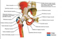
Psoas Major: Origin, Insertion, Action, Nerve, and Artery
|

Origin: Transverse processes and bodies of T12-L5 vertebra
Insertion: Lesser trochanter of femur (with Iliacus forms iliopsoas) Action: Flexes hip Nerve: Femoral Artery: Lumbar branch of iliolumbar |
|
|
Psoas minor: Origin, Insertion, Action, Nerve, and Artery
|

Origin: Vertebral margins of T12-L1 vertabrae
Insertion: Pectineal line, iliopectineal eminence Action: Assists in hip flexion (weak, present in ~50% of people) Nerve: L1 ventral ramus Artery: Lumbar branch of iliolumbar |
|
|
Iliacus: Origin, Insertion, Action, Nerve, and Artery
|

Origin: Iliac fossa / sacral ala
Insertion: Lesser trochanter, capsule of hip joint, body of femur (with psoas major forms iliopsoas) Action: Flexes hip Nerve: muscular branches of Femoral n Artery: Iliac branch of iliolumbar |
|
|
Tensor fascia latae: Origin, Insertion, Action, Nerve, and Artery
|

Origin: Anterior part of outer lip of iliac crest, anterior border of ilium
Insertion: Middle third of thigh along iliotibial tract Action: Assists in flexing, abducting, and internally rotating the hip; counteracts backward pull of gluteus maximus Nerve: Superior gluteal n Artery: Superior gluteal artery, lateral femoral circumflex artery |
|
|
Gluteus medius: Origin, Insertion, Action, Nerve, and Artery
|

Origin: Outerial surface of ilium, between anterior and posterior gluteal lines
Insertion: Posterior of greater trochanter Action: Abducts and internally rotates thigh Nerve: Superior gluteal Artery: Deep branch of superior gluteal Trendelenburg gait if muscle is out |
|
|
Gluteus minimus: Origin, Insertion, Action, Nerve, and Artery
|

Origin: Outer surface of ilium between anterioir and inferior gluteal lines
Insertion: Anterior border of greater trochanter of femur Action: Abducts and internally rotates the thigh Nerve: Superior gluteal nerve Artery: Deep branch of superior gltueal |
|
|
Gluteus maximus: Origin, Insertion, Action, Nerve, and Artery
|
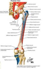
Origin: Posterior gluteal line, tendon of sacrospinalis, dorsal surface of sacrum and coccyx, sacrotuberous ligament
Insertion: Gluteal tuberosity of femur, iliotibial tract Action: Extends and externally rotates thigh Nerve: Inferior gluteal n Artery: Superior gluteal, inferior gluteal, 1st perforating branch of profunda femoris Must be split in posterior approach to the hip |
|
|
Obturator externus: Origin, Insertion, Action, Nerve, and Artery
|

Origin: Outer margin of obturator foramen, outer surface of obturator membrane
Insertion: Trochanteric fossa of femur Action: Adducts and externally rotates thigh Nerve: Posterior branch of obturator n Artery: Obturator, medial femoral circumflex Inserts at start point of IM nail |
|
|
Piriformis: Origin, Insertion, Action, Nerve, and Artery
|

Origin: Anterior sacrum
Insertion: Superior border of greater trochanter Action: Externally rotates thigh, abducts thigh when limb is flexed Nerve: N. to piriformis Artery: Superior gluteal, inferior gluteal, internal pudendal |
|
|
Superior gemellus: Origin, Insertion, Action, Nerve, and Artery
|

Origin: Outer surface of ischial spine
Insertion: Medial surface of greater trochanter, blends with obturator internus tendon Action: Externally rotates thigh Nerve: N. to obturator internus Artery: Inferior gluteal |
|
|
Obturator internus: Origin, Insertion, Action, Nerve, and Artery
|
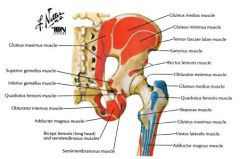
Origin: Ischiopubic rami, obturator membrane
Insertion: Medial greater trochanter Action: Adducts and externally rotates the hip Nerve: N. to obturator internus Artery: Muscular branches of internal pudendal, superior gluteal Exits through lesser sciatic foramen |
|
|
Inferior gemellus: Origin, Insertion, Action, Nerve, and Artery
|

Origin: Upper part of ischial tuberosity
Insertion: Medial surface of greater trochanter, blends with obturator internus tendon Action: Externally rotates thigh Nerve: N. to quadratus femoris Artery: Inferior gluteal |
|
|
Quadratus femoris: Origin, Insertion, Action, Nerve, and Artery
|

Origin: Lateral margin of ischial tuberosity
Insertion: Quadrate tubercle of femur, linea quadrata Action: Adducts and externally rotates thigh Nerve: N. to quadratus femoris Artery: Medial femoral circumflex |
|
|
Articularis genus: Origin, Insertion, Action, Nerve, and Artery
|

Origin: Distal anterior femoral shaft (arises deep to V. intermedius)
Insertion: Synovial capsule Action: Pulls capsule of knee joint superiorly when in extension Nerve: Femoral Artery: Lateral femoral circumflex |
|
|
Sartorious: Origin, Insertion, Action, Nerve, and Artery
|

Origin: Anterior superior iliac spine, upper half of iliac notch
Insertion: Proximal medial tibia (pes anserinus) Action: Flexes and externally rotates hip Nerve: Muscular branches of femoral (L2-L4) Artery: Muscular branches of femoral can avulse from ASIS |
|
|
Rectus Femoris: Origin, Insertion, Action, Nerve, and Artery
|

Origin:
Straight head: Anterior Inferior Iliac Spine Reflected head: Superior acetabular rim Insertion: Upper border of patella, by ligamentum patellae into tibial tuberosity Action: Extends leg, flexes thigh Nerve: Femoral Artery: Lateral femoral circumflex can avulse from AIIS |
|
|
Vastus lateralis: Origin, Insertion, Action, Nerve, and Artery
|

Origin: Greater trochanter, lateral linea aspera
Insertion: Lateral border of patella, lateral tibial tuberosity Action: Extends leg Nerve: Muscular branches of femoral (L2-L4) Artery: Lateral femoral circumflex Oblique fibers can affect Q angle |
|
|
Vastus intermedius: Origin, Insertion, Action, Nerve, and Artery
|
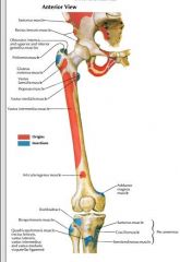
Origin: Proximal femoral shaft
Action: Patella / tibial tubercle Nerve: Muscular branches of femoral Artery: Lateral femoral circumflex Covers articularis genus |
|
|
Vastus medialis: Origin, Insertion, Action, Nerve, and Artery
|

Origin: Intertrochanteric line, medial linea aspera
Insertion: medial border of patella, tibial tubercle Action: Extends leg and draws patella medially Nerve: Muscular branches of femoral Artery: Muscular branches of femoral, muscular branches of profunda femoris, genicular branches of popliteal weak in many patellofemoral disorders |
|
|
Obturator externus: Origin, Insertion, Action, Nerve, and Artery
|

Origin: Outer margin of obturator foramen, outer surface of obturator membrane
Insertion: Trochanteric fossa of femur Action: Adducts thigh, externally rotates thigh Nerve: Posterior branch of obturator Artery: Obturator, medial femoral circumflex Insertion at start point of IM nail |
|
|
Adductor longus: Origin, Insertion, Action, Nerve, and Artery
|

Origin: Body of pubic pubis (inferior), in angle between between crest and symphysis
Insertion: Middle half of medial lip of linea aspera Action: Adducts thigh and assist in flexing it Nerve: Anterior branch of obturator Artery: Medial femoral circumflex, obturator Tendon can ossify |
|
|
Adductor brevis: Origin, Insertion, Action, Nerve, and Artery
|

Origin: Outer surface of inferior surface of pubic ramus
Insertion: Line extending from lesser trochanter to linea aspera Action: Adducts thigh and assists in flexing it Nerve: Anterior branch of obturator Artery: Medial femoral circumflex, obturator a Deep to pectineus |
|
|
Adductor magnus: Origin, Insertion, Action, Nerve, and Artery
|

Origin:
1. Pubic ramus 2. Ischial tuberosity Insertion: Linea aspera, adductor tubercle Action: Adducts and flex/extend thigh Nerve: 1. Obturator 2. Sciatic Artery: Medial femoral circumflex, perforating branches of profunda femoris, muscular branches of popliteal Muscle has two separate parts |
|
|
Gracilis: Origin, Insertion, Action, Nerve, and Artery
|
Origin: Lower half of pubic symphysis, upper half of pubic arch
Insertion: Upper part of medial surface of tibia Action: Flexes and medially rotates leg, adducts thigh Nerve: Anterior branch of obturator Artery: Muscular branches of profunda femoris, obturator, medial femoral circumflex Used in ligament reconstruction |
|
|
Pectineus: Origin, Insertion, Action, Nerve, and Artery
|

Origin: Pectineal line of pubis
Insertion: Pectineal line of femur Action: Adducts, flexes, and internally rotates thigh Nerve: Muscular branches of femoral Artery: Medial femoral circumflex, obturator |
|
|
Semitendinosus: Origin, Insertion, Action, Nerve, and Artery
|

Origin: Ischial tuberosity with tendon of biceps
Insertion: Proximal medial tibia (pes anserinus) Action: Flexes leg, extends thigh, internally rotates leg when knee is semi-flexed Nerve: Sciatic (tibial) Artery: Perforating branches of profunda femoris, superior muscular branches of popliteal |
|
|
Semimembranosus: Origin, Insertion, Action, Nerve, and Artery
|

Origin: Upper and lateral facet of ischial tuberosity
Insertion: Medial posterior surface of medial condyle of tibia Action: Flexes leg, extends thigh, internally rotates leg when knee is semiflexed Nerve: Sciatic Artery: Perforating branches of profunda femoris, superior muscular branches of popliteal |
|
|
Bicep Femoris: Origin, Insertion, Action, Nerve, and Artery
|

Origin:
Long head: ischial tuberosity Short head: Linea aspera / supracondylar line Insertion: Long head: Head of fibula Short head: Fibula, lateral tibia Action: Extends thigh, flexes leg Nerve: Long head: sciatic (tibial) Short head: sciatic (peroneal) Artery: Perforating branches of profunda femoris, superior muscular branches of popliteal |
|
|
Tibialis Anterior: Origin, Insertion, Action, Nerve, and Artery
|
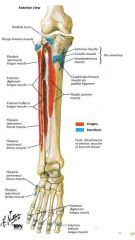
Origin: Lateral condyle of tibia, upper two-thirds of lateral surface of tibia, interosseous membrane, lateral IM septum
Insertion: Medial and plantar surface of medial cuneiform bone, base of 1st metatarsal bone Action: Dorsiflexes and inverts foot Nerve: Deep peroneal Artery: Muscular branches of anterior tibial Tests L4 motor function |
|
|
Extensor hallucis longus: Origin, Insertion, Action, Nerve, and Artery
|

Origin: Middle half of anterior surface of fibula, adjacent interosseous membrane
Insertion: Base of distal phalanx of great toe Action: Extends great toe, continued action dorsiflexes foot Nerve: Deep peroneal Artery: Muscular branches of anterior tibial Tests L5 motor function |
|
|
Extensor digitorum longus: Origin, Insertion, Action, Nerve, and Artery
|

Origin: Lateral condyle of tibia, upper three fourths of anterior fibula, interosseous membrane, IM septa
Insertion: dorsal surface of middle and distal phalanges of lateral 4 toes Action: Extends phalanges of lateral 4 toes, continued action dorsiflexes foot Nerve: Deep peroneal Artery: Muscular branches of anterior tibial Sindle tendon divides into 4 tendons |
|
|
Peroneus tertius: Origin, Insertion, Action, Nerve, and Artery
|

Origin: Lower anterior surface of fibula, adjacent interosseous membrane
Insertion: Dorsal surface of base of 5th metatarsal bone Action: Dorsiflexes and everts foot Nerve: Deep peroneal n. Artery: Muscular branches of anterior tibial commonly know as the 5th tendon of extensor digitorum longus. Often adjoined to the EDL. |
|
|
Peroneus longus: Origin, Insertion, Action, Nerve, and Artery
|

Origin: Lateral condyle of tibia, head and upper two-thirds of lateral surface of fibula, IM septa
Insertion: Lateral side of medial cuneiform bone, base of 1st metatarsal bone Action: Plantarflexes and everts foot Nerve: Superficial peroneal Artery: Muscular branches of anterior tibial, muscular branches of peroneal Tests S1 motor function, runs under the foot |
|
|
Peroneus brevis: Origin, Insertion, Action, Nerve, and Artery
|

Origin: Lateral two-thirds of lateral surface of tibia
Insertion: Lateral side of base of 5th metatarsal bone Action: Plantarflexes and everts foot Nerve: Superficial peroneal Artery: Muscular branches of peroneal Can cause avulsion fx at base of 5th metatarsal; has most distal muscle belly |
|
|
Gastrocnemius: Origin, Insertion, Action, Nerve, and Artery
|

Origin: Lateral (lateral head) and medial femoral (medial head) condyles
Insertion: Into calcaneus via achilles tendon Action: Plantarflexes foot, Nerve: Tibial Artery: Sural branches of popliteal Tests S1 motor function, fabella is tendon of lateral head |
|
|
Soleus: Origin, Insertion, Action, Nerve, and Artery
|
Origin: Posterior fibular head/soleal line of tibia
Insertion: Into calcaneus by achilles tendon Action: Plantar flexes foot Nerve: Tibial Artery: Posterior tibial, peroneal, sural branches of popliteal |
|
|
Plantaris: Origin, Insertion, Action, Nerve, and Artery
|

Origin: Lateral supracondylar line of femur, oblique popliteal ligament of knee joint
Insertion: Medial side of posterior part of calcaneus via achilles tendon Action: Plantarflexes foot (weak) Nerve: Tibial Artery: Sural branches of popliteal Long tendon can be harvested for tendon reconstruction |
|
|
Popliteus: Origin, Insertion, Action, Nerve, and Artery
|

Origin: Lateral condyle of femur, oblique popliteal ligament of knee
Insertion: Triangular area on posterior surface of tibia above soleal line Action: Flexes leg, internally rotates tibia/knee in beginning of flexion (i.e. during swing phase) Nerve: Tibial Origin is inraarticular; primary restratint to external rotation of the knee Artery: Genicular branches of popliteal |
|
|
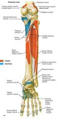
Flexor hallicus longus: Origin, Insertion, Action, Nerve, and Artery
|

Origin: Lower two-thirds of posterior tibia, interosseous membrane and IM septum
Insertion: base of distal phalanx of great toe Action: Plantarflexes great toe, continued action aids in plantarflexing toe Nerve: Tibial Artery: Muscular branches of peroneal Tests S1 motor function |
|
|
Flexor digotorum longs: Origin, Insertion, Action, Nerve, and Artery
|

Origin: Posterior surface of middle three fifths of tibia, fascia covering tibialis posterior
Insertion: Plantar surface of base of distal phalanx of lateral four toes Action: Plantarflexes lateral 4 toes Nerve: Tibial Artery: Posterior tibial At ankle, tendon is just anterior to tibial artery |
|
|
Tibialis posterior: Origin, Insertion, Action, Nerve, and Artery
|

Origin: Posterior tibia, fibula, interosseous membrane
Insertion: Tuberosity of navicular bone, plantar surface of all cuneiform bones, plantar surface of 2-4 metatarsal bones, cuboid bone, sustentaculum tali Action: Plantarflexes and inverts foot Nerve: Tibial Artery: Peroneal Tendon rupture / degeneration can cause acquired flat foot |
|
|
Abductor Hallucis: Origin, Insertion, Action, Nerve, and Artery
|
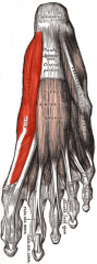
Origin: Medial process of calcaneus, calcaneal tuberosity, laciniate ligament, platnar aponeurosis
Insertion: Medial side of base of proximal phalanx of great toe Action: Abducts great toe Nerve: Medial plantar toe Artery: Medial plantar toe Fascia can entrap nerve to ADM |
|
|
Flexor digitorum brevis: Origin, Insertion, Action, Nerve, and Artery
|
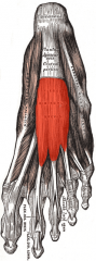
Origin: Medial process of calcaneus, plantear aponeurosis, adjacent IM septa
Insertion: middle phalanx of lateral 4 toes Action: flexes lateral 4 toes, Nerve: Medial plantar Artery: Medial plantar supports longtitudinal arch |
|
|
Abductor digiti minimi: Origin, Insertion, Action, Nerve, and Artery
|
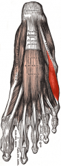
Origin: Lateral and medial process of calcaneus, calcaneal fascia, adjacent IM septum
Insertion: Latearl side of base of proximal phalanx of pinky toe Action: Abducts pinky toe, assists in flexing it Nerve: Lateral plantar Artery: Lateral plantar nerve can be entrapped by abduction hallucis fascia |
|
|
Quadratus plantae: Origin, Insertion, Action, Nerve, and Artery
|

Origin: Medial head: from medial surface of calcaneus and medial border of long plantar ligament
Lateral border: from lateral border of plantar surface of calcaneus and lateral border of long plantar ligament Insertion: Tendons of flexor digotorum longus Action: Flexes terminal phalanges of lateral 4 toes Nerve: Lateral plantar Artery: Lateral plantar |
|
|
Lumbricals: Origin, Insertion, Action, Nerve, and Artery
|

Origin: arise from tendons of flexor digitorum longus
Insertion: bases of terminal phalanges of 4 lateral toes along with extensor digitorum long and interossei Action: flexes MTP joint and extends IP joint Nerve: 1. medial plantar, 2-4. lateral plantar Artery: Plantar metatarsal |
|
|
Flexor Hallucis Brevis: Origin, Insertion, Action, Nerve, and Artery
|

Origin: Medial part of plantar surface of cuboid bone, adjacent portion of lateral cuneiform bone, prolongation of tendon of tibalis posterior
Insertion: Medial and lateral sides of proximal phalanx of great toe Action: Flexes great toe Nerve: Proper digital nerve of great toe of medial plantar nerve Artery: First plantar metatarsal Sesamoid bones are within the tendons |
|
|
Adductor hallucis: Origin, Insertion, Action, Nerve, and Artery
|
Origin:
Oblique head: from bases of 2-4 metatarsal bones Transverse head: from capsule of 2-5 metatarsophalangeal ligaments Insertion: Lateral side of base of proximal phalanx of sole Action: adducts great toe, assists in flexing it Nerve: Deep branch of lateral plantar Artery: First plantar metatarsal 2 heads have different orientations, contributes to hallux valgus deformity |
|
|
Flexor digit minimi brevis: Origin, Insertion, Action, Nerve, and Artery
|
Origin: Base of 5th metatarsal
Insertion: Base of proximal phalanx of pinky toe Action: Flexes pinky toe Nerve: Lateral plantar Artery: Lateral plantar small, relatively insignificant muscle |
|
|
Plantar interossei: Origin, Insertion, Action, Nerve, and Artery
|
Origin: Medial 3-5 metatarsals
Insertion: medial proximal phalanges of toes 3-5 Action: adducts toes, flex MTPJ; extends LJ, attachment to MT is medial for all 3 Nerve: Lateral plantar Artery: Plantar metatarsal |
|
|
Dorsal interossei: Origin, Insertion, Action, Nerve, and Artery
|
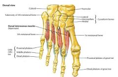
Origin: adjacent MT shafts
Insertion: medial proximal phalanx (2nd toe), Lateral proximal phalanx (toes 2-4) Action: Abducts toes Nerve: Lateral plantar Artery: Dorsal metatarsal Larger than the planter interossei (bipennate) |
|
|
Extensor hallucis brevis: Origin, Insertion, Action, Nerve
|

Origin: Dorsolateral calcaneus
Insertion: bases of proximal phalanx of great toe Action: extends great toe at MCPJ Nerve: deep peroneal assists EHL with its action |
|
|
Extensor digititorum brevis: Origin, Insertion, Action, Nerve, and Artery
|
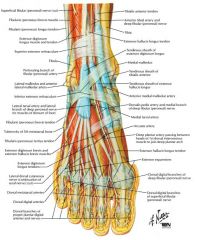
Origin:Dorsolateral calcaneus
Insertion: Base of proximal phalanx of toes 2-4 Action: Extends lesser toes at MCPJ Nerve: deep peroneal no tendon to small toe |