Reading...
![]()
Play button
![]()
Play button
![]()
Use LEFT and RIGHT arrow keys to navigate between flashcards;
Use UP and DOWN arrow keys to flip the card;
H to show hint;
A reads text to speech;
42 Cards in this Set
- Front
- Back
|
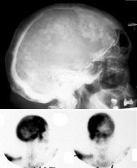
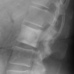
Paget’s Disease
Cotton Wool appearance The osteolytic phase is called osteoporosis circumscripta and appears as multiple geographic, well-demarcated regions of bone resorption that may be mistaken for metastases |
|
|
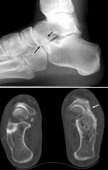
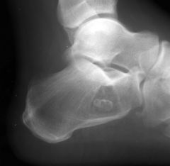
Calcaneonavicular Coalition
Anteater Nose |
|
|
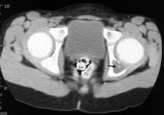
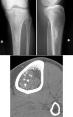
Aneurysmal Bone Cyst
|
|
|
Osteopetrosis
Bone in Bone sign |
|
|
EG
Button Sequestrum Could also be ostomyelitis |
|
|
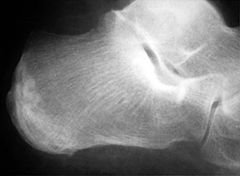
Intraosseous Lipoma
A classic appearance of intraosseous lipoma of the calcaneus is the presence of a well defined lytic lesion with a central calcification resembling a cockade. A cockade is badge, usually in the form of a rosette, or knot, and generally worn upon the hat |
|
|
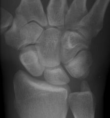
Rotatory subluxation of scaphoid
Signet ring sign |
|
|
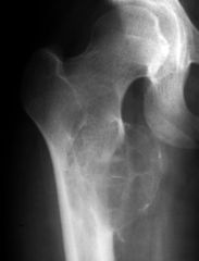
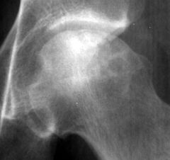
AVN
Air Crescent Sign Fractures that occur in the subchondral bone may be recognized by a crescentic lucent zone that separates the fragment from the remainder of the femur |
|
|
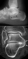
Middle Subtalar Coalition
Lateral radiograph of the foot revealing a classic C sign which is a C-shaped line formed by the medial outline of the talar dome and the inferior outline of the sustentaculum tali |
|
|
Unicameral Bone Cyst
|
|
|
Hemolytic Anemia
Hair on End Appearance Thallasemia vs. SCD The ‘hair’ represents the accentuated trabeculae extending between the inner and outer skull tables in the expanded diploic marrow spaces |
|
|
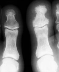
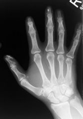
Psoriatic Arthritis
Ivory Phalanx |
|
|
Ivory Vertebra
most common causes of ivory vertebrae are typically metastatic disease, and Paget’s Lymphoma Infection Degenerative Oseoid Osteoma |
|
|

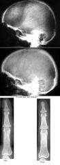
Thanatophoric Dwarf
The is no gas in the lungs. The ribs are very short, as are the limb bones. The rib ends and metaphyses are flared. The femora are curved, like an old telephone handset. The ilia are small and square. |
|
|
CDH
|
|
|
Osteoid Osteoma
|
|
|
Stress Fracture
|
|
|
Multiple Enchondromas
|
|
|
Fibrous Cortical Defect
|
|
|
Chondrosarcoma
|
|
|
Rickets
|
|
|
Scapholunate ligament tear
Terry Thomas sign |
|
|
Osteonecrosis
|
|
|
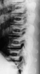
Vertebra Plana
MELT metastasis, multiple myeloma eosinophilic granuloma lymphoma trauma, tuberculosis |
|
|
Aneurysmal Bone Cyst
The list of entities that involve the epiphysis or apophysis is relatively short, and includes: chondroblastoma infection giant cell tumor aneurysmal bone cyst (ABC) enchondroma Geode |
|
|
Rickets
widening of the metaphyses with ragged, "frayed" margins Two main causes of osteomalacia: problems with vitamin D metabolism problems with renal tubular phosphate loss |
|
|
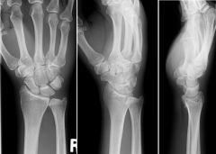
Madelung Deformity
Findings: increased width between the distal radius and ulna. relatively long ulna compared to radius (positive ulnar variance). decreased carpal angle. triangularization of the distal radial epiphysis. wedging of the carpus between the deformed radius and the protruding ulna, with the lunate at the apex of the wedge Post-traumatic extension injuries to radial epiphysis excessive or repetitive loading of an immature joint young gymnasts Dysplastic multiple hereditary exostosis syndrome dyschondrosteosis (a mesomelic variety of dwarfism) onycho-osteodysplasia syndrome (HOOD syndrome: nail-patella syndrome) Genetic Turner's syndrome Idiopathic |
|
|
Paget’s Disease
3 major phases: 1) lytic 2) mixed lytic-sclerotic 3) sclerotic |
|
|
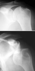
Anterior Instability / Chronic Dislocation
Hill Sachs impaction fracture seen in the humeral head due to anterior dislocation |
|
|
Calcaneal Stress Fracture
|
|
|
GCT of Apophysis
DDx ABC |
|
|
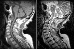
Rheumatoid Arthritis
60-70% of patients with RA develop cervical spine symptoms Erosion of the dens occurs in 14-35% of patients with RA A potentially devastating complication of RA is atlantoaxial subluxation |
|
|
FCD
Fibroxanthoma / NOF / FCD |
|
|
Metastatic RCC
Thyroid mets can look similar DDX ABC / Telangiectatic osteosarcoma GCT Chondroblastoma Brown Tumor |
|

|
Tumoral Calcinosis
Idiopathic Tumoral Calcinosis mass-like calcific deposits about joints usually normal or slightly elevated serum calcium and phosphate ST Calcifications Renal Osteodystrophy amorphous visceral calcifications in heart, lungs, muscle, stomach and kidneys amorphous nonvisceral calcifications in eyes, skin, arteries, and periarticular areas -- these may occasionally be globular and mass-like metastatic calcification if the calcium-phosphate product is elevated chondrocalcinosis if secondary hyperparathyroidism is present Calcinosis universalis Associated with scleroderma or dermatomyositis Calcification is usually in thin, plaque-like and in skin and subcutaneous tissues No large, lobular masses are usually not seen Calcinosis circumscripta 40% are associated with scleroderma, dermatomyositis or Raynaud's Calcifications are thin, and occur in fingertips and "toe-tips" Milk-alkali syndrome Large, calcified periarticular masses History of milk, antacid consumption in huge quantities elevated serum calcium, azotemia, alkalosis Calcification also of lung, kidneys, vessels Hypervitaminosis D Due to toxic level intake (4 - 18 million Units/day) Elevated serum levels of calcium and Vitamin D Metastatic calcification (hyperparathyroidism and chronic renal disease) Serum PTH and calcium levels abnormal Soft tissue calcifications are usually fine and punctate Heterotopic ossification Scleroderma Usually thin calcifications of hands, feet Dermatomyositis Usually associated with fine, reticular calcifications |
|
|
GCT
DDX Giant cell tumor Aneurysmal bone cyst Chondroblastoma Infection Mets |
|
|
Cysticercosis
|
|
|
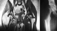
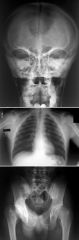
Cleidocranial Dysplasia
Autosomal Dominant Findings Clavicular dysplasia Wormian bones Narrow iliac wings Pubic Bone dysplasia Coxa vara |
|
|
Fx Hamate / Dorsal Dislocation of 4th MC
|
|
|
Hyperparathyroidism
"salt and pepper" appearance DDx of initial film Paget’s Myeloma Reversal of findings strongly suggests hyperparathyroidism |
|
|
Scleroderma
acro-osteolysis. soft tissue calcinosis. soft tissue atrophy, especially at fingertips (sclerodactyly). Differential diagnosis for acro-osteolysis Scleroderma Hyperparathyroidism Thermal injury Psoriasis Neuroarthropathy Trauma diabetes mellitus leprosy etc. |
|
|
Enchondroma
Less likely low grade chondrosarcoma |