![]()
![]()
![]()
Use LEFT and RIGHT arrow keys to navigate between flashcards;
Use UP and DOWN arrow keys to flip the card;
H to show hint;
A reads text to speech;
119 Cards in this Set
- Front
- Back
|
What are two general terms to describe lightening of the skin? |
Leukoderma, hypopigmentation |
|
|
Hypomelanosis is classified into what two groups? How do they differ? |
1. Melanocytopenic - partial or total absence of melanocytes 2. Melanopenic - normal # of melanocytes, but failure to produce or transfer melanin |
|
|
What wavelength is UV light (Wood's lamp)? |
365 nm |
|
|
What histologic stain can detect melanocytes with tyrosine activity? |
Dihydroxyphenylalanine (DOPA) |
|
|
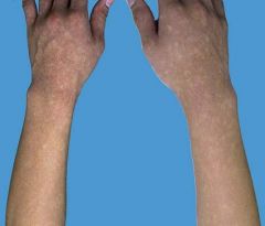
What is the pathogenesis of vitiligo? |
Progressive loss of functional melanocytes |
|
|
Pigmented disorder that is a result of progressive loss of functional melanocytes? |
Vitiligo |
|
|
Which amino acid is crucial to the synthesis of melanin? |
Tyrosine |
|
|
Symptomatology in vitiligo? |
Usually asymptomatic, but pruritus is sometimes noted |
|
|
Facial vitiligo typically involves what areas? |
Perioral, periocular |
|
|
Vitiligo on the extremities tends to favor which areas? |
Elbows, knees, fingers/toes, flexor wrists, dorsal ankles, shins i.e. sites that have repeated trauma, pressure, or friction! |
|
|
Is hair depigmentation a common or uncommon finding in vitiligo? |
Uncommon (10 to 60% incidence); follicular melanocytes are often spared in vitiligo |
|
|
Prognosis of hair depigmentation in vitiligo? |
Poor; spontaneous repigmentation does not occur in general |
|
|
Variant of vitiligo in which multiple small (confetti-like) discrete amelanotic macules seen, sometimes superimposed on a hyperpigmented macule? |
Vitiligo ponctue |
|
|
Variant of vitiligo in which erythema is observed at the margins of depigmented lesions? |
Inflammatory vitiligo |
|
|
Variant of vitiligo in which there is a uniform hypopigmented zone between normal and completely depigmented skin? |
Trichrome vitiligo There is also quadrichrome and pentachrome vitiligo (depending on # of different hypopigmented shades) ! |
|
|
Variant of vitiligo in which depigmented lesions are seen in sites of trauma? |
isomorphic Koebner phenomenon (IKP) Only a minimal threshold of injury is required for IKP to occur |
|
|
What are the two major classifications for vitiligo? |
1. Segmented (does not cross midline) 2. Non-segmental (i.e. vitiligo vulgaris) |
|
|
Are segmental and non-segmental forms of vitiligo part of a spectrum, or two distinct entities? |
Two distinct forms of vitiligo (as suggested by genetic and clinical data) |
|
|
What is the clinical appearance of vitiligo vulgaris? |
Scattered depigmented patches that are widely distributed |
|
|
Which classification of vitiligo would acrofacial vitiligo fall into? |
Non-segmental |
|
|
What is the most significant ocular abnormality associated with vitiligo? |
Uveitis |
|
|
In what syndrome is the most severe form of uveitis seen, in the setting of vitiligo? |
Vogt-Koyanagi-Harada (VKH) syndrome uveitis + aseptic meningitis + hearing abnormalities + vitiligo of the head/neck + poliosis (absence of melanin in hair on head/brow/eyelashes) |
|
|
Syndrome composed of uveitis, aseptic meningitis, hearing abnormalities, vitiligo of the head/neck, and poliosis? |
Vogt-Koyanagi-Harada (VKH) syndrome |
|
|
Syndrome composed of whitening of scalp hair/eyebrows/eyelashes + depigmentation of face + ipsilateral visual changes (decreased visual acuity, atrophic iris)? |
Alezzandrini syndrome |
|
|
What is Alezzandrini syndrome? |
whitening of scalp hair/eyebrows/eyelashes + depigmentation of face + ipsilateral visual changes (decreased visual acuity, atrophic iris) |
|
|
What are some auto-immune diseases commonly associated with vitiligo (6)? |
1. Autoimmune thyroid disease 2. Pernicious anemia 3. Addison's disease 4. Lupus erythematosus 5. Rheumatoid arthritis 6. Adult-onset diabetes mellitus |
|
|
Which form of vitiligo is seen in significantly higher incidence in children compared to adults? |
Segmental vitiligo |
|
|
In the differential diagnosis of vitiligo, which drug must be ruled out? |
Imatinib; known to cause drug-induced leukoderma |
|
|
Repigmentation from successful treatment of vitiligo occurs in what pattern? |
Perifollicular pattern; also from the periphery to inward |
|
|
When using topical steroids to treat vitiligo, after how long with no response would you discontinue using the steroid? |
After 2 months |
|
|
What is the phototherapy of choice in vitiligo? |
NBUVB |
|
|
When using phototherapy to treat vitiligo, energy of the light is increased until what is visualized? |
mild asymptomatic erythema |
|
|
Which psoralen used in PUVA is less phototoxic, 8-MOP or 5-MOP? |
5-MOP (i.e. bergapten) Unfortunately it is not approved for use in the US... |
|
|
What wavelength of excimer laser is used to treat vitiligo? |
308 nm |
|
|
What is the efficacy of the excimer laser in treating vitiligo? |
20-50% of lesions achieve > 75% repigmentation |
|
|
What are some potential surgical therapies for vitiligo treatment? |
Minigrafting Suction blister epidermal grafting Grafting of cultured autologous melanocytes or non-cultured epidermal cell suspensions |
|
|
What is the fundamental difference b/w vitiligo and oculocutaneous albinism?
|
Albinism --> absence of melanin pigment w/in melanocytes; # of melanocytes is normal |
|
|
Most common inherited disorder that leads to diffuse hypomelanosis?
|
Oculocutaneous albinism |
|
|
Inheritance pattern in oculocutaneous albinism?
|
Autosomal recessive |
|
|
What is the defective enzyme in albinism? |
Tyrosinase |
|
|
Ocular manifestations of oculocutaneous albinism (2 groups)?
|
2. Strabismus, nystagmus, lack of binocular vision |
|
|
Which OCA1 subtype has reduced tyrosinase activity?
|
OCA1B
|
|
|
Which OCA1 subtype has absent tyrosinase activity?
|
OCA1A
|
|
|
Which OCA type is considered to have a "tyrosinase-positive" phenotype?
|
OCA2 |
|
|
What causes the ocular manifestations of OA and OCA (2)?
|
2. Misrouting of optic nerve fibers during development |
|
|
Management of albinism?
|
2. Early and frequent ophthalmologic evaluation |
|
|
What is the inheritance pattern for piebaldism? |
Autosomal dominant |
|
|
Clinical presentation of piebaldism? |
(due to absence of melanocytes in involved areas) |
|
|
Etiology of piebaldism?
|
Mutations in the KIT proto-oncogene |
|
|
Percentage of cases of piebaldism that present with a white forelock? |
80-90% of cases |
|
|
Characteristic distribution of the leukoderma in piebaldism? |
Central forehead/midfrontal portion of scalp, central anterior trunk, mid extremities |
|
|
Characteristic appearance of the leukoderma in piebaldism? |
Irregular well-circumscribed milk-white lesions that have normally pigmented / hyperpigmented macules and patches within them |
|
|
Inheritance pattern of Waardenburg syndrome? |
Can be either autosomal dominant or recessive |
|
|
Characteristics of Waardenburg syndrome (5)? |
1. Achromia of hair/skin in same pattern as piebaldism 2. Congenital deafness 3. Partial or total heterochromia irides 4. Medial eyebrow hyperplasia (synophrys) 5. Dystopia canthorum (will not see nasolacrimal caruncle) |
|
|
Which Waardenburg syndrome type will not have dystopia canthorum? |
Type 2 |
|
|
Which Waardenburg syndrome type is associated with limb abnormalities? |
Type 3 |
|
|
What other disease is Waardenburg syndrome type 4 associated with? |
Hirschsprung disease |
|
|
What is the genetic mutation in Waardenburg syndromes type 1 & 3? |
PAX3 transcription factor |
|
|
What is the genetic mutation in Waardenburg syndrome type 4? |
SOX10 transcription factor |
|
|
What is the most frequently observed pigmentary abnormality in Waardenburg syndrome? |
White forelock (seen in 20-60% of cases) |
|
|
Characteristics of Tietz syndrome? |
Diffuse pigmentary dilution of the skin/hair + deafness + hypoplasia of eyebrows |
|
|
Do you see absence of melanocytes or melanin in Waardenberg syndrome? |
Absence or minimal # of melanocytes |
|
|
Do you see absence of melanocytes or melanin in piebaldism? |
Absence or minimal # of melanocytes |
|
|
What is the inheritance pattern in Hermansky-Pudlak syndrome? |
Autosomal recessive |
|
|
Most forms of Hermansky-Pudlak syndrome are caused by mutations in genes that encode what? |
BLOC; i.e. biogenesis of lysosome-related organelles complexes |
|
|
Clinical features of Hermansky-Pudlak syndrome (5)? |
1. Pigmentary dilution of skin/hair/eyes 2. Ocular disease (nystagmus, reduced acuity) 3. Bleeding tendency 4. Interstitial pulmonary fibrosis and granulomatous colitis 5. Recurrent bacterial infections |
|
|
Most common cause of premature death in Hermansky-Pudlak syndrome? |
Progressive pulmonary fibrosis |
|
|
Inheritance pattern for Chediak-Higashi syndrome? |
Autosomal recessive |
|
|
Clinical features of Chediak-Higashi syndrome? |
1. Oculocutaneous albinism 2. Bleeding diathesis 3. Progressive neurologic dysfunction 4. Severe immunodeficiency due to abnormal lytic granules in lymphocytes, NK cells, neutrophils |
|
|
What is the histopathologic hallmark of Chediak-Higashi syndrome? |
Giant lysosome-related organelles (melanosomes, platelet dense granules, neutrophil granules) |
|
|
Clinical features of tricho-hepato-enteric syndrome? |
1. Diffuse pigmentary dilution of skin/hair 2. Platelet defects 3. Immunodeficiency 4. Brittle hair, facial dysmorphism 5. Intractable diarrhea during infancy |
|
|
Other name for tricho-hepato-enteric syndrome? |
Phenotypic diarrhea of infancy |
|
|
Inheritance pattern of Griscelli syndrome and Elejalde syndrome? |
Autosomal recessive |
|
|
Clinical presentation of Griscelli syndrome Type 1 (3)? |
1. Pigmentary dilution of skin 2. Silvery-gray hair 3. Neurologic impairment |
|
|
Clinical presentation of Griscelli syndrome Type 2 (3)? |
1. Pigmentary dilution of skin 2. Silvery-gray hair 3. Immune abnormalities |
|
|
Inheritance pattern of tuberous sclerosis complex? |
Autosomal dominant |
|
|
Classic triad seen in tuberous sclerosis? |
1. Facial angiofibromas ("adenoma sebaceum") 2. Seizures 3. Mental deficits |
|
|
What is the characteristic pattern of hypomelanotic white macules seen in tuberous sclerosis? |
Lance-ovate (rounded at one end, pointed at the other) |
|
|
When are the hypomelanotic macules seen in tuberous sclerosis? |
At birth, or during the neonatal period |
|
|
Most common location of hypomelanotic macules in tuberous sclerosis? |
Posterior trunk; also commonly seen on the lower extremities |
|
|
Difference in the color of the "white macules/patches" in tuberous sclerosis vs vitiligo? |
Dull white color (hypomelanotic) vs pure-white color (amelanotic) |
|
|
What causes the hypomelanosis in tuberous sclerosis? |
Decreased size of melanosomes + poor melanization of melanosomes # of melanocytes is not affected |
|
|
Genetic mutations responsible for tuberous sclerosis (2)? |
TSC1 (hamartin) and TSC2 (tuberin) They negatively regulate mTOR signaling |
|
|
What # of hypomelanotic macules in an infant should raise suspicion of tuberous sclerosis? |
3 or more |
|
|
Clinical features of hypomelanosis of Ito? |
Hypopigmentation along Blaschko's lines Look for hypopigmented streaks and whorls |
|
|
Clinical features of nevus depigmentosus? |
Hypomelanotic patches with irregular but well-defined borders that break apart into smaller macules at the periphery |
|
|
Name of the pattern of hypomelanosis that occurs in patients with mosaic forms of trisomy 13? |
Phylloid hypomelanosis |
|
|
Pattern of hypomelanosis seen in kwashiorkor disease? |
Erythematous skin with marked desquamation --> leads to hypomelanosis and patchy post-inflammatory hypermelanosis |
|
|
Pathogenesis of post-inflammatory hypomelanosis (2)? |
1. Dysregulation of growth factors and cytokines regulating keratinocyte-melanocyte interactions 2. Severe local inflammation causing loss of functional melanocytes or even melanocyte death |
|
|
Two most common inflammatory disorders that can cause complete depigmentation? |
1. Severe atopic dermatitis 2. Discoid lupus erythematosus |
|
|
Treatment of post-inflammatory hyperpigmentation? |
Sun exposure/UVB phototherapy |
|
|
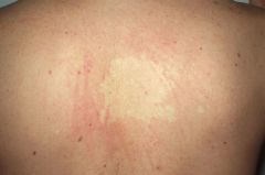
Most common location of involvement of pityriasis alba? |
Face, especially the malar region |
|
|
Clinical presentation of pityriasis alba? |
Erythematous macules/patches that fade and leave a whitish, dry, powdery appearance |
|
|
Cutaneous neoplastic disorder that can cause hypomelanotic macules on the trunk/extremities? |
Hypomelanotic mycosis fungoides A variant of early-stage MF most frequently observed in darkly pigmented patients |
|
|
Characteristics of the hypomelanosis that can be seen in scleroderma? |
Circumscribed areas of complete pigment loss except for perifollicular and supravenous retention of pigment |
|
|
Clinical presentation of pityriasis (tinea) versicolor? |
Round, oval hypomelanotic macules with fine scale; typical coalesce into patches centrally |
|
|
Differences in the hypomelanosis seen in lepromatous leprosy vs tuberculoid leprosy? |
Lepromatous --> multiple small, ill-defined hypomelanotic macules Tuberculoid --> discrete large (up to 30cm) hypopigmented patches; surface will be dry/mildly scaly |
|
|
What is the pattern of hypomelanosis seen in secondary syphilis? |
"Necklace of Venus" - small (1-2mm) mypomelanotic macules scattered within a larger area of hyperpigmentation on the neck |
|
|
What is the pattern of hypomelanosis seen in onchocerciasis? |
"Leopard skin" Small hypomelanotic macules on shin --> coalescence to form amelanotic patches --> persistence of pigmented macules within the patches gives rise of leopard skin |
|
|
After what age would the finding of a halo nevus be UNCOMMON? |
Over 40 years of age Presence of halo nevi in patient over 40 should raise suspicion for cutaneous and/or ocular melanoma |
|
|
Most common location for a halo nevus? |
Trunk, particularly the back |
|
|
What are the four stages in the evolution of a halo nevus? |
1. Appearance of halo 2. Loss of pigment within the central nevus 3. Disappearance of the nevus 4. Disappearance of the halo |
|
|
What causes the "halo" in halo nevi? |
Destruction of melanocytes by T cells; these T cells also destroy nevus cells, causing disappearance of the halo nevi |
|
|
Management of halo nevi? |
No treatment other than reassurance |
|
|
Pathogenesis for melanoma-associated vitiligo-like leukoderma? |
Immune reaction against antigenic determinants shared by normal and malignant melanocytes |
|
|
What are the 3 major groups of depigmenting compounds? |
1. Phenols/catechols 2. Sulfhydryls 3. Miscellaneous |
|
|
What are the most widely used "bleaching" agents? |
Hydroquinone and its derivatives (MMEH, MBEH) |
|
|
What common in-clinic treatment has a side effect of hypomelanosis, particularly in darkly pigmented individuals? |
Intralesional steroids Can also be seen with topical steroids |
|
|
What chemotherapeutic agent is noted to cause hypomelanosis? |
imatinib mesylate (Gleevec) |
|
|
Characteristics of Gleevec-associated hypomelanosis (3)? |
1. Generalized lightening of skin 2. Hypopigmentation of distal digits 3. Progression of vitiligo These changes are reversible and dose-dependent |
|
|
Very common (80% in ppl over 70) disorder seen in elderly patients that presents with well-circumscribed asymptomatic porcelain white macules? |
Idiopathic guttate hypomelanosis (IGH) |
|
|
Most common location of involvement in idiopathic guttate hypomelanosis? |
Extensor forearms, shins |
|
|
Most common (3) histologic features in idiopathic guttate hypomelanosis? |
1. Flattening of the dermal-epidermal junction 2. Moderate-marked reduction or absence of melanin granules in basal layer 3. Basket-weave hyperkeratosis |
|
|
Common skin disorder in young women from tropical climates that presents with poorly defined, nummular, non-scalp hypopigmented macules and small patches on the trunk? |
Progressive macular hypomelanosis Confluence of lesions may occur centrally |
|
|
What person is most commonly affected by progressive macular hypomelanosis? |
Young women who reside in or are from tropical climates |
|
|
Clinical presentation of nevus anemicus? |
Pale area of variable size (3-6 cm) with an irregular, "broken-up" outline |
|
|
Pathogenesis of nevus anemicus? |
Decreased blood flow through capillaries in the dermal papillae due to localized hypersensitivity to catecholamines |
|
|
What makes nevus anemicus more noticeable? |
Episodes of vasodilation in the surrounding skin (i.e. heat, emotional stress) |
|
|
What are Bier spots? |
Pale macules on the extremities caused by localized vasoconstriction Most often in young women; may be induced by dependent position or use of tourniquets |