Reading...
![]()
Play button
![]()
Play button
![]()
Use LEFT and RIGHT arrow keys to navigate between flashcards;
Use UP and DOWN arrow keys to flip the card;
H to show hint;
A reads text to speech;
28 Cards in this Set
- Front
- Back
- 3rd side (hint)
|
What affects the appearance of normal lung?
|
Species and Breed
Age Body condition - obese, thin degree of ventilation radiographic technique body wall/rib structures |
|
|
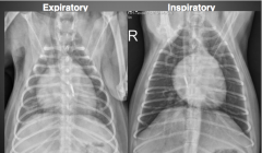
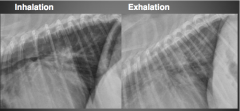
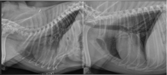
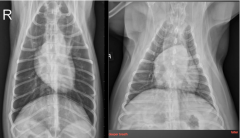
Lung inflation changes
|
|
|
|
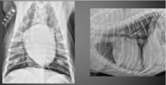
Chest Conformation
|
deeper chest, obese, barrel chest
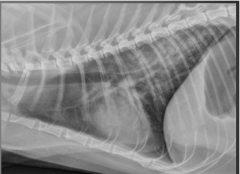
normal lungs: white tree branches on dark background (pulmonary vessels in everything else filled with gas) |
|
|
|
Lung Patterns
|
Interstitial - unstructured
Alveolar Bronchial (Vascular) |
|
|
|
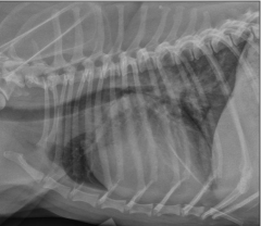
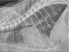
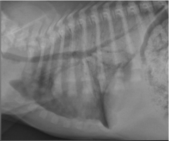
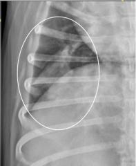
(Unstructured) Interstitial Pattern
|
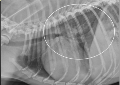
See pulmonary vessels, but margins indistinct (blurry)
increased lung opacity -perihilar, diffuse (CHF) |
|
|
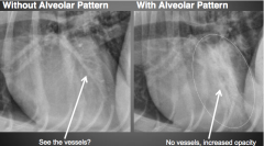
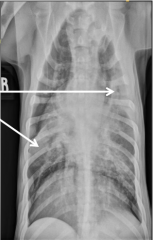
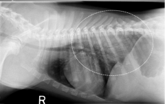
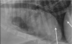
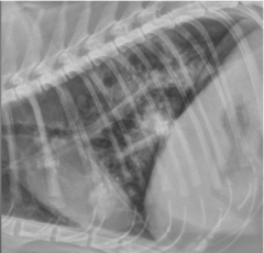
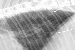
Alveolar Pattern
|
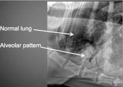
Can not see lung vessels at area of pattern
+/- air bronchograms present +/- lobar sign present increased lung opacity caused by completely non-aerated portion of lung if no air in lung, the pulmonary vessels will silhouete with lungs and become non-visible |
caused by lung disease (no gas in alveoli because they're filled with fluid or cells) (assume this first)
caused by atalectasis (no gas in alveoli because they're collapsed) - determine incidental cause of lung volume loss vs pathologic cause of volume loss or lobar airway obstruction |
|
|
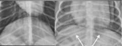
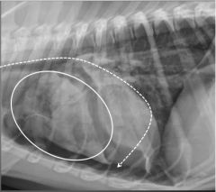
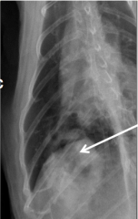
Air Bronchograms
|
Air bronchogram is when gas remains in bronchus within a region of lung with an alveolar pattern
"black tree branches on white background" hallmark of alveolar pattern -absence of seeing vessel outer margins |
will disappear if bronchus becomes filled with fluid/cells or becomes collapsed
|
|
|
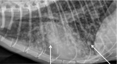
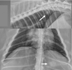
Lobar Sign
|
Sharp delineartion at the margin of an alveolar pattern and some adjacent aerated lung lobe
useful to draw attention to aleolar pattern indicated process is focal to affected lobe, not diffuse |

|
|
|
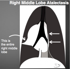
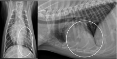
Atalectasis
|
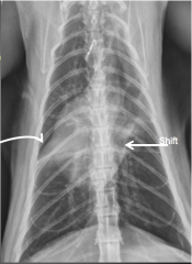
changes in lung volume - decreased size
(vs. diseased lobes will be normal or increased) on DV/VD view 1. mediastinal shift towards the collapsed lobe 2. direct visualization of lobe margins with decreased lobe sign (commonly right middle lobe) |
|
|
|
Bronchial Pattern
|
accentuation (thickening) of airway walls (bronchial walls)
caused by fluid or cells gathering around or within airway wall often used as indicator of disease particular to the airway - seen end on (doughnuts) or seen side on (parallel lines/train tracks) diffuse exaggeration of airway markings concurrent interstitial pattern often also present (broncho-interstital pattern) |

|
|
|
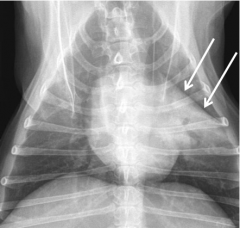
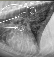
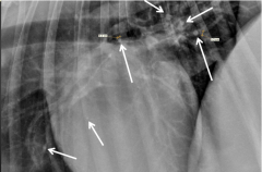
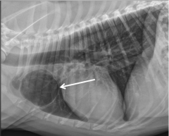
Lung Nodules
|
sphere of soft tissue (4-5mm) - faint soft tissue opaque - smallest visible nodule
round structure in lung <4-5mm: either end on vessel or nodule made of mineral (benign osseous metaplasia/osteoma or mineralized granuloma) |
|
|
|
End on vessels
|
often misdiagnosed by students as nodules
represent a length of vessel should be same length as overlying vessel should be more opaque than overlying vessel |
|
|
|
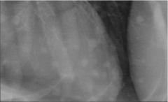
Benign Pulmonary Osteomas
|
mostly 1-2mm
as opaque or more opaque than ribs |
|
|
|
Miliary Pattern
|
lung apperance suggests many very small nodules difficult to seperate (like grains of sand) - summation of many small nodules
e.g. lymphoma |
|
|
|
Bronchopneumonia
|
one of the most common lung diseases
bacterial infection enters through the airways typically settles to the gravity dependent potion (ventral) variable signs of inflammation, variable cough, commonly caused by aspiration of GI contents from vomiting or regurgitation (aspiration pneumonia) may occur secondary to viral insult or immune compromise -primary rule out for ventrally distributed alveolar pattern -lag behind clinical symptoms |

|
|
|
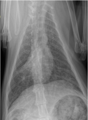
Cardiogenic Pulmonary Edema
|
fluid displaced into the lung interstitium and alveolar spaces from left congestive heart failure
typically see radiographic left heart enlargement and pulmonary venous congestion dogs: perihilar and caudodorsal lung regions interstitial to alveolar with severity cats: distribute anywhere (ventrally, diffusely, multifocal, can have prominent bronchial component) |
|
|
|
Feline Cardiogenic Edema
|
commonly not appearing to predominate perihilar region, seen throughout thorax
often broncho-interstitial distribution often accompanied by pleural and pericardial effusion uncommonly localized to perihilar region |
|
|
|
Non-cardiogenic Pulmonary Edema
|
numerous causes (seizures, head trauma, choking, strangulation, electrocution, systemic inflammation, pancreatitis, CKD)
large component of acute lung injury -worst form is ARDS (acute respiratory distress syndrome) ultimately results in leaky lung vessels, fluid often more proteinaceous than cardiogenic edema, responds more slowly to diuretic therapy distributed in caudodorsal lungs, often bilaterally symmetric, may extend to whole lung if severe, often no sign of heart or pulmonary vessel enlargement, history may increase presumptive diagnosis |
|
|
|
Pulmonary Contusion
|
bleeding/bruising from direct trauma
often unilateral in auto injury, can be bilateral if crushed or bite injury can occur anywhere lung can be traumatized interstitial or alveolar pattern, will worse with severity often have hx or see other radiographic evidence of trauma |
|
|
|
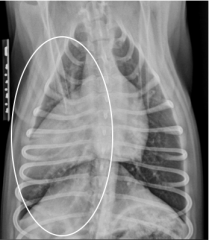
Bronchial Disease
|
inflammatory diseases of the lower airways (bronchi)
often breed specific name cats = feline asthma dogs = chronic bronchitis, allergic pneumonitis horse = summer pasture associated obstructive pulmonary disease (heaves) may be allergic in nature or infectious hx of cough, may have expiratory effort, open mouth breathing (cats) indicates lower airway predilection of disease, may be combined bronchial and interstitial pattern, cats with asthma may have hyper-inflated lungs Feline Asthma |
|
|
|
Pulmonary Hemorrhage
|
could be caused by coagulopathy (rodenticide toxicity, immune mediated causes)
should be differential for abnormal lung apperance when clinical and lab findings suggest bleeding (hemoptysis, bleeding/brusing, prolonged coagulation times) interstitial or alveolar pattern, one or multiple areas of lung, any distribution any area of lung, no classic apperance |
|
|
|
Pulmonary Thromboemoblism (PTE)
|
infarcted area of lung secondary to thrombus
often secondary to heartworm disease can be secondary to hypercoagulable state (Cushing's, DIC, neoplasia, PLN) typically patient will have acute respiratory distress/dyspnea, typically responds to O2 therapy Affects focal areas of lung, 2 very different patterns 1. heartworm disease: focal interstitial or alveolar pattern 2. not HWD: hyper lucent lung (decreased vessel size) |
|
|
|
Lung Nodules
|
2 major differentials:
Neoplasia Granulomatous disease most common cause of single soft tissue lung nodule in dogs and cats -> primary lung neoplasia most common cause of multiple soft tissue lung nodules -> pulmonary metastatic neoplasia |
bronchogenic carcinoma with cavitary lung lesion
|
|
|
Granulomatous diseases
|
Fungal diseases (endemic areas): Histoplasmosis, blastomycosis, coccidoides
Eosinophilic granulomas: eosinophilic bronchopneumonopathy, allergic pneumonitis, heartworm disease Parasitic granulomas: paragonimus, heartworms mineralized nodules are usually benign (old, osteoma); rarely some osteosarcoma or MLO metastasis is partially mineralized |
|
|
|
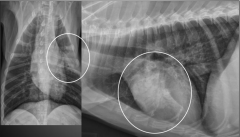
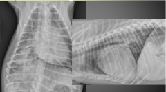
Lung Neoplasia
|
nodules (< 3 cm), masses (> 3 cm)
primary: Bronchogenic carcinoma, squamous cell carcinoma, alveolar carcinoma high differential for nodule in middle or peripheral portion of lung in older cat |
mineralized nodules are usually benign (old, osteoma); rarely some osteosarcoma or MLO metastasis is partially mineralized
raise index of suspicion for carcinoma DDX: granulomatous disease or benign (broncholithiasis, mineralized alveolar mucous glands) |
|
|
Cavitary Lung Lesions
|
Gas filled lung lesion
thin walled: pulmonary bulla (in parenchyma) or bleb (immediately under surface) -acquired or congenital -can be incidental, can also rupture and cause pneumothorax thick walled: neoplasia, abscess, granulmoa(Paragonimus), hemorrhaging bulla(trauma) |
Paragonimus granulmoas
dx: fecal flotation |
|
|
Pulmonary lymphoma
|
wide variety of radiographic appearances
-mild to severe unstructured interstitial pattern -alveolar pattern or bronchial pattern -nodules, masses +/- pleural effusion or lymphadenomegaly severe interstitial pattern |
|
|
|
Idiopathic Pulmonary Fibrosis of Cats
|
middle age to older cats
can be concurrent with neoplasia has poor to grave prognosis wide variety of radiographic appearances: mild to severe interstitial pattern, alveolar or bronchial, nodules, masses (don't biopsy cat lung) |
|