Reading...
![]()
Play button
![]()
Play button
![]()
Use LEFT and RIGHT arrow keys to navigate between flashcards;
Use UP and DOWN arrow keys to flip the card;
H to show hint;
A reads text to speech;
35 Cards in this Set
- Front
- Back
|
What are the functions of the kidneys?
|
To remove toxins from the bloodstream and produce urine.
To conserve salts, glucose, proteins and water, and through doing, regulate BP, hemodynamics and the acid-base balance of the body (renin and EPO) |
|
|
Why are the kidneys located "retro"peritoneally?
|
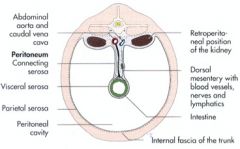
Only the ventral surface of each kidney is covered by peritoneum.
|
|
|
Which vertebrae are the right and left kidney located near?
What size are they in the dog/cat? |
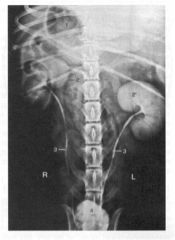
R ~ T10-T13
L ~ T13 - L2 In dog, the kidney is 2.5 to 3.5 x the length of L2. In cat, the kidney is 2-3 x the length of L2. |
|
|
What vessels lead to and away from the kidneys?
|
Renal a. goes to the kidney; renal v. drains blood from it.
Renal lnn. are at the hilus. The ureter carries urine away to the bladder. |
|
|
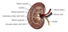
Define the fibrous capsule, renal hilus, renal sinus, renal pelvis, renal crest, cortex, and medulla of the kidneys.
|
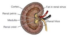
The fibrous capsule surrounds the kidney.
The hilus is the entrance/exit of the renal artery, vein, ureter and lymphatics. The renal sinus is the cavity containing the renal pelvis, fat, vessels, lymphatics and nerves. The renal pelvis is the funnel shaped structure receiving urine from the collecting ducts to pass to the ureter. The pelvis has p in it. Get it? The renal crest is the ridge projecting longitudinally into the pelvis. The cortex is peripherally located with the renal corpuscles and convoluted tubules. The medulla contains the collecting ducts, renal pyramids, and renal crest. |
|
|
What is meant by pseudopyramidal?
|
It means that the pyramids have fused to be one longitudinally. In the cat and dog, our longitudinal cut showed no pyramids, but a sagitall cut looked like this, where the individual pyramids were evident.
|
|
|
What is the pelvic recess?
|
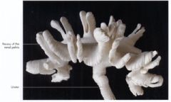
It is the extension of the renal pelvis between the renal pyramids.
|
|
|
Which kidneys are more easily palpated dog or cat?
|
Cat.
|
|
|
Do the kidneys of the dog or cat have subcapsular veins?
|
The cat kidneys have subcapsular veins draining into the renal v.
|
|
|
What shape is the renal crest in the dog? In the cat?
|
The renal crest is longitudinal in the dog and cone shaped in the cat.
|
|
|
What are the functions of the adrenal glands?
|
The cortex produces mineralocorticoids, corticosteroids, and sex hormones.
The medulla produces epinephrine and norephinephrine. |
|
|
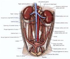
Where are the adrenal glands located? What vessels are they located between?
|
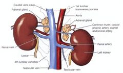
They are located cranial to the kidneys.
The phrenicoabdominal artery is dorsal to them and the phrencioabdominal vein is ventral to them. |
|
|
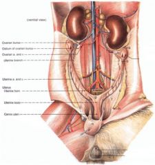
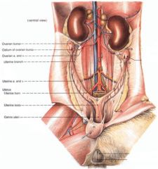
Let's go from Ovary to Vulva.
|
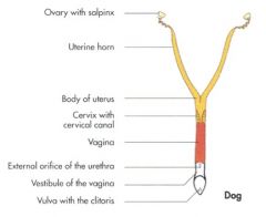
Ovary to uterine tube to uterus, which includes the horn, body and cervix, then onto the vajayjay to the vestibule and then the vulva.
|
|
|
What are the connecting tissues of the broad ligament?
|
The mesometrium (uterus), mesovarium (ovary), and the mesosalpinx (uterine tube).
|
|
|
What's released from the ovarian bursa?
|
Ova
|
|
|
In relation to the kidneys, where are the ovaries located?
|
The ovaries are dorsocaudal to the kidneys. The right ovary is more cranial than the left (just like the kidneys!)
|
|
|
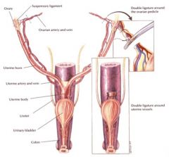
What vessels course within the mesovarium covering the ovary? What happens to the mesovarium in a spay procedure?
|
The ovarian artery and vein course within the mesovarium.
They are ligated during a spay. |
|
|
What ligaments are associated with the ovaries? Where do they course between? Which one gets broken down during an ovariohysterectomy (spay)?
|
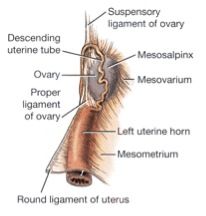
The proper ligament of the ovary - course between the ovary and the tip of the uterine horn.
The suspensory ligament of the ovary - courses between the ovary and the body wall/last rib. The suspensory ligament is broken down during an ovariohysterectomy by strumming. |
|
|
What is the ovarian bursa? What forms it? Is the opening to the ovarian bursa larger in the dog or cat?
|
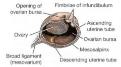
The ovarian bursa is the peritoneal sac that encloses the ovary.
It is formed by the mesovarium and mesosalpinx. The opening to the ovarian bursa is larger in the cat. |
|
|
What else is the uterine tube known as?
Where does it course between? What supports it? |

The uterine tube is also known as the oviduct, fallopian tube, or salpinx.
It courses between the ovarian bursa and the uterine horn, first cranially and then caudally along the lateral side of the ovarian bursa. It's supported byt he mesosalpinx. |
|
|
Where does fertilization occur?
|
In the ampulla of the uterine tube
|
|
|
What are the three regions of the uterine tube?
|
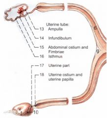
The infundibulum - funnel shaped, fimbria around it's edge "catch" the ovum. The palm with fingers.
Abdominal ostium - the wrists - located between the infundibulum and ampulla. The ampulla - the forearms - the longest part, where fertilization typically occurs. The isthmus - the tubouterine junction, between the uterine tube and uterine horn. |
|
|
What are the regions of the uterus?
|
Uterine horns, uterine body and the cerivx.
|
|
|
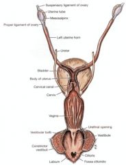
Identify C. What is it a remnant of? Where does it course between? Does it get broken down in spays?
|
C is the round ligament. It's a remnant of the gubernaculum. It courses between the uterine horn and the vaginal process.
It does get broken down in spay procedures. |
|
|
Where is the cervix located?
What is the passageway within the cervix called? What is the internal uterine ostium? What is the external uterine ostium? |

The cervix is located at the caudal portion of the uterus.
The cervical canal is the passageway within the cervix. The internal uterine ostium is the opening of the cervix into the body of the uterus. The external uterine ostium is the opening of the cervix into the vagina. |
|
|
What is the vagina the region between?
What is the vaginal fornix? Where does it extend from and to? |

The vagina is the region between the cervix and vestibule.
The fornices of the vagina are the deepest portions of the vagina, extending into the recesses created by the extension of the cervix into the vaginal space. The word 'fornix' is Latin for 'arch'. The vaginal fornix extends cranial to the cervix ventrally. |
|
|
Where does the region of the vestibule extend in the female reproductive organs?
What structure drains into it? What type of muscle exists in the vestibule? |
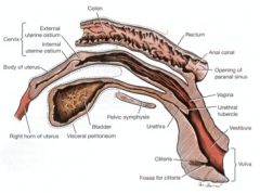
The vestibule is the region of the reproductive tract between the vagina and vulva.
The urethra drains into it at the urethral tubercle by way of the external urethral orifice. Skeletal muscle is in the wall of the vestibule. |
|
|
Is the urethra in the female located ventrally or dorsally?
|
Ventrally
|
|
|
Is the clitoris located ventrally or dorsally?
|
Ventrally
|
|
|
Is the fossa for the clitoris located ventrally or dorsally?
|
Ventrally
|
|
|
Is the vaginal fornix located ventrally or dorsally?
|
Ventrally
|
|
|
Define the vulva.
|
It is the labia with dorsal and ventral commissures (corners)
|
|
|
What arteries go into and veins from the ovaries? Where do they come from/go to, respectively?
|
The ovarian arteries supply the ovaries, the ovarian bursa, uterine tube, and uterine horns. It's a branch off of the aorta.
The right ovarian vein drains into the caudal vena cava. The left ovarian vein drains into the left renal vein. |
|
|
Which ovary could be harder to exteriorize in an ovariohysterectomy and why?
|
The right, because it is more cranial
|
|
|
What is pyometra
|
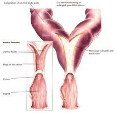
Pus in the uterus
|