![]()
![]()
![]()
Use LEFT and RIGHT arrow keys to navigate between flashcards;
Use UP and DOWN arrow keys to flip the card;
H to show hint;
A reads text to speech;
17 Cards in this Set
- Front
- Back
|
Difference between primary and secondary congenital aphakia |
Primary results from failed induction of the surface ectoderm during embryogenesis (PAX6 mutation, severe developmental abnormalities) whereas secondary results from resorption or extrusion of the lens before or during birth (congenital infections e.g. rubella) |
|
|
Ocular and systemic features of Alport syndrome |
Ocular: Anterior or posterior lenticonus, posterior polymorphous corneal dystrophy, anterior polar cataract, dot and fleck retinopathy retinal and iris neovascularization; Systemic: Haemorrhagic nephritis, deafness |
|
|
Systemic associations of posterior lenticonus |
Alport syndrome, oculocerebrorenal syndrome of Lowe |
|
|
Features of the oculocerebrorenal syndrome of Lowe |
Systemic acidosis, renal rickets, hypotonia, posterior lenticonus and congenital cataracts |
|
|
Histological features of phacoanaphylactic uveitis |
Zonal granuloma (central nidus of degenerating lens material surrounded by concentric layers of inflammatory cells: inner layer of multinucleated giant cells and neutrophils, intermediate layer of lymphocytes and plasma cells, outer layer of fibrovascular connective tissue) |
|
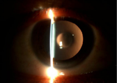
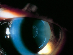
Identify this clinical sign |
Oil droplet reflex (anterior and posterior lenticonus) |
|
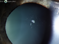
Identify this clinical sign and describe its histology |
Glaukomflecken: patches of white dots below the lens capsule, focal areas of necrotic lens epithelial cells |
|
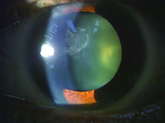
Identify the pathology and describe its histology |
Anterior subcapsular fibrous plaque: metaplastic transformation into fibroblast-like cells |
|
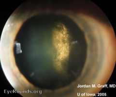
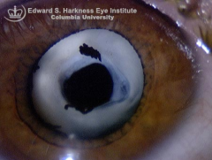
Identify the pathology and describe its histology |
Posterior subcapsular cataract: epithelial disarray at the equator, posterior migration of the lens epithelial with enlargement to form bladder cells of Wedl |
|
|
Risk factors for posterior subcapsular cataract |
Chronic intraocular inflammation, diabetes mellitus, ionizing radiation exposure, smoking, prolonged corticosteroid use |
|
Identify the pathology and describe its histology |
Elschnig pearls: collections of proliferating epithelial cells |
|
Identify the pathology and describe its histology
|
Soemmering ring: sequestration of proliferating lens fibers in the equatorial region |
|
|
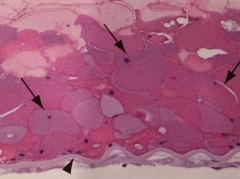
Morgagnian globules |
Eosinophilic bodies that accumulate in slitlike spaces between the lens fibers in cortical degeneration |
|
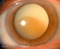
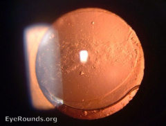
Identify this pathology |
Morgagnian cataract |
|
|
Risk factor for exfoliation of the lens capsule |
Prolonged ocular exposure to infrared radiation (glass and steel workers) |
|
Describe these clinical signs |
Central disc: thin, homogeneous white central deposit on the anterior lens capsule, inrolled edge surrounded by a relatively clear zone; Peripheral band: coarse, granular, “hoarfrost” material on the outer third of the anterior lens surface (pseudoexfoliation syndrome) |
|
|
Describe corneal endothelial changes in PEX |
Naumann's sign: small flakes or clumps of pseudoexfoliative material (PEXM) and usually a diffuse, nonspecic melanin pigment deposition on the corneal endothelial surface and reduced endothelial density, polymegathism, pleomorphism, endothelial cell damage, cell detritus, intraendothelial inclusions, and retroendothelial accumulations |