![]()
![]()
![]()
Use LEFT and RIGHT arrow keys to navigate between flashcards;
Use UP and DOWN arrow keys to flip the card;
H to show hint;
A reads text to speech;
154 Cards in this Set
- Front
- Back
|
Sialolithiasis |
1- Stone(s) in the salivary duct. 2- Can occur in all 3 main ducts (Parotid, submandibular m, sublingual) 3- Single stones most common in submandibular gland (Whartons duct) 4- Presentation pre or periprandial pain with swelling of salivary gland 5- Cause dehydration or trauma 6- Treatment 1- NSAID 2- Gland massage 3- Warm compression 4- Sour candy (promote salivary flow) |
|
|
Sialadenitis |
Inflammation of the salivary gland due to obstruction, infection or immune mediated mechanism |
|
|
Salivary gland tumors |
1- Most are benign and commonly affects parotid gland (80-85%) 2- Nearly half submandibular gland neoplasms and most sublingual and minor salivary gland tumors are malignant 3- Present with painless mass/swelling 4- Facial palsy/pain suggest malignant involvement |
|
|
Pleomorphic adenoma (benign mixed tumor) |
1- Most common type of tumor of the salivary gland 2- Composed of chrondromyxoid stroma and epithelium 3- Recur if incompletely excused or rupture intraoperativly 4- May undergo malignant transformation |
|
|
Mucoepidermoid carcinoma |
1- Most common malignant tumor of the salivary gland 2- Has mucus and squamous components |
|
|
Warthin tumor (papillary cystadenoma lymphomatosum) |
1- Benign cystic tumor of the germinal center 2- Seen in smokers 3- 10% bilateral 10% Multifocal 10% Malignant |
|
|
Achalasia |
1- Failure of relaxation of LES due to degeneration of inhibitory neurons (containing NO and VIP) in the myenteric (Auerbach) plexus of esophageal wall 2- Associated with increase risk of esophageal cancer 3- Pseudoachalasia (2’ Achalasia) due to Chagas’ disease (Trypanosoma cruzi) or extra esophageal malignancy (mass effect or paraneoplastic) 4- Diagnosis 1- Manometry 1- Uncoordinated or absent peristalsis with high LES resting pressure ( progressive dysphagia of solid and liquid) 2- Barium swallow 1- Dilated esophagus with a distal area of stenosis (bird beak appearance) 5- Treatment 1- Medical 1- Ca channel blocker 2- Vasodilator (NO) 3- Botox 2- Surgical 1- Myotomy |
|
|
Diffuse esophageal spasm |
1- Spontaneously, non-peristaltic (uncoordinated) contraction of the esophagus with normal LES pressure 2- Presents with dysphagia and angina like chest pain 3- Barium swallow corckscrew esophagus 4- Manometry diagnostic 5- Treat with CCB and Nitrate |
|
|
Eosinophilic esophagitis |
1- Infiltration of eosinophils in the esophagus often seen in atopic patients 2- Present with food allergens- dysphagia and food impaction 3- Esophageal rings and liner furrows on endoscopy 4- Unresponsive to GERD therapy |
|
|
Esophageal perforation |
1- Most commonly iatrogenic due to esophageal instrumentation 2- Non-iatrogenic cause 1- Spontaneous rupture 2- Foreign material ingestion 3- Trauma 4- Malignancy 3- Presents with pneumothorax and subcutaneous emphysema due to dissecting air 4- Boerhave syndrome 1- Transmural tear of the distal esophagus due to violent retching |
|
|
Esophageal strictures |
1- Associated with 1- Acid reflux 2- Caustic ingestion 3- Esophagitis |
|
|
Esophageal varies |
1- Dilated submucosal vein in lower 1/3 of esophagus 2’ to portal hypertension 2- Common in cirrhosis 3- May cause life threatening hematemesis |
|
|
Esophagitis |
1- Associated with 1- acid reflux 2- Infection in immunocompromised (Candida(white pseudomembrane), HSV-1( punched out ulcer) CMV (liner ulcer ) 3- Caustic ingestion 4- Pill induced esophagitis (bisphosphate, tetracycline, NSAID, iron, and potassium chloride)
|
|
|
Gastroesophageal reflux disease |
1- Presents with 1- Heartburn 2- Regurgitation 3- Dysphagia 2- May also presents with 1- chronic cough 2- Hoarseness (laryngophayngeal reflux) 3- Associated with Asthma 4- Transient decrease in LES tone |
|
|
Mallory Weiss syndrome |
1- Longitudinal laceration of the Gastroesophageal junction 2- Partial thickness confined to mucosa and submucosa 3- Due to severe vomiting, found in alcoholics and bulimia 4- Present with hematemesis |
|
|
Plummer Vinson syndrome |
1- Triad of 1- Dysphagia 2- Iron deficiency anemia 3- Espoohageal webs 2- Increase risk of esophageal squamous cell carcinoma 3- Assoviated with glossitis |
|
|
Schatzki ring |
1- Ring formed at Gastroesophageal junction 2- Due to chronic acid reflux 3- Can present with dysphagia |
|
|
Scleroderma esophageal dysmotility |
1- Esophageal smooth muscle atrophy 2- Decrease LES pressure and dysmotility 3- Acid reflux and dysphagia 4- Stricture, Barrett’s esophagus and aspiration 5- Part of CREST |
|
|
Barrett esophagus |
1- Specialized metaplasia 2- Replacement of non-keratinized stratified squamous epithelium to non-ciliated columnar epithelium with goblet cells (intestinal epithelium) 3- Due to chronic GERD 4- Increase risk esophageal adenocarcinoma |
|
|
Esophageal cancer |
1- Presents with progressive dysphagia (solid then liquid) and weight loss 2- Aggressive cause lack of serous of the esophageal wall allowing rapid extension 3- Poor prognosis due to advanced disease at presentation |
|
|
Adenocarcinoma of the esophageal |
1- Lower 1/3 2- Risk factors 1- Chronic GERD 2- Barrett esophagus 3- Obesity 4- Smoking 5- Alcohol 3- Most common in AMERICA |
|
|
Adenocarcinoma of the esophageal |
1- Lower 1/3 2- Risk factors 1- Chronic GERD 2- Barrett esophagus 3- Obesity 4- Smoking 5- Alcohol 3- Most common in AMERICA |
|
|
Squamous cell carcinoma of the esophagus |
1- Upper 2/3 2- Risk factor 1- Achalasia 2- Caustic strictures 3- Hot liquid 4- Smoking 5- Alcohol 3- Most common worldwide |
|
|
Acute gastritis |
1- Erosion causes by 1- NSAIDs - decrease PDE2- decrease gastric mucosa protection 2- Burns (curling ulcer)- hypovolemia - mucosal ischemia 3- Brain injury (Cushing ulcer)- increase vagal stimulation- increase ACH- increase H secretion 2- Common in alcoholics and patients taking NSAIDs daily |
|
|
Chronic gastritis |
1- Mucosal inflammation often leads to atrophy (hypochlorite- hypergastrinemia) and intestinal metaplasia (increase risk of gastric cancer |
|
|
H pylori causing gastritis |
1- Most common cause of gastritis 2- Increase risk of peptic ulcer disease and MALT lymphoma 3- Occurs in antrum first then spread to the body of the stomach |
|
|
Autoimmune disease causing gastritis |
1- Autoantibodies afffect H/K ATPase pump on parietal cells and intrinsic factor 2- Increase risk of pernicious anemia 3- Occurs in the body/fundus of stomach |
|
|
Menetrier disease |
1- Hyperplasia of gastric mucosa- Hypertrophic rugae 2- Cause excess mucosal production with resultant protein loss and parietal cell atrophy with decrease acid production 3- Precancerous 4- Presentation (WAVEE) 1- Weight loss 2- Anorexia 3- Vomiting 4- Epigastric pain 5- Edema (due to protein loss) |
|
|
Gastric cancer |
1- Most common 1- Gastric adenocarcinoma 2- Lymphoma 3- GI stromal tumor 4- Carcinoid 2 Early aggressive local spread with nodal/liver metastasis 3- Presents late with 1- weight loss 2- Abdominal pain 3- Early satiety 4- Acanthosis nigricans or leser-Trelat sign |
|
|
Types of gastric cancer |
1- Intestinal 1- Associated with H. Pylori 2- Nitrosamines (smoked foods) 3- Tobacco smoking 4- Achlorhydria 5- Chronic gastritis 6- Occur on lesser curvature 7- Ulcer with raised margins |
|
|
Types of nodes and tumor in gastric cancer |
1- Virchow node 1- Left supraclavicular node by metasyfrom stomach 2- krukenberg tumor 1- Bilateral metastasis to the ovary 2- Abundance mucin secreting signet ring cells 3- Sister Mary Joseph nodes - subcutaneous periumbilical lymph nodes due to metastasis 4- Blummer shelf 1- Palpable mass on digital rectal examination due to metastasis to pouch of Douglas |
|
|
Gastric ulcers |
1- Pain increase with meals - weight loss 2- Associated with H-pylori 70% 3- Decrease mucosal protection against gastric secretion 4- Also caused by NSAIDs 5- Increase risk of malignancy 6- Biopsy margin to rule out malignancy |
|
|
Duodenal ulcer |
1- Pain decrease with meal- weight gain 2- Associated with H.pylori 90% 3- Decrease mucosal protection against acid secretion or increase gastric acid 4- Other cause Zollinger Ellison syndrome 5- Cancer benign |
|
|
Ulcer complications |
1- Hemorrhage 1- Gastric, Duodenal (posterior >anterior) 2- Most common compilation 3- Rupture of gastric ulcer on lesser curvature- bleeding from left gastric artery 4- Rupture of duodenal ulcer at posterior wall- bleeding from gastroduodenal artery 2- Obstruction 1 Pyloric channel edema 2- Duodenal 3- Perforation 1- Duodenal (anterior>posterior) 2- Anterior duodenal ulcer perforates into anterior abdominal cavity causing pneumoperitonium 2- Air under the diaphragm causing referred pain to the shoulder due to irritation of phrenic nerve |
|
|
Upper GI bleed |
1- From the mouth to proximal ligament of treitz 2- Present with coffee ground hematemesis (altered blood by digestive enzymes) and melena (dark foul smelling stool) 3- Associated with 1- Peptic ulcer disease 2- Ruptured actives |
|
|
Lower GI bleed |
1- Distal to ligament of treitz to anus 2- Presents with hematochezia (bright red blood) 3- Associated with 1- Hemorrhoids 2- Anal fissure 3- Diverticulosis 4- Angiodysplasia 5- Cancer 6- IBD |
|
|
Celiac disease |
1- Gluten sensitive enteropathy (celiac sprue) 2- Autoimmune mediated intolerance of gliadin (gluten protein found on wheat) 3- Malabsorption and steatorrhea 4- Associated with 1- HLA-DQ2 and HLA-DQ8 2- Northern European descents 3- Dermatitis herpetiform 4- Decrease bone density 5- Decrease mucosal absorption primarily affecting distal duodenum and proximal jejunum 6- Findings 1- IgA anti-tissue transglutaminase (IgA tTG) 2- Anti-endomysial 3- Anti- deamidated gliadin peptide antibodies 4- Villous atrophy 5- Crypts hyperplasia 6- Intraepithelial 7- Moderately increase risk of malignancy 7- Treatment- Gluten free diet 8- Increase risk of T cell lymphoma |
|
|
Pancreatic insufficiency |
1- Caused by 1- Chronic pancreatitis 2- Cystic fibrosis 3- Obstructing cancer 2- Causes malabsorption of fat and fat soluble vitamins (ADEK) and vitamins B12 3- Decrease duodenal HCO and Fecal elastase |
|
|
Tropical sprue |
1- Similar findings to celiac sprue (affect small bowel can affect the ileum) 2- Associated with megaloblastic anemia due to folate deficiency and later B12 deficiency 3- Seen in residence or recent travel to tropics 4- Respond to antibiotics |
|
|
Whipple disease |
1- Cause by tropheryma whipplei (Intracellular gram positive) 2- PAS +, Foamy macrophages in intestinal lamina propria, mesenteric nodes 3- Affect older men 4- Presentation 1- Cardiac findings 2- Arthralgia 3- Neurological findings 4- Diarrhea 5- Steatorrhea |
|
|
The result of a lactose hydrogen breath test is positive if post- lactose breath hydrogen rises how much above baseline |
More than 20 ppm |
|
|
D-Xylose test in pancreatic insufficiency |
Normal |
|
|
What Fecal finding can be used in screening for malabsorption syndrome |
Fecal fat (using Sudan stain) |
|
|
Crohn’s disease |
1- Entire portion of the GI tract, skip lesions and rectum is spared 2- Gross 1- Transmural inflammation - fistula 2- Cobblestone appearance 3- Creeping fat 4- Bowel wall thicken (string sign on barroom swallow x ray) 5- Linear ulcer 6- Fissures 3- Microscopy 1- Non-caseating granuloma 2- Lymphoid infiltrate 3- Th1 mediated 4- Complication 1- Malabsorption 2- Malnutrition 3- Colorectal cancer (increase risk of pancolitis) 4- Fistula (Enterovesical recurrent UTI and pneumoturia) 5- Phlagem/abscess 6- Stricture 7- Perianal disease 5- Intestinal manifestation 1- Diarrhea with or without blood 2- Abdominal pain 6- Extraintestinal manifestation 1- Eye 1- Episcleritis 2- Uveitis 2- Oral ulcer (Apthous stomatitis) 3- Rash 1- Pyodermoa gangrenosum 2- Erythema nodosa 4- Arthritis 1- Polyarthritis 2- Ankylosis spondylitis 5- Kidney stone (calcium oxalate) 6- Gallstone 7- Positive for anti-Saccharomyces cerevisiae antibody (ASCA) 7- Treatment 1- Antibiotics (Ciprofloxacin, metronidazole) 2- Azathioprine 3- Biologics (infliximab, adolimumab) 4- Corticosteroids |
|
|
Ulcerative colitis |
1- Colonic inflammation, continuous colonic lesions, always involve rectum 2- Gross 1- Mucosal and submucosal inflammation 2- Loss of haustra (lead pipe appearance) 3- Microscopy 1 Crypt abscess and ulcer 2- Bleeding, no granuloma 3- Th2 mediated 4- Complication 1- Malabsorption 2- Malnutrition 3- Colorectal cancer (increase risk of pancolitis) 4- Toxic megacolon 5- Fulminant colitis 6- Perforation 5- Intestinal manifestation 1- Bloody diarrhea 2- Abdominal pain 6- Extraintestinal manifestation 1- Eyes 1- Episcleritis 2- Uveitis 2- Oral ulcer -Apthous stomatitis 3- Rash 1- Pyoderma gangrenosum 2- Erythema nodosum 4- Arthritis 1- Inflammatory polyartritis 2- Ankylosis spondylitis 4- 1’ sclerosis cholangitis associated with pANCA 7- Treatment 1- 5- aminosalicylic preparation (mesalamone) 2- 6- mercaptopurine 3- Inflixamab 4- Colectony
|
|
|
Microscopic colitis |
1- Inflammation of the colon that cause chronic watery diarrhea 2- More common in older female 3- Colonic mucosa normal on endoscopy 4- Histology 1- Inflammatory infiltrates in lamina propria with thickened subepithelial bands and intraepithelial lymphocytes |
|
|
Irritable bowel syndrome |
1- Recurrent abdominal pain occurring with >2 of the following 1- Related to defecation 2- Change in stool consistency 3- Change in stool frequency 2- No structural abnormality 3- More common in middle age females 4- Chronic symptoms can be diarrhea predominant, constipation predominant or mixed 5- Treatment 1- Lifestyle changes 2- Dietary changes |
|
|
Appendicitis |
1- Inflammation of the appendix due to obstruction by fecolith (adults) or lymphoid hyperplasia (children) 2- Presentation 1- RUQ pain 2- Positive McBurney point (1/3 from right ASIS to umbilicus) 3- Nausea/vomiting 4- Fever 5- Perforation 1- Rebound tenderness 2- Rigidity 3- Guarding 4- Positive psoas, obturator and rovsing sing 3- Pathophysiology Proximal obstruction of appendiceal lumen causing a close loop obstruction — Increase intraluminal pressure- Stimulationnof visceral afferent nerve fibers of T8-T10 — initial diffuse periumbilical pain- inflammation extends to serous and irritate parietal peritoneum 4- Differential diagnosis 1- Ectopic pregnancy (BHCg to rule out) 2- Pseudoappendicitis 3- Diverticulosis 5- Treatment 1- appendectomy |
|
|
Diverticulum |
1- Out-pouching of the alimentary tract that communicates with the lumen of the gut 2 Most (esophageal, stomach, duodenal and colonic) are acquired and are false diverticula 3- True diverticulum 1- Involves all the layers of the gut 4- False diverticulum - 1- Involves mucosa and submucosa 2- Occurs especially where vasa recta perforates muscularis externa |
|
|
Diverticulosis |
1- Many false diverticula of the colon 2- Commonly in the sigmoid colon 3- Common in ~50% of people >60 years old 4- Cause by increase intraluminal pressure and focal weakness in colonic wall 5- Often asymptomatic or Associated with vague discomfort 6- Associated with obesity, low fiber diet or diet high in fat/red meat 7- Complication 1- Diverticular bleeding (painless hematochezia) 2- Diverticulitis |
|
|
Diverticulitis |
1- Inflammation of diverticula with wall thickening 2- Presentation 1- LLQ pain 2- Fever 3- Leukocytosis 3- Complication 1 Fistula (colovesical fistula- pneumoturia) 2- Obstruction (inflammatory stenosis) 3- Perforation - Peritonitis 4- Treatment- Antibiotics |
|
|
Zenker diverticulum |
1- Pharyngoesophageal false diverticulum 2- Esophageal dysmotility causes herniation of mucosal tissue at killian triangle (between thyropharngeal and cricopharyngeal Part of inferior constrictor) 3- Presentation 1- Dysphagia 2- Obstruction 3- Bad breath 4- Aspiration 5- Gargling 6- Neck mass 4- More common in older men |
|
|
Mekels diverticulum |
1- True diverticulum 2- Persistence of the vitelline (omphalomesenteric) duct 3- May contain ectopic 1- Acid secreting gastric mucosa 2- Pancreatic mucosa 4- Most common congenital anomaly of the GI tract 5- Presentation 1- RLQ pain 2- Hematochezia/melena 3- Obstruction 4- Intussusception 5- Vulvulus 6- Contrast to omphalmesenteric cyst- cystic dilation of vitelline duct 7-Diagnosis 1- T99 petehnate scan (aka meckel scan) uptake of heterotropic gastric mucosa |
|
|
Rules of 2 meckel diverticulum |
1- 2 times more common in males 2- 2 Inches in length 3- 2 ft from ileocecal valve 4- 2% of the population 5- Present 2 years of life 6- 2 mucosa 1- Gastric 2- Pancreatic |
|
|
Hirschsprung disease |
1- Congenital megacolon characterized by lack of ganglionic cells/enteric nerve plexus (Auerbach and Meissner plexus) in dia segment of colon 2- Associated with loss of function in RET 3- Presentation 1- Bilious vomiting 2- Failure to pass meconium wishing 2 days- chronic constipation 3- Abdominal distention 4- Squirt sign- Explosive expulsion feces on DRE- empty colon 4- Normal portion of the colon proximal to the aganionic segment is dilated resulting in transition zone 5- Increase risk with Down syndrome 6- Diagnosis 1- Absent of ganglionic cells on rectal suction biopsy 7- Treatment - resection |
|
|
Malrotation |
1- Anomaly of midgut rotation during fetal development 2- Improper positioning of the bowel (small bowel of clipped up on the right side) 3- Formation of fibrous bands (Ladd bands) 4- Can lead to 1- Volvulus 2- Duodenal obstruction |
|
|
Intussusception |
1- Telescoping of the proximal bowel segment into the distal segment commonly at ileocecal junction 2- Commonly idiopathic or can be due to lead points 3- Compromised blood supply 1- Intermittent sever abdominal pain 2- Red current jelly dark stool 4- Commonly in infants 6month-2years 5- Common cause of lead point Infants - Meckels diverticulum Adults- intraluminal mass/tumor 6- Physical examination 1- Fetal position to ease pain 2- Sausage shape mass in palpating 7- Diagnosis U/S or CT- target sign 8- Associated with 1- IgA vasculitis (HSP) 2- Recent viral infection (adenovirus, Peyer patches hypertrophy can crest lead points) 9- Treatment - Enema |
|
|
Volvulus |
1- Twisting of a portion of the bowel around its mesentery 2- Can cause obstruction and infarction 3- Can occur throughout GI tract 4- Midgut volvulus more common in infants and children 5- Sigmoid volvulus more common in elderly (coffee bean sign) |
|
|
Angiodysplasia |
1- Tortuous dilation of veins 2- Presents with hematochezia 3- Commonly affect right side of colon 4- Common in older patients 5- Diagnosis with angiography 6- Associated with 1- End stage renal failure 2- Von willibran disease 3- Aortic stenosis |
|
|
Ileus |
1- Intestinal hypomotility without obstruction 2- Causes 1- Constipation 2- Decrease faltus 3- Abdominal pain/distention 4- Decrease bowel sounds 3- Associated with 1- abdominal surgery 2- Opiods 3- Hypokalemia 4- Sepsis 4- Treatment. 1- Bowel rest 2- electrolyte correction 3- cholinergic drugs |
|
|
Necrotizing enterocolitis |
1- In premature, formula- fed infants with immature immune system 2- Can lead to 1- pneumatosis intestinalis 2- Pneumoperitoneum 3- Portal venous gas 3- Location 1-Terminal ileum 2- proximal colon |
|
|
Meconium ileus |
1- Obstruction of intestine 2- Failure to pass stool at birth 3- Associated with cystic fibrosis |
|
|
Most common cause of small bowel obstruction |
1- Adhesions |
|
|
Colonic ischemia |
1- Reduced blood flow to the colon 2- Common in elderly 3- Cramping abdominal pain followed by hematochezia 4- Commonly at watershed areas (splenic flexure and rectosigmoid junction) 5- Thumbprint sign |
|
|
Chronic mesenteric ischemia |
1- intestinal angina 2- Pain after eating and weight loss |
|
|
Which artery is often blocked in patients with acute mesenteric ischemia |
SMA |
|
|
Cause of mesenteric ischemia |
1- Emboli |
|
|
Non-neoplastic polyps |
1- Harmartomas 2- Hyperplastic 3- Inflammatory 4- Mucosal 5- Submucosal |
|
|
Malignant polyps |
1- Adenomatous 2- Serrated |
|
|
Harmartomas polyps |
1- Solitary lesions with no significant risk of transformation 2- Normal colonic mucosa with distorted architecture 3- Associated with peutz- jeghers syndrome 2- Juvenile polyposis |
|
|
Hyperplastic polyps |
1- Most common 2- Small located at rectosigmoid junction 3- Evolve into serrated polys and more advance lesion |
|
|
Inflammatory polyps |
1- Due to mucosal erosion in inflammatory bowel disease |
|
|
Mucosal polyps |
1- Small <5mm 2- Look similar to normal mucosa 3- Clinically insignificant |
|
|
Submucosal polyps |
1- Include 1- Lipoma 2- Lieomyoma 3- Fibroma 4- Other lesions |
|
|
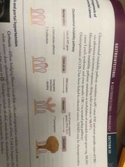
Adenomatous polyps |
1- Neoplastic bud chromosomal instability pathway and mutation in APC and KRAS 2- Usually asymptomatic May present with occult bleeding 3- 3 types 1- Tubular - less risk of malignant transformation 2- Villous - most risk of malignant transformation 3- Tubulovillous - Intermediate risk of malignant transformation |
|
|
Serrated polyps |
1- Neoplastic, Characterized by CpG island methylated phenotype (CIMP, cystine bases with guanin linked by phosphodiesterase binds) 2- Defect can silent MMR gene (DNA mismatch repair) expression 3- Mutation leads to microsatellite instability and mutation in BRAF (saw tooth pattern crypts on biopsy) 4- 20% sporadic |
|
|
Familial Adenomatous polyposis |
1- Autosomal dominant mutation of APC tumor suppressor genetic in chromosome 5q22 2- 2 hit hypothesis 3- Thousands if polyps develop after puberty, pancolitis always involve rectum 4- Prophylaxis collecting and 100% risk of CRC |
|
|
Gardner syndrome |
1- FAP + osseous and soft tissue tumor (eg osteoma of the skull or mandible) 2- Congenital hypertrophy of the retinal pigment epithelium 3- Impacted/supernumerary teeth |
|
|
Turcot syndrome |
1- FAP or lynch syndrome + malignant CNS tumor |
|
|
Peutz jeghers syndrome |
1- Autosomal dominant 2- Numerous polyps throughout the GI tract 3- Hyperpigmentation of the mouth, lips, hands and genitalia 4- Increase risk of breast and GI cancer |
|
|
Juvenile polyposis |
1- Autosomal dominant syndrome in children <5 years old 2- Numerous polys in the colon, stomach and small bowel 3- Associated with increase risk of CCR |
|
|
Lynch syndrome |
1- Previously called hereditary non-polyposis colorectal cancer (HNPCC) 2- Autosomal dominant mutation of mismatch repair genes (MLH1, MSH2) with subsequent microsatellite instability 3- 80% progress to CRC 4- Proximal colon always involved 5- Associated with 1- Endometrial cancer 2- Ovarian cancer 3- Skin cancer |
|
|
Epidemiology of colorectal cancer |
1- Age >50 years old 2- 25% familial |
|
|
Presentation of colorectal cancer |
1- Rectosigmoid> Ascending>Descending 2- Right side - (cecum and ascending colon) occult bleeding 3- Left side - (rectosigmoid) obstruction and hematochezia 4- Ascending 1- Exophytic mass 2- Iron deficiency anemia 3- Weight loss 5- Descending 1- Infiltrative mass 2- Colic pain 3- Hematochezia 4- Partial obstruction 6- May present with S.bovis bacteremia/endocarditis or an episode of diverticulitis |
|
|
Diagnosis of colorectal cancer |
1- Iron deficiency anemia in men and postmenopausal women >50 years old 2- Screening 1- Low risk - Screening at age 50 with colonoscopy every 10 years 2- Flexible sigmoidoscopy every 5 years 3- Fecal occult blood every year 4- Fecal immunochemistry test (FIT) every year 5- FIT- Fecal DNA every year 6- CT colonography 2- Patients with first degree relative who has colon cancer - Screening at age 40 or 10 year prior to relative presentation 3- Patients with IBD- distance screening protocol 3- Apple core lesion on barium enema X ray 4- CEA- monitor recurrence |
|
|
Risk factor for colorectal cancer |
1- Adenomatous and serrated polyps 2- Family History 3- IBD 4- Tobacco use 5- Diet height in processed meat and low fiber |
|
|
Molecular pathogenesis of colorectal cancer |
1- Chromosomal instability pathway with mutation of APC tumor suppers gene causing FAP and most sporadic CRC Decrease APC, increase KRAS 2- Adenoma-carcinoma sequence 3- Microsatellite instability pathway with mutation or methylation of mismatch repair gene causing lynch syndrome and some sporadic CRC 4- Serrated polyp pathway 5- Overexpression of COX 2 have been linked to CRC 6- NSAIDs are chemoprotective |
|
|
Chromosomal instability pathway |
Back (Definition) |
|
|
Cirrhosis |
1- Diffuse bridging fibrosis and nodular regeneration that disrupts the normal architecture of the liver 2- Increase risk of hepatocellular carcinoma 3- Causes 1- Alcohol 2- Non- alcoholic steatohepatitis 3- Chronic hepatitis 4- Autoimmune disease 5- Biliary disease 6- Genetic and metabolic disease |
|
|
Portal hypertension |
1- Increase pressure in the portal venous system 2- Causes 1- Cirrhosis 2- Venous obstruction 1- Budd- Chianti syndrome 2- Portal vein obstruction 3- Schistosoma |
|
|
Cirrhosis of the reproductive system |
1- Gynecomastia 2- Testicular atrophy 3- Amenorrhea 4- Due to increase estrogen |
|
|
What cells are responsible for fibrosis of the liver |
Hepatic stallet cell (ito) |
|
|
Spontaneous bacterial peritonitis |
1- Also called primary bacterial peritonitis 2- Common and potential fetal in patients with cirrhosis and ascites 3- Often asymptomatic can present with 1- Fever 2- Chills 3- Abdominal pain 4- ileus 5- Worsen encephalopathy 4- Commonly due to gram negative bacterial (E. Coli or Klebsiella) less common gram positive streptococcus 5- Diagnosis- Paracenthesis with ascitic fluid WBC >250 cells/mm and positive blood culture 6- Treatment - Ceftriaxone |
|
|
Enzymes related to liver damage |
1- Aspartate aminotransferase (AST) and Alanine aminotransferase (ALT) 2- Alkaline phosphatase 3- y glutamyl transpeptidase |
|
|
Aspartate aminotransferase and alanine aminotransferase AST and ALT |
1- In liver disease ALT> AST 2- In alcoholic liver disease AST> ALT 3- If AST>ALT in non-alcohol liver disease suggests progression to advances fibrosis or cirrhosis |
|
|
Alkaline phosphatase ALP |
1- Increase in 1- Biliary tree disease 2- Infiltrative disorders (placenta) 3-Bone disease |
|
|
Y glutamyl transpeptidase GGT |
1- Increase in liver and biliary disease 2- Associated with alcohol use |
|
|
Functional liver markers |
1- Albumin - Decrease in advance liver disease (market of livers bio synthetic function) 2- Bilirubin - Increase in liver disease 3- Prothrombin - Increase in liver disease (decrease production of coagulation factors, marker of liver biosynthetic function) 4- Platelets- 1- Decrease in advance liver disease (decrease thrombopoietin, liver sequestration) 2- Portal hypertension (splenic sequestration/Splenomegaly) |
|
|
Most common cause of increase AST |
Acetaminophen overdoses |
|
|
Reye syndrome |
1- Rare, fetal childhood hepatic encephalopathy 2- Associated with viral infection (VZ and Influenza) that has been treated with aspirin 3- Aspirin metabolic decrease beta oxidation by reversibly inhibiting mitochondrial enzymes 4- Findings 1- Mitochondrial symptoms 2- Fatty liver (microvesicular fatty changes) 3- Hypoglycemia 4- Hepatomegaly 5- Vomiting 6- Coma 5- Avoid aspirin in children unless they have Kawasaki disease |
|
|
Alcoholic liver disease |
1- Hepatic steatosis 2- Alcoholic hepatitis 3- Alcoholic cirrhosis |
|
|
Hepatic steatosis |
1- Macrovascular fatty changes that may be reversible with alcohol cessation |
|
|
Alcoholic hepatitis |
1- Require sustainable, long term consumption 2- Swollen and necrotic hepatocytes with neutrophilic infiltration 3- Mallory bodies (intracytoplasmic eosinophilic inclusions of damage keratin filaments) |
|
|
Alcoholic cirrhosis |
1- Final and usually irreversibly form 2- Sclerosis around central vein may be seen in early disease 3- Regenerative nodules surrounded by fibrous band in response to chronic liver injury 4- Portal hypertension and end stage liver disease |
|
|
Non-alcoholic fatty liver disease |
1- Metabolic syndrome (Insulin resistant) — obesity— fatty infiltrates in hepatocytes — cellular ballooning and eventually necrosis 2- May cause cirrhosis and HCC 3- Independent of alcohol use 4- ALT> AST |
|
|
Autoimmune hepatitis |
1- Chronic inflammation of the liver 2- More common in female 3- Usually asymptomatic May present with 1- Fatigue 2- Nausea 3- Pruritus 4- Associated with positive 1- Antinuclear antibodies (ANA) 2- Anti smooth muscle antibodies (anti-SMA) 3- Anti liver/kidney microsomal 1 antibody (anti- LKM1) 5- Labs increase AST and ALT 6- Histology 1- Portal and periportal lymphoplasmocytic infiltrate |
|
|
Hepatic encephalopathy |
1- Cirrhosis— portosystemic shunt — decrease ammonia metabolism- neuropsychiatric dysfunction 2- Reversible neuropsychiatric dysfunction ranges from disoriented/asterixis (mild) to difficult arosal or coma 3- Triggers 1- Increase ammonia production or absorption 1- Infection 2- GI bleeding 3- Constipation 2- Decrease ammonia removal 1- Renal failure 2- Diuretics 3- Post TIP procedure 4- Treatment 1- Lactulose (Increase ammonia generation) 2- Rifoximin (decrease ammonia producing gut bacteria) |
|
|
Hepatocellular carcinoma |
1- Most common primary malignant tumor of the liver in adults 2- Causes 1- HBV, HCV 2- Alcoholic/non-alcoholic liver disease 3- Autoimmune hepatitis 4- Wilson disease 5- Hemochromatosis 6- Aflotoxin from Aspergillus 3- May lead to bud chiari syndrome 4- Findings 1- Tender Hepatomegaly 2- Ascites 3- Anorexia 4- Jaundice 5- Polycythemia 5- Spread hematogenously 6- Diagnosis 1- Increase alpha feta protein 2- U/S or Contrast CT/MRT 3- Biopsy |
|
|
Angiosarcoma |
1- Malignant tumor of endothelial original 2- Associated with exposure to 1- Arsenic 2- Vinyl chloride |
|
|
Cavernous hemangioma |
1- Most common benign tumor of the liver (venous malformation) 2- Biopsy contraindicated due to risk of hemorrhage 3- Age 30-50 years |
|
|
Hepatic adenoma |
1- Rare benign tumor of the liver 2- Associated with OCP or anabolic steroid use 3- Risk of spontaneous regression or rupture ( abdominal pain or shock) |
|
|
Which viral factor for HCC can cause malignancy without underlying cirrhosis |
HBV |
|
|
Budd Chiari syndrome |
1- Thrombosis or compression of hepatic vein with centrilobular congestion and necrosis 2- Congestive liver disease features 1- Ascites 2- Abdominal pain 3- Varices 4- Hepatomegaly 5- Liver failure 3- Absence of JVP 4- Associated with 1- Hypercoagulablity state 2- Postpartum state 3- polycythemia 4- HCC 5- May lead to nutmeg liver (mottled appearance) |
|
|
Alpha 1 antitrypsin deficiency |
1- Misfolded gene product protein aggregates in the hepatocellular ER- cirrhosis with PAS + globulesin the liver 2- Codominant trait 3- Presents in young patients with liver damage and Dyspnea without history of smoking 4- In lungs - decrease alpha 1 antitrypsin— uninhibited elastase in alveoli — decrease elastic tissue — panacinar emphysema |
|
|
Jaundice |
1- Yellowing of the skin and/or sclera due to bilirubin deposition 2- Hyperbilirubinemia can be due to increase production or decrease clearance 3- Examplse 1- Hemolysis 2- Obstruction 3- Tumor 4- Liver failure |
|
|
Conjugated (direct) hyperbilirubinemia |
1- Biliary tract obstruction 1- Gallstone 2- Chilangiocarcinoma 3- Pancreatic or liver tumor 4- Liver flukes 2- Biliary tract disease 1- Primary sclerosing cholangitis 2- Primary biliary cholangitis 3- Excretion defects 1- Dublin-Johnson syndrome 2- Rotor syndrome |
|
|
Unconjugated (indirect) hyperbilirubinemia |
1- Physiological (newborns) 2- Hemolytic 3- Crigler- Nijjar syndrome 4- Gilbert syndrome |
|
|
Mixed hyperbilirubinemia |
1- Hepatitis 2- Cirrhosis |
|
|
Physiological neonatal jaundice |
1- At birth, immature UDP glucuronosyltransferase- Unconjugated hyperbilirubinemia- jaundice and/or kernicterus 2- Kernicturus- Deposition of Unconjugated lipid soluble bilirubin in the brain, particularly the basal ganglia 3- Present in the first 24hrs of life, resolved within 1-2 weeks without treatment 4- Treatment 1- Phototherapy (convert insoluble Unconjugated bilirubin to its water soluble form) |
|
|
Biliary atresia |
1- Most common cause for pediatric liver transplantation 2- Fibro-obliterative destruction of extrahepatic duct- cholestasis 3- Presents in newborn with 1- Persistent jaundice after 2 weeks of life 2- Dark urine 3- Pale stone 4- Hepatomegaly 3- Labs increase direct bilirubin and GGT |
|
|
Gilbert syndrome |
1- Milder decrease in UDP- Glucuronosyltransferase conjugation and decrease bilirubin uptake 2- Asymptomatic or mild jaundice with 1- Stress 2- Illness 3- Fasting 3- Increase Unconjugated bilirubin 4- Relatively common and benign |
|
|
Crigler- Najjer syndrome type 1 |
1- Absent of UDP-glucuronosyltransferase 2- Present at birth, may not have neurological symptoms until late in life 3- Features 1- Jaundice 2- Kernicturus 3- Unconjugated bilirubin 4- Treatment 1- Plasmapheresis 2- Phototherapy 3- Liver transplant m- curative 5- Type 2 is less severe and respond to phenobarbital with increase liver enzyme synthesis |
|
|
Dublin johnson syndrome |
1- Conjugated hyperbilirubinemia due to defective liver excretion 2- Grossly black (dark) liver due to impaired excretion of epinephrine metabolites 3- Benign |
|
|
Rotor syndrome |
1- Similar to Rubin johnson syndrome defective liver excretion 2- Milder in presentation without black liver 3- Due to impaired hepatic uptake and excretion |
|
|
Pattern of inheritance for hereditary hyperbilirubinemia |
Autosomal recessive |
|
|
Wilson disease |
1- Also called hepatolenticular degeneration 2- Autosomal recessive mutation in hepatocytes copper- transporting ATPas (ATP7B gene, chromosome 13) 3- Decrease copper incorporation in apoceruloplasm and excretion in bile - decrease serum ceruloplasm 4- Copper accumulate in 1- Brain 2- Cornea 3- Kidney 4- Liver 5- Increase urine copper
|
|
|
Presentation of Wilson disease |
1- Less than 40 years old with liver failure 2- Neurological symptoms (basal ganglia) 3- Psychological symptoms (behavioral) 4- Kayser- fleischer rings (deposit of copper in basement membrane of cornea) 5- Hemolytic anemia 6- Renal failure (fanconi syndrome) |
|
|
Treatment of Wilson disease |
1- Chelation with penicillamine or trientine, oral zinc 2- Liver transplant if acute liver failure related to Wilson disease |
|
|
Hemochromatosis |
1- Autosomal recessive 2- Of HFR gene chromosome 6 3- Associated with HLA- A3 4- Leads to abnormal iron sensing and increase intestinal absorption (increase ferritin and iron, decrease TIBC- increase transferrin saturation) 5- Iron overload can be 2’ chronic transfusion therapy (beta thalassemia major) 6- iron accumulate in 1- Pituitary 2- Heart 3- Liver 4- Pancreas 5- skin 6- Joints 7- Gonads
|
|
|
Diagnosis of hemochromatosis |
1- Hemosiferin (iron) on liver MRI 2- Biopsy with Prussian blue stain |
|
|
Presentation of hemochromatosis |
1- < 40 years old with total body iron >20 g, later in female 2- Restrictive cardiomyopathy ( classic) dilated cardiomyopathy (reversible) 3- Triad 1- Cirrhosis 2- Diabetes Mellitus 3- Skin pigmentation ( bronze diabetis) 4- Hypogonadism 5- Arthropathy (calcium pyrophosphate deposition in joins, especially MCP joint) 6- HCC major cause of death |
|
|
Treatment of hemochromatosis |
1- Phlebotomy 2- Iron chelation with deferoxamine, deferassisa, deferiprone |
|
|
Primary sclerosing cholangitis |
1- Unknown cause of concentric “onion skin” bile duct fibrosis 2- Alternating strictures and dilatation (beading) of intra and extrahepatic bile duct on ERCP and MRCP 3- Affect middle aged men with ulcerative colitis 4- Associated with ulcerative colitis 1- PANCA + 2- Increase IgM 5- Complication 1- Secondary biliary cholangitis 2- Cholangiocarcinoma or gallbladder cancer |
|
|
Primary biliary cholangitis |
1- Autoimmune reaction 1- lymphocytic infiltrate +/- granuloma 2- Destruction of lobular bile duct 2- Affect middle aged women 3- Associated with other autoimmune conditions 1- Hashimoto thyroiditis 2- Rheumatoid arthritis 3- Celiac disease 4- Anti mitochondria antibody positive and increase IgM 5- Treatment - urosodiol |
|
|
Secondary biliary cirrhosis |
1- Extrahepatic bile duct obstruction - increase pressure in intrahepatic duct - injury/fibrosis and bile stasis 2- In patients with know obstructive lesions 1- Gallstones 2- Strictures 3- Pancreatic tumor 3- May be complicated by ascending cholangitis |
|
|
Biliary tract disease |
1- Presentation 1- Jaundice 2- Pruritus 3- Dark urine/ pale stool 4- Hepatosplenomegaly 2- Labs 1- Increase conjugated bilirubin 2- Increase cholesterol 3- increase ALP 4- Increase GGT |
|
|
Cholangiocarcinoma |
1- Malignant tumor of the bile duct epithelium 2- Risk factors 1- Primary sclerosisng cholangitis 2- Liver flukes 3- Presents late with 1- Fatigue 2- Weight loss 3- Abdominal pain 4- Jaundice 4- Imaging May show bilary tract obstruction 5- Histology- Infiltrative neoplastic gland with desmoplastic stroke |
|
|
Choleithiosis |
1- Stone in the gallbladder 2- 2 types 1- Cholesterol 1- 80% of stones 2- Radiolucent with 10-20% radio-opaque 3- Risk factors 1- Obesity 2- Crohn’s disease 3- Advanced age 4- Estrogen 5- Mutilparity 6- Rapid weight loss 7- Native Americans 2- Pigmented stones 1- 1- Black (radio-opaque, Ca-bilirubinate, hemolysis ) 2- Brown (Bacterial breakdown) 2- Risk factors 1- Crohn’s disease 2- Chronic hemolysis 3- Alcoholic cirrhosis 4- Advance age 5- Biliary infection 6- TPN 3- Most common complications 1- cholecystitis 2- Acute pancreatitis 3- Ascending cholangitis 4- Diagnosis - U/S 5- Treatment - Elective cholecystectomy if symptomatic |
|
|
Biliary colic |
1- Dull RUQ pain associated with nausea and vomiting after meals 2- Neurohormal activity (eg CCK) trigger contraction of the gallbladder causing stone to move in cystic duct 3- Labs normal 4- U/s show cholelithiasis |
|
|
Choledocholithiasis |
1- Gallstone in the bile duct 2- Labs 1- Increase conjugated bilirubin 2- Increase ALP 3- Increase GGT 4- Increase AST/ALT |
|
|
Calculus cholecystitis |
1- Gallstone in the impacted at the cystic duct cause inflammation of the gallbladder and thickening of the gallbladder wall 2- May produce secondary infection 3- Most common type |
|
|
Acalculous cholecystitis |
1- Due to 1- Gallbladder stasis 2- Hypoperfusion 3- Infection (CMV) 2- Seen in critically I’ll patients |
|
|
Cholecystitis |
1- Inflammation of the gallbladder 2- Positive Murphy’s sign - 1- Inspiratory pause on palpating of RUQ due to pain 2- Pain radiates to the shoulder due to irritation of phrenic nerve 3- Increase ALP if bile duct involve 4- Diagnosis - U/S or HIDA scan (if gallbladder not visible on HIDA scan May suggest obstruction) |
|
|
Gallstone ileus |
1- Fistula between the gallbladder and GI tract 2- Gallstone enters GI lumen and cause obstruction at ileocecal valve 3- Air in the biliary tree (pneumobilia) 4- Rigler triad 1- Air in biliary tree (pneumobilia) 2- Small bowel obstruction 3- Gallstone (usually iliac fossa) |
|
|
Porcelain gallbladder |
1- Calcification of the gallbladder due to chronic cholecystitis 2- Found incidentally on imaging 3- Treatment Prophylaxis cholecystectomy due to increase risk of gallbladder cancer (adenocarcinoma) |
|
|
Ascending cholangitis |
1- Infection of the biliary tree due to obstruction that leads to stasis and bacterial overgrowth 2- Charcot triad 1- RUQ pain 2- Fever 3- Jaundice 3- Reynolds’s pentad 1- Charcot triad 2- Altered mental status 3- Shock (hypotension) |
|
|
4 risk factors for development cholelithiasis |
Fat Fertile Female Forty |
|
|
Acute pancreatitis |
1- Inflammation of the pancreas 2- Auto-digestion of the pancreas by pancreatic enzymes surrounded by edema 1- Cause 1- Idiopathic 2- Gallstone (40%) 3- Ethanol (30%) 4- Trauma 5- steroid 6- Mumps 7- Autoimmune 8- Scorpion sting 9- Hypercalcemia/Hypertriglyceridemia (1-4%) 10- ERCP 11- Drugs (sulfa, NRTI, protease inhibitors) 4- Diagnosis (2 of 3) 1- Acute epigastric pain radiating to the back 2- Increase serum amylase and lipase (more specific) 3x the upper limit of normal 3- Imaging (CT or MRI with contrast) 5- Complications 1- Pseudocysts (surrounded by granulation tissue not epithelium) 2- Abscess 3- Hemorrhage 4- Necrosis 5- Infection 6- Hypocalcemia/Hyperglycemia 7- End organ failure (SIRS, ALF, renal failure) 8- Chronic pancreatitis
|
|
|
Chronic pancreatitis |
1- Chronic inflammation, atrophy and calcification of the pancreas 2- Cause 1- alcohol abuse 2- Genetics (cystic fibrosis) 3- Idiopathic 3- Serum amylase and lipase may not be elevated 4- Complication 1- Pseudocysts 2- Pancreatic insufficiency 5- Pancreatic insufficiency (<10% pancreatic function) leads to 1- Steatorrhea 2- Fat soluble vitamins deficiency 3- Diabetis Mellitus |
|
|
Pancreatic adenocarcinoma |
1- Malignant tumor arises from the pancreatic duct 2- Disorganized cellular structure with cellular infiltrate 3- Often metastasis at present 4- More common in the head of the pancreas 5- Associated with CA-19-9 and CEA markers 6- Survival ~ 1 year after diagnosis |
|
|
Risk factor for pancreatic adenocarcinoma |
1- Age > 50 years 2- Chronic pancreatitis (>20 years) 3- Diabetes Mellitus 4- Tobacco use 5- Jewish and African-American males |
|
|
Presentation of pancreatic adenocarcinoma |
1- Abdominal pain radiating to back 2- Weight loss (malabsorption and anorexia) 3- Migratory thrombophlebitis- redness and tenderness of the extremity trousseau syndrome 4- Courvoisiers sign- palpable painless gallbladder |
|
|
Treatment of pancreatic adenocarcinoma |
1- Whipple’s procedure (pancreaticoduodenectomy) 2- Chemotherapy 3- Radiation therapy |