![]()
![]()
![]()
Use LEFT and RIGHT arrow keys to navigate between flashcards;
Use UP and DOWN arrow keys to flip the card;
H to show hint;
A reads text to speech;
73 Cards in this Set
- Front
- Back
|
Endocrine System |
Plays a big role in maintaining homeostasis -The organs of this system are the endocrine glands that secrete messengers called hormones directly in to the bloodstream and then carried to their target organ, tissues or cells |
|
|
Hypersecretion |
if a gland secretes too much hormone--target will overreact |
|
|
Hyposecretion |
The gland secretes too little hormone--target will not be active |
|
|
Organs of Endocrine System |
|
|
Pituitary Gland |
-also known as the hypophysis is located inferior to the hypothalamus and is connected to the hypothalamus by a narrow stalk of tissue called the infundibulum -It is divided into two lobes: the anterior and posterior -These lobes function independently of one another -Called the master gland because of its role of regulating other endocrine glands |
|
|
The following are hormones secreted by Pituitary Gland: |
none |
|
|
From the Anterior Pituitary:
GH |
TARGET: Bones, muscles, and other tissues ACTION: Stimulates Cell Growth and division |
|
|
ACTH |
TARGET: Adrenal Cortex ACTION: Stimulates release of Adrenal Cortex Hormones |
|
|
TSH |
TARGET: Thyroid Gland ACTION: Stimulates release of Thyroid Hormones |
|
|
FSH |
TARGET: Gonads ACTION: Stimulates development of ova and sperm |
|
|
LH |
TARGET: Gonads ACTION: Stimulates secretion of sex hormones |
|
|
PRL |
TARGET: Mammary Glands ACTION: Stimulates milk production |
|
|
MSH |
TARGET: Melanocytes ACTION: Stimulates Melanin production |
|
|
Posterior Pituitary:
|
TARGET: Renal Tubules ACTION:Stimulates water reabsorption |
|
|
OXT |
Target: Uterus and Mammery Glands ACTION: Stimulates Uterine contractions during labor and delivery and the release of milk |
|
|
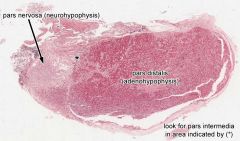
Posterior Lobe: |
Also called the neurohypophysis and composed of neurons that originate in the hypothalamus PARS NERVOSA |
|
|
Anterior Lobe |
Also called the Adenohypophysis Composed of granular tissue and regulated by the hypothalamus PARS DISTALIS |
|
Thyroid |
Function Independently of one another Hormones: T4 and T3: Secreted by Follicle Cells Target: essentially every cell of the body Action: Work together to regulated oxygen utilization and rate of metabolism of the cells CT: Secreted by parafollicular cells Target: bone and kidneys
|
|
|
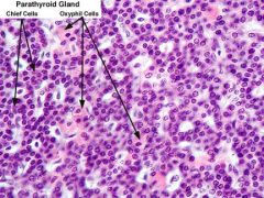
Parathyroid: |
There are Four Pea Like thyroid glands located on the posterior surface of the thyroid gland however they are completely independent of the thyroid--they are parathyroid glands composed of chief cells and oxyphil cell The chief cells secrete the Parathyroid Hormone (PTH): Target: Bone and Kidneys Action:Raises blood calcium levels by causing calcium to be removed from bone and reabsorbed by the kidney |
|
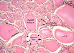
THYROID SLIDE |
Parathyroid Slide |
|
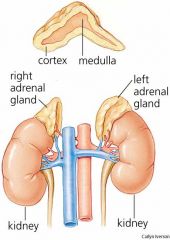
Adrenal Glands
|
Located above each kidney--function independently of the kidney -Is subdivided into two regions with different functions: the outer cortex and the inner medulla. It produces many steroid hormones called corticosteroid hormones. From the Cortex: Mineralcorticoids: Target: Kidney Action: Increases renal absorption of sodium
Glucocorticoids: Target: Most cells Action: affects rate of glucose metabolism |
|
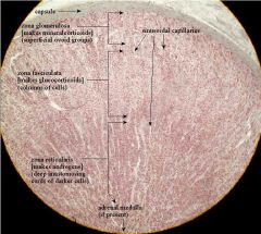
Capsule Zona Glomerulosa Zona Fasciculata Zona Reticularlis Adrenal Medulla--secretes epinephrine and nonepinephrine |
none |
|
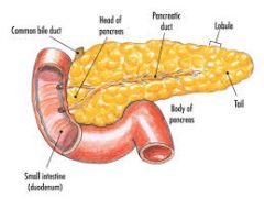
Pancreas |
The pancreas is unique in that it is both an exocrine and endocrine gland. The exocrine cells are arranged into pancreatic acini. They secrete digestive enzymes and buffers into the pancreatic duct that carries them into duodenum to aid in the digestion of food in the intestine. The endocrine cells are visible clumps of cells scattered throughout the pancreas called pancreatic islets
|
|
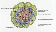
Outer-Exocrine pancreatic acini Inner- Endocrine Pancreatic Islets Along with Alpha and Beta Cells |
Hormones : Insulin: Target-essentially every cell of the body Action: lowers blood glucose levels by stimulating the uptake of glucose from the bloodstream by the cells of the body
Glucagen: Targets: Liver Action: Raises blood glucose level by stimulating liver to break down glycogen and release glucose back into the bloodstream. |
|
|
Thymus Gland |
Is located in the superior mediastinum of the thoracic cavity posterior to the sternum. It is large in infants and children and virtually disappears as an adult. Secrets Thymosin which plays a role in the maturation in the T Lymphocytes |
|
|
Pineal Gland |
Located on the posterior wall of the Thalamus -Secrete Melatonin, which regulates the body's 24 hour clock
|
|
|
Testes |
Male Gonads and produce male gametes or sperm along with testosterone
|
|
|
Ovaries |
Female Gonads which produce female gametes or ova. -Secrete estrogen and progesterone |
|
|
Hypersecretion of Growth Hormone |
Gigantism-bones grow too fast |
|
|
Hyposecretion of Growth Hormone |
Dwarfism |
|
|
Hyposecretion of Antidiurtec Hormone |
Diabetes Insipidus- the kidneys are not able to reabsorb water when the body needs to conserve water
|
|
|
Hyposecretion of Parathyroid Hormone |
Tetany- low blood level of calcium, leading to muscle and nerve irritability |
|
|
Hypersecretion of Parathyroid Hormone |
Recklinghausen's Disease- the bones lose calcium and blood calcium level is too high
|
|
|
Hyposecretion of Insulin |
Diabetes Mellitus- There are high levels of glucose in the bloodstream |
|
|
Hypersecretion of Epinephrine |
Pheochromocytoma- increased heart rate and blood pressure |
|
|
Hypersecretion of Androgens |
Virilism-The appearance of male secondary sexual characteristics in a female |
|
|
Hyposecretion of T3 and T4 |
Hashimoto's Disease- low metabolic rate, weakness, mental and physical sluggishness |
|
|
Hypersecretion of Insulin |
Hypoglycemia-low levels of glucose in the bloodstream, producing fatigue and fainting |
|
|
Cardiovascular Physiology |
In order to pump blood, the heart alternates between relaxation phase DIASTOLE during which the heart chambers FILL up with blood. And a contraction phase SYSTOLE during which the heart EJECTS blood. |
|
|
Cardiac Cycle |
The alternation between diastole and systole. Each heart beat lasts one cardiac cycle.
As the surge of blood that has been ejected from the heart moves through the blood vessels it can be palpated as the pulse in arteries |
|

Heart Valves |
One pair of valves- the Antrioventicular valves--the TRICUSPID on the right side and the MITRAL valve on the left) are found between the atria and the ventricles. They prevent blood in the ventricles from flowing back into the atria when the ventricles contract. The second pair of valves are SEMILUNAR with the PULMONARY valve on the right and the AORTIC on the left) these prevent blood that has just been ejected into the great arteries from flowing back into the ventricles when they relax. The two sounds LUB (longer and louder) AND DUB (shorter and shaper) occur when these valves close
|
|
|
Blood Pressure |
Is the pressure exerted by blood against the walls of the blood vessels.
-Blood pressure rises and falls as the heart contracts and relaxes. So it is necessary to report it as two numbers such as 120/70 The higher number represents SYSTOLIC -produced by the left ventricle contracts to eject a fresh bolus of blood into the Aorta. The lower number represents the DIASTOLIC Pressure or when the ventricles and fill with blood |
|
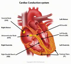
Conduction System |
is a network of modified, autorhythmic heart muscles cells that are ableto stimulate a heartbeat. |
|
|
1. SA NODE |
The pacemaker of the heart -Contractile cells that initiate stimulus that results in heart contraction -Stimulates internodal pathway |
|
|
2. Internodal Pathway |
-Conductile Cells -receives stimulus from the SA Node -distributes stimulus throughout atria -Stimulates atrial contraction and AV node |
|
|
3. AV Node |
-Receives stimulus from internodal pathway -Stimulates AV Bundle -contractile cells may initiate stimulus if SA Node does not |
|
|
4. Right Bundle Branch |
-Conductile cells that carry stimulus to apex of the right ventricle -Stimulates Purkinje fibers |
|
|
5. Purkinje Fibers |
-network in each ventricle wall -carry stimulus to ventricular cardiac muscle cells |
|
|
6. Left Bundle Branch |
-Conductile cells that carry stimulus to apex of left ventricle |
|
|
AV Bundle |
Located in Atrioventicular Septum -receives stimulus from AV NODE -Conductile cells carry stimulus to bundle branches -Also called bundle of His |
|
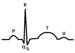
QRS Complex |
Q-small downward deflection R-Very Tall upward deflection S-small to medium downward deflection Represents: Ventricular Depolarization Followed By: Ventricular Systole
|
|
|
P Wave |
A small, upward deflection Represents: Atrial Depolarization Followed by: Atrial Systole |
|
|
T Wave |
Medium, Upward deflection Represents: Ventricular Repolarization Followed By: Ventricular Diastole |
|
|
Arrythmias |
Is a general term that refers to a change in the rhythm of the heartbeat |
|
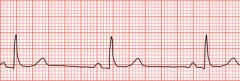
Bradycardia |
The ECG wave is normal, but the heart rate is usually less than 60 bpm. This occurs when the SA Node does not initiate the wave of stimulation often enough |
|
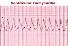
Ventricular Tachycardia |
The ECG shows multiple QRS complexes without visible P or T waves. This rhythm indicates damage to the ventricles. |
|
Atrial Flutter |
-The atria are being stimulated at a very fast rate. This results in a quivering in the atrial heart muscle. The ECG shows several small P waves before each QRS |
|
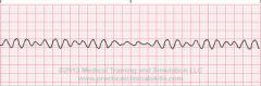
Ventricular Fibrillation |
There is no organized wave of stimulation. -Since there is no organized contraction of th ventricles blood is not effectively ejected from the heart |
|
|
Respiratory System Ventilation |
The process of moving air in and out of the lungs |
|
|
Inhalation |
Brings fresh air into the AVEOLI of the lungs where OXYGEN is loaded onto the red blood cells for delivery to the cells of the body. |
|
|
Exhalation |
Expels stale air containing CARBON DIOXIDE that was removed from the blood stream |
|
|
Inhalation and exhalation are the processes by which the body brings in oxygen and expels carbon dioxide. The breathing process is aided by a large dome-shaped muscle under the lungs called the diaphragm. When you breathe in, the diaphragm contracts downward, creating a vacuum that causes a rush of fresh air into the lungs. The opposite occurs with exhalation, where the diaphragm relaxes upwards, pushing on the lungs, allowing them to deflate. |
none |
|
|
VT TIDAL VOLUME |
Volume of Air moving into or our of lungs during quiet breath |
|
|
IRV Inspiratory Reserve Volume |
Maximum volume of air that can be inhaled above tidal volume |
|
|
ERV Expiratory Reserve Volume |
Maximum volume of air that can be exhaled below tidal volume |
|
|
RV Residual Volume |
Volume of Air that remains in respiratory system after maximal exhilaration |
|
|
IC Inspiratory Capacity |
VT + IRV
|
|
|
FRC Functional Residual Capacity |
ERV + RV |
|
|
VC Vidal Capacity |
IRV + VT + ERV |
|
|
TLC Total Lung Capacity |
IRV + VT + ERV +RV |
|
|
Hyperventilation |
Breathing Faster and Deeper increases the amount of Carbon Dioxide expelled from the body |
|
|
Rebreathing |
Increases the concentration of carbon dioxide in inhaled air |
|
|
Exercising |
Increases the rate of cellular respiration, for powering muscle, thereby increasing the amount of carbon dioxide waste produced |