![]()
![]()
![]()
Use LEFT and RIGHT arrow keys to navigate between flashcards;
Use UP and DOWN arrow keys to flip the card;
H to show hint;
A reads text to speech;
41 Cards in this Set
- Front
- Back
|
What is the small intestine the site of? |
The small intestine is the site of terminal digestion of food |
|
|
What does the ileum consist of? |
Itconsists of three parts: the duodenum, the jejunum and the ileumwhich have common histological and topographical features aswell as distinctions allowing each region to be identified |
|
|
Which sections are retroperitoneal and which are intraperitoneal? |
Theduodenum is, for the most part, a retroperitoneal structure whilethe jejunum and ilium are intraperitoneal. |
|
|
What does the inner lining of the small intestine form? |
Throughout its length the inner lining forms a series of ridges, theplicae circularis, which are covered with tall finger-like projectionsof the mucosa to form villi |
|
|
What are between these tubular glands? |
Between these are the tubular glandsor crypts of Lieberkuhn which extend to or beyond the musculariscoat |
|
|
What is the epithelium of the small intestine? |
The epithelium of the small intestine is simple columnar(enterocytes) with goblet cells |
|
|
What are the simple columnar cells? |
The simple columnar cells are theabsorptive cells and have a brush border to increase their surfacearea. |
|
|
What features of the small intestine enhance it's surface area? |
The small intestine hasa folded mucosal coat,a series of villi and anepithelium with a brushborder. All thesefeatures greatlyincrease the surfacearea. |
|
|
Why do the ducts of the gall bladder and pancreas open into the duodenum? |
The ducts of the gall bladder and pancreas open into the duodenum bringing digestiveenzymes, bicarbonate (to counter the acidic chyme entering the duodenum from thestomach and bile salts from the liver to emulsify the fats |
|
|
How does the membrane change from the jejunum to the ileum? |
There is a decrease inthe folding of themucous membraneand an increase inlymphoid tissue fromjejunum to ileum |
|
|
What are the layers of the small intestine? |
The layers of thesmall intestine followthe same pattern asthe rest of the GIT inhaving a mucosa, asubmucosa, amuscular layer anda serosa. |
|
|
What types of cells does the epithelium consist of? |
The epithelium consistsof 5 types of cells: 1. Enterocytes 2. Goblet 3. Paneth 4. Endocrine 5. Stem |
|
|
What are the enterocytes? |
The enterocytes are thetall columnar cell withbasely placed nucleiwhich contain between 2-3000 microvilli and,which in turn, are coatedwith glycoproteinscontaining a number ofenzymes. The enterocytes are theabsorptive cells. |
|
|
How are Glucose and galactose absorbed by the enterocyte? |
Glucose and galactose are absorbedby the enterocyte by active transportusing Na+ dependent glucosetransporters (SGLT1) |
|
|
How is fructose absorbed by the enterocyte? |
Fructose is absorbed by theenterocyte by active transport usingNa+ dependent glucose transporters (GLUT5) |
|
|
How do the absorbed monosaccharides pass through the basal membrane? |
The absorbed monosaccharidespass through the basal membraneusing GLUT2 glucose transporters |
|
|
How are proteins digested and absorbed in the small intestine? |
Protein digestion starts in thestomach with pepsin whichhydrolyses proteins into largepolypeptides. Pancreatic proteolytic enzymesacting in the small intestine breakdown the polypeptides into freeamino acids which are transportedinto the cell and from there into theunderlying capillaries. |
|
|
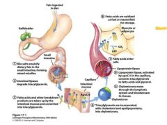
Describe the Digestion & Absorption of Lipids |
|
|
|
What are the goblet cells of the epithelium of the small intestine? |
These are situated amongst the enterocytes and in thesmall intestine are as numerous as the enterocytes. |
|
|
What are the paneth cells of the epithelium? |
Paneth cells are found in the lower third of the cryptsand have basal nuclei and prominent granules. Theyrelease digestive enzymes, immunoglobulins andlysozyme. Paneth cells secretions therefore help todestroy parasites and bacteria |
|
|
what are the endocrine cells? |
These are usually found in the lower 1/3rd of thecrypts and secrete a number of hormones |
|
|
What are the functions of the endocrine cells? |
1. Regulate water and electrolyte metabolism andenzyme secretion. 2. Regulation of gastrointestinal motility and mucosalgrowth. 3. Stimulation of the release of other hormones |
|
|
What does the lamina propria consist of? |
The lamina propria consists of loose connective tissue with bloodand lymph vessels, nerve fibres and smooth muscle |
|
|
What types of tissue can be seen indicative of the lamina propria? |
The quantityof lymphoid tissue increases and in the ileum Peyer’s patches areclearly seen. There are also varying amounts of glandular tissue |
|
|
What do the peyer's patches contain? |
Peyer’s patches contain a germinal centre and a dome and arecovered by M cells. The dome cells are B lymphocytes and thegerminal cells are IgA positive B cells. |
|
|
What does the submucosa contain in the duodenum? |
The submucosa contains the Brunner gland in the duodenum. |
|
|
What are the features of the muscularis? |
Themuscularis is well developed and composed of an inner circularand outer longitudinal layers of smooth muscle. |
|
|
What is the distinctive feature of the duodenum? |
The distinctive feature of the duodenum is thesubmucosal Brunner glands which are mostly of themucous type which aid in neutralising the acid chymeextruded from the pylorus |
|
|
Where can the brunners glands be seen? |
Beneath the surfacevilli the Brunnerglands can clearlybe seen. Thesediminish in numberalong the length ofthe duodenum. Theglands secreteapprox. 200ml perday. |
|
|
What are the features of the jejunum? |
The jejunum has the tallest villi, there are no BrunnerGlands and few lymphoid follicles |
|
|
How can the ileum be identified? |
The ileum is easily identified due to its aggregations oflymphoid tissue, Peyer’s patches |
|
|
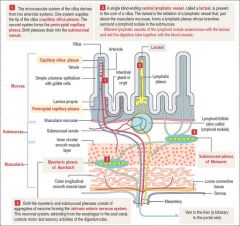
What is the Blood, Lymph & Nerve Supply to the Small Intestine? |
|
|
|
What are the parts of the large intestine? |
The parts of thecolon are thecaecum, vermiformappendix,ascending,transverse anddescending colon,the sigmoid colon,the rectum andanal canal |
|
|
What is the function of the colon? |
The colon acts to resorb fluid andelectrolytes and form the faecalmass. |
|
|
What is one of the more obvious external features of the Colon? |
One of the more obviousexternal features of the largeintestine is the taenia coli which arethe three ribbon like thickenings ofthe longitudinal muscle. They givethe large intestine its distinctive‘puckered’ appearance |
|
|
Internally, what does the mucous membrane contain? |
Internally the mucous membranecontain a large number of glands tolubricate the colon to ease thepassage of the increasingly drymass |
|
|
Why are there large numbers of lymphocytes in the colon? |
There are large numbers oflymphocytes both in small groupsand large aggregates to counter thebacteria found in this region. |
|
|
What are the features of the layers of the colon? |
There are no villi in the colon. Thesurface cells are columnar with thecrypts containing large quantities ofgoblet cells |
|
|
What is the morphology of the enterocytes in the colon? |
The morphology of the enterocytes issame as for the small intestine andreabsorption is accomplished by thesame Na+/K+ activated ATPase driventransporter system |
|
|
Why do the goblet cells continually secrete? |
The goblet cellscontinually secrete to lubricate the bowelto facilitate the passage of theincreasingly solid contents |
|
|
Why is there considerable reabsorption in the colon? |
There is considerable reabsorption asseveral litres of fluid are added duringthe digestive process which does notinclude fluid taken in with the food e.g.1litre from saliva, 1litre from thepancreas, 500ml from bile production |