Reading...
![]()
Play button
![]()
Play button
![]()
Use LEFT and RIGHT arrow keys to navigate between flashcards;
Use UP and DOWN arrow keys to flip the card;
H to show hint;
A reads text to speech;
28 Cards in this Set
- Front
- Back
|
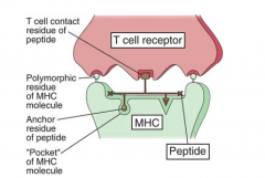
Antigen presentation
|
- For effective presentation, an antigen fragment is displayed within the groove of special antigen presenting molecules
- These antigen presenting molecules are proteins encoded by the MAJOR HISTOCOMPATIBILITY COMPLEX (MHC) |
|
|
Antigen presenting cells
|
- Dendritic cells (most efficient during primary response)
- Macrophages - B cells |
|
|
Immature dendritic cells
|
Express:
- High concentrations of FcγR - Low concentrations of both class II MHC and costimulatory molecules (e.g. B7-1, B7-2) |
|
|
Dendritic cells
|
- Derived from CD34+ bone marrow stem cells
- Can be generated in vitro from blood monocytes or from CD34+ hematopoietic stem cells (HSC) in the presence of GM-CSF and other cytokines - Express CD4 - Capture antigen in the periphery and transport it to the appropriate lymphoid tissue |
|
|
CD4
|
- 1 receptor for the human immunodeficiency virus, type 1 (HIV-1)
- Infection with HIV-1 is initiated by interaction of viral envelope proteins with (at least) two receptors, the CD4 molecule and a chemokine receptor, CCR5 and/or CXCR4 |
|
|
Macrophages
|
- For primary immune responses the macrophage is NOT as effective in antigen presentation as the dendritic cell
- Express CD4 and the chemokine receptors CCR5 and/or CXCR4 - Macrophage activation upregulates Class II MHC and costimulatory molecules |
|
|
B cells
|
- B cells recognize antigen via antibodies
- Antigen recognition by B cells is so specific, B cells can bind antigen even when it is present in low concentration - B cells serve as the predominant antigen presenting cells when antigen is limiting |
|
|
Class I MHC molecules
|
All nucleated cells express class I MHC molecules
|
|
|
Class II MHC molecules
|
Only professional antigen presenting cells express MHC II
|
|
|
MHC
|
- Located on chromosome 6
- In humans, the class I MHC molecules are encoded by three loci (A, B, and C) - Class II MHC molecules are encoded by a “D” locus subdivided into 3 loci: DP, DQ, and DR |
|
|
General Features Polymorphism
|
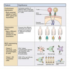
- Genes encoding each MHC locus are present in multiple forms (alleles) within the population
- The likelihood that 1 of the MHC alleles present in individuals within a population will bind to, and present, an antigen fragment of any particular microorganism to T cells, is increased by the existence of numerous alleles - Differences in susceptibility to infection are believed to be a reflection of unique presentation of antigen to T cells by the MHC of susceptible versus resistant individuals |
|
|
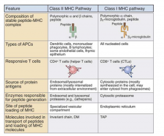
Role of Class II MHC Molecules and Class II MHC Restriction
|

- Class II MHC molecules present antigen to CD4+ T cells
- CD4+ T cells ONLY recognize antigenic peptides if they are displayed with Class II MHC molecules present on the surface of so-called “professional” antigen presenting cells - CD4+ T cells are RESTRICTED to “see” antigen in association with class II MHC = class II MHC restriction |
|
|
Structure of Class II MHC molecules
|
- Heterodimer consisting of two transmembrane polymorphic polypeptide chains (~29 kd each) in non-covalent association
- DP, DQ, and DR, are structurally similar and each binds antigenic fragments that vary between 15 to 30 amino acids in length |
|
|
Invariant chain - MHC II
|
- Transiently associates with class II MHC molecules in the endoplasmic reticulum, forming a trimeric complex (class II MHC heterodimer and Ii).
- This invariant chain blocks the binding of superfluous peptides that the MHC heterodimer might encounter in the endoplasmic reticulum. - Serves to direct the transport of class II MHC molecules through the golgi, from where they are released within endosomes |
|
|
Features of MHC I & II
|
|
|
|
Codominance of MHC II
|
Codominant expression of HLA molecules means that one copy of each HLA (DP, DQ, and DR) inherited from each parent will be expressed. Therefore a minimum of six different class II molecules will be expressed on the cell surface providing the parents are genetically unrelated.
|
|
|
Class I MHC molecules: Role of Class I MHC Molecules and Class I MHC Restriction
|
- Infected cells are identified for destruction by CD8+ T cells following expression of a complex on the target cell surface, composed of an antigen fragment displayed in association with class I MHC molecule.
|
|
|
Structure of Class I MHC
|
- Each class I MHC molecule is composed of a transmembrane polymorphic polypeptide “heavy” chain (~45 kd) non-covalently associated with a smaller polypeptide (~12 kd), termed β2−microglobulin (nonpolymorphic molecule)
|
|
|
Codominance of I MHC
|
- In humans, the class I MHC molecules are HLA-A, HLA-B, and HLA-C, each of which is codominantly expressed on all nucleated cells
- Codominant expression of HLA molecules means that one copy of each HLA (A, B, and C) that is inherited from each parent will be transcribed and translated into protein |
|
|
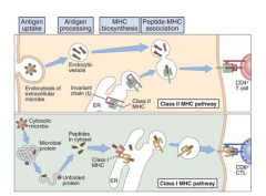
Exogenous Antigen Processing and Presentation by Class II MHC
|

1. Uptake of antigen by antigen presenting cells
2. Fusion of the endocytotic vesicle with lysosomes that release their contents into the endosome. 3. 2nd fusion process creating a chimeric endosome - class II MHC/Ii complex is exposed to lysosomal enzymes, 4. Li chain is degraded, and an antigenic peptide binds in the newly exposed groove of the class II MHC. 5. The chimeric endosome migrates to, and fuses with, the cell membrane 6. When these complexes are recognized by T cell receptors present on CD4+ T cells - activated to secrete cytokines |
|
|
Endogenous Antigen Processing and Presentation by Class I MHC
|

- Cytosolic proteins are targeted for degradation following the attachment of ubiquitin, a small peptide found in the cytosol of all cells.
- Peptide fragments generated by the proteasome, in turn, bind to the transporter of antigen processing (TAP) proteins, and are transported into the endoplasmic reticulum, where they contact coassembled class I MHC molecules - Complexes are released from the GOLGI into vesicles that fuse with the cell membrane |
|
|
Cross Presentation: Exogenous Antigens and Presentation by Class I MHC
|
- Exogenous antigens normally processed in phagolysosomes and presented on the cell surface, are instead presented by Class I MHC molecules
- Mediated primarily by dendritic cells - Exogenous antigens can be presented with Class I MHC, to CD8+ T cells |
|
|
Viral Sabotage of Antigen Processing & Presentation by Class I MHC Molecules
|
Sabotaging the expression of the peptide/class I MHC complexes on the cell surface effectively protects the virus and the infected cells can serve as veritable “factories and reservoirs” for viruses.
|
|
|
Herpes simplex virus (HSV)
|
- Neurons express little cell surface class I MHC molecules, and so they provide a relatively safe haven for viruses
- Recurrent infections of non neuronal tissues can occurs because HSV also produces an immediate early protein that binds to the cytosolic portion of the TAP transporter. - Inhibits TAP-mediated translocation of peptides from the cytosol into the ER - In the absence of peptide transport to ER, antigenic peptide/class I MHC complexes do not form, and the infection (by virus) in the cell remains undetected |
|
|
Epstein Barr virus (EBV)
|
- One of the mechanisms by which EBV thwarts the immune system is by inhibiting the activity of proteasomes
- Virus prevents hydrolysis of viral proteins into peptide size fragments - Only peptides (antigenic fragments) of a suitable size can bind in the “groove” of class I MHC molecules - Whole virus, or even whole viral protein, is too large to bind in this groove - EBV remains safely sequestered in the B cell |
|
|
Cytomegalovirus (CMV)
|
- Member of the herpes family, infects immunocompetent individuals without causing symptoms
- In immunocompromised individuals, infection with CMV may cause numerous disorders including encephalitis, pneumonia, hepatitis, retinitis or blindness - CMV encodes proteins that redirect newly synthesized class I MHC molecules from the ER back into the cytoplasm where they themselves are degraded by the proteasome |
|
|
Nonresponsiveness
|
Determinant Selection is a term used to describe a phenomenon in which the MHC molecules present on the cell do not bind a particular peptide and so that peptide cannot be presented to T cells
|
|
|
CD1 molecules as Antigen Presenting Molecules
|
- CD1 molecules are glycoproteins that present lipid and glycolipid antigens (e.g., mycobacterial phospholipids, glycolipids) to subsets of T cells
- Structurally the CD1 molecules are similar to Class I MHC in that they form a complex with β2 microgloblin - Consists of 5 members that can be divided into two groups CD1a, CD1b, CD1c, and CD1e - All CD1 proteins have a group of hydrophobic amino acids in hydrophobic pockets which can interact with hydrophobic molecules (e.g., lipids) |