![]()
![]()
![]()
Use LEFT and RIGHT arrow keys to navigate between flashcards;
Use UP and DOWN arrow keys to flip the card;
H to show hint;
A reads text to speech;
72 Cards in this Set
- Front
- Back
|
Describe the directions/orientation for the human body. |
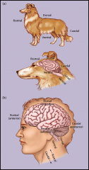
Superior - top - dorsal |
|
|
What are the 3 different section/cuts of the brain?
|
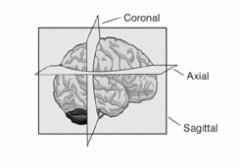
Sagittal section: divides the brain into right and left (hemispheres) Coronal, frontal transverse section: divides brain into front and back Horizontal (axial) section: divides brain into upper and lower |
|
|
What cut produces a a view of the inner medial surface of the brain? |
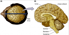
a mid-sagittal cut |
|
|
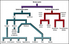
What are the divisions of the nervous system and their functions? (simple version) |
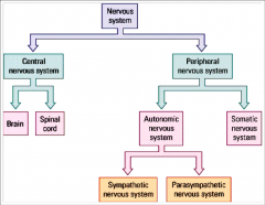
Nervous system: composed of nerves and specialized cells that send signals throughout the body Central nervous system: made up of the brain and spinal cord, controls most functions of the body and mind Brain: all functions Spinal cord: transmission of neural signals Peripheral nervous system: connects the CNS to the limbs and organs, communication relay Autonomic nervous system: regulates functions of internal organs. Not aware of this, cannot control, involuntary Sympathetic nervous system: fight or flight response Parasympathetic nervous system: responsible for rest and digest, or feed and breed. Somatic nervous system: skeletal and muscle movement. We are aware of this, it is voluntary. |
|
|
How does the sympathetic nervous system change the body in terms of the fight or flight response? (10 things) |
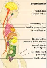
1. Pupils dilate
2. Salvation inhibited 3. Increased respiration 4. Bronchial passages dilated 5. Increased heart rate 6. Digestion inhibited 7. Secretion of adrenal hormones 8. Increased secretion by sweat glands 9. Hair follicles raised/goosebumps 10. Bladder relaxed |
|
|
How does the parasympathetic nervous system change the body in terms of "rest and digest"? (7 things) |
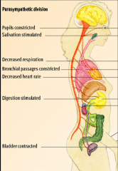
1. Pupils constricted 2. Salvation stimulated 3. Decreased respiration 4. Bronchial passages constricted 5. Decrease heart rate 6. Digestion stimulated 7. Bladder contracted |
|
|
What are the 4 types of spinal nerves and their functions? |
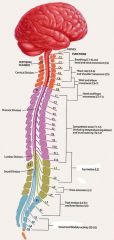
Cervical: muscles of your neck, shoulders, arms and hands, and diaphragm. Thoracic: trunk muscles and muscles involved with breathing. Lumbar: hip leg and foot muscles Sacral: Anal and urethral sphincters |
|
|
What are dermatomes? |
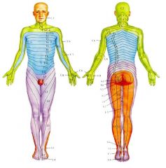
Dermatomes represent specific regions of nerve reception of sensory impulses. They are an area of the skin supplied by nerves from a single spinal root.
|
|
|
Describe the anatomy of the spinal cord. |
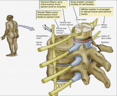
- Spinal nerves connect to spine - Dorsal root/fibres (front) carries sensory information from body to spinal cord (AFFERENT) - Ventral root/fibres (back) carries motor information from spinal chord to muscles (EFFERENT) - interneurons in the spine (grey as an X, white surrounding) - covered by layers of dura matter |
|
|
What are the 3 major divisions of the brain? |
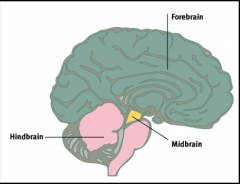
1. Forebrain 2. Midbrain 3. Hindbrain. |
|
|
What does the Hindbrain (Rhombencephalon) consist of? |
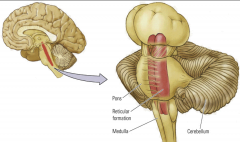
(Metencephalon "across brain") (Mylencephalon "spinal brain") - Pons (nuclei that relay signals from forebrain to cerebellum) - Cerebellum (balance, coordination) - Part of the Reticular Formation (sleep and consciousness) - Medulla (involuntary things) |
|
|
What is the function of the cerebellum? What is it composed of? |
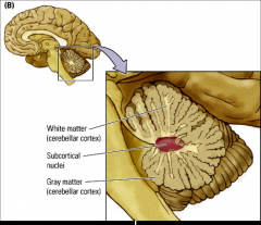
- Has to do with coordination, motor tasks, balance, etc. - Composed of white matter, subcortical nuclei, gray matter. |
|
|
What is the midbrain (mesencephalon) composed of? Further describe these two areas. |
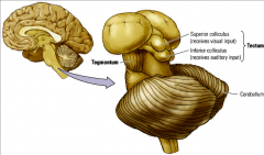
1. Tegmentum (floor): contains RF system. It is also the locus of different nuclear groups that synthesize neurotransmitters. 2. Tectum (roof): Superior colliculus which makes us turn towards visual stimulus. Inferior colliculus which makes us turn our head towards auditory stimulus. |
|
|
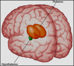
What are the two areas in the forebrain? What are these two made up of? |
1. Diencephalon - Thalamus - Hypothalamus - Epithalamus - Pineal Body - Habenula - Striamedullaris ofthe thalamus. 2. Telencephalon - Neocortex/Cortex - Basal Ganglia - Limbic System - Olfactory Bulbs |
|
|
What are the functions of the structures within the diencephalon? |
Thalamus (inner chamber): made up of many nuclei. It relays motor and sensory signals to the cerebral cortex. Hypothalamus (lower chamber): links the nervous system to the endocrine system via the pituitary gland Epithalamus: linkslimbic system components in the forebrain with other parts of the brain. Pineal Body: secretes melatonin, regulates sleep and wakefullness cycle. Habenula: a nucleus that projects to the midbrain. Regulates eating and drinking. |
|
|
What are the functions of the structures within the telencephalon? |
Neocortex/cortex: higher brain functions, separated into 4 lobes; frontal, occipital, parietal, temporal Basal Ganglia: many voluntary movements Limbic System: emotions and memory Olfactory Bulbs: sense of smell |
|
|
What are the structures of the thalamus and what are their functions? |
Dorsomedial Nucleus (connects to frontal lobe): circadian rhythms, eating, sleeping, body weight, energy consumption, etc. LGN: relay center in the thalamus for the visual pathway. It receives a major sensory input from the retina. The LGN is the main central connection for the optic nerve to the occipital lobe. MGN: part of the auditory thalamus and represents the thalamic relay between the inferior colliculus (IC) and the auditory cortex (AC) |
|
|
What are relay nuclei and what do they do? |
Nuclei found in the thalamus. Sensory signals generated in all types of receptors are projected via complex pathways to specific relay nuclei in the thalamus. Either receive info, or project to certain areas of the brain.
|
|
|
What are the different relay nuclei in the thalamus? |
- Sensory - Auditory - Visual - Somatosensory - Motor - Association: where information is integrated and combined. primary, secondary, and then association. - Limbic |
|
|
What are the different areas of the thalamus? (9 areas) Where do they project to/from in the cortex and what are they responsible for? What area has no connections and why? |
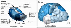
1. Anterior (A): Mammillary Bodies -> emotionalregulation, processing, fear, anxiety, etc. part of the OCD. PROJECTS OUT TO Cingulate Gyrus -> emotions and behaviour. 2. Dorsomedial (DM): Amygdala -> emotions and memory. Caudate -> storing/learning from memories. Frontal cortex -> executive functions such as planning for the future, judgment, decision-making skills, attention span, and inhibition. 3. Posterior (P): Areas 17 & 18 -> primary visual cortex.lSuperior collicullus -> turn head towards visual stimulus. 4. Lateral posterior (LP): its own area -> positioning in space 5. Medial Geniculate Nucleus (MGN): its own area -> auditory relay 6. Lateral Geniculate Nucleus (LGN): its own area -> visual relay 7. Ventrolateral posterior (VLP): its own area -> somatosensory relay 8. Ventrolateral (VL): Cerebellum -> balance and coordination. Basal Ganglia -> voluntary movements, habits, eyemovements, etc. Substantia nigra -> reward, addiction, and movement as it produces dopamine. 9. Ventral anterior (VA): basal ganglia --- The anterior temporal lobe has no connections becuase it has to do with object recognition and voice recognition, semantic knowledge which does not require relay to other areas. |
|
|
What are the two types of nuclei in the hypothalamus? What are their functions? |
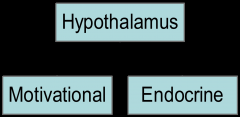
1. Motivational Nuclei: responsible for the 4 Fs (fighting, fleeing, feeding, and FORNICATION) 2. Endocrine: all hormones that effect everything in your body. |
|
|
What else does the hypothalamus contain that is important? |
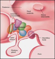
Contains smaller commissures that allow for information to pass from both hemispheres to the other side. This is important in split brain patients as it allows them to still function normally. |
|
|
Explain the more complex diagram of the nervous system. |
|
|
|
What are the 3 areas that make up the telencephalon? What are their functions? |
1. Neocortex: higher function such as sensory perception, conscious thought, etc. 2. Basal Ganglia: (Parkinson's Disease) Caudate nucleus -> learning from past memories. Putamen -> regulate movement and influence learning. Globus Pallidus -> primarily inhibits movements 3. Limbic System: emotion and memory. direct connection to olfactory system which is why smell is so strong in triggering memories. |
|
|
What are the 7 areas of the Limbic system? Describe their functions. |
1. Cingulate Gyrus: linking behavioural outcomes to motivation, executive function, and respiratory control. 2. Fornix: C-shaped bundle of fibres. recall memory. 3. Hippocampus: long term memory 4. Mammillary Bodies: recollective memory. 5. Amygdala: emotions 6. Olfactory Bulb: sense of smell 7. Septum: |
|
|
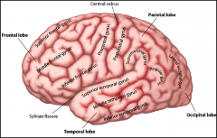
What is the neocortex? what is its function? what is it made up of? |
The part of the brain you can see. Highercognition functions, planning of behavior, attention, memories, language etc. made up of gyri (bumps) and sulci/fissures (grooves). takes up 80% of brain volume. |
|
|
Name the major gyri of the cerebral cortex. |
|
|
|
Name the major primary cortexes of the brain and where they are located. |
1. Primary somatosensory cortex 2. Primary motor cortex 3. Primary visual cortex 4. Primary auditory cortex |
|
|
What are the functions of the post and pre central gyri? |
Precentral - motor strip, primary motor cortex. Postcentral - somatosensory cortex. |
|
|
How did Broadmand come up with the brain map? |
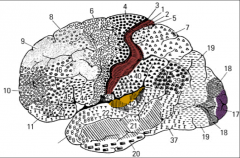
He looked at cell densities of different areas of the brain under a microscope. Changing densities indicated different brain areas with different function. |
|
|
Explain the difference in the relative thicknesses of the layers of the motor vs. somatosensory cortex. |
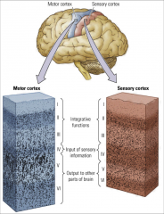
6 layers in total. 1-3 are integrative functions, 4 is input of sensory information, and 6-7 are output to other parts of the brain. 1-3 are the same for both. 4 is double in thickness for the sensory cortex. 6 and 7 are two times thicker in motor than sensory. |
|
|
What are two areas in the corpus collosum where specific information crosses? |
1. at the back (Spleenium) visual info crosses 2. at the front (Genu (the knee)) motor info crosses. |
|
|
What is the number, name, and function of each of the 12 cranial nerves? |

1. Olfactory: smell 2. Optic: vision (retina to cortex) 3. Oculomotor: eye movement 4. Trochlear: eye movement 5. Trigemenial: chewing and facial sensation 6. Abducens: eye movements 7. Facial: facial movements 8. Auditory Vestibular: hearing and balance 9. Glossopharyngeal: tongue and pharynx movement and sensation 10. Vagus: heart, blood vessels, viscera, movement of larynx and pharynx. 11. Spinal accessory: neck muscles 12. Hypoglossal: tounge muscles |
|
|
What are meninges? Name the 3 layers (skull inward) |
The meninges are the protective sheath around the brain and spinal cord. Dura matter, arachnoid matter, pia matter. |
|
|
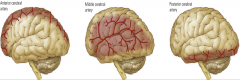
Describe the blood flow through the different arteries in the brain. |
1. Internal carotid arteries supply the anterior area of the brain. 2. This branches off into the R & L anterior cerebral arteries and middle cerebral arteries 3. The Middle cerebral arteries branch into the striate arteries. 4. Vertebral arteries supply the posterior areas of the brain. 5. join to form one basilar artery under the brain stem. 6. this branches into the R & L posterior cerebral arteries. |
|
|
What areas of the brain do the different arteries supply? |
ICA: anterior areas of the brain ACA: medialand inferior frontal lobes, medial parietal lobes, and corpus callosum MCA: lateralsurfaces of the frontal, temporal and parietal lobes, & some of theinferior surfaces of the frontal lobes, and medial surfaces of the temporallobes SA: internal capsule VA: posterior areas of the brain BA: PCA: occipitallobes, areas of the medial & inferior temporal lobes, hippocampus |
|
|
What is the Circle of Willis? What is its purpose? |
Atthe base of the brain in the subarachnoid space, the anteriorcommunicating artery(only one) and the R & L posterior communicating arteries connect the internal carotid andvertebral artery systems to form the Circle of Willis.
Offers the brain some protection incase one of the arteries get blocked, as blood can flow around this circle and reach certain areas of the brain. |
|
|
Where do aneurysms and subarachnoid haemorrhages usually take place in the brain? |
Circle of Willis. |
|
|
How does blood leave the brain? |
1. the Veins empty out into the venous sinuses in the dura matter. 2. these empty into the internal jugular vein. |
|
|
What are the ventricles of the brain? |
There are three, the lateral (centre of each hemisphere), 3rd (midbrain) and 4th (brainstem). Ventricles contain cerebrospinal fluid made by the choroid plexus (special glial cells). CSF circles around the brain in the subarachnoid space spreading around nutrients and removing waste. |
|
|
What does the blockage of a ventricle cause? |
Hydrocephalus occurs, where the ventricles enlarge. Most common in babies right after birth. People with cognitive problems also tend to have enlarged ventricles. |
|
|
What are the 8 basic principles of functional organization in the brain? |
1. Thesequence of brain processing is “In-Integrate-Out”
2. Sensoryand motor divisions exist throughout the nervous system 3. Thebrain’scircuits are crossed 4. Thebrain Is both symmetrical and asymmetrical 5. Thenervous system works through excitation and inhibition 6. TheCentral Nervous System has multiple levels of function 7. Brainsystems are organized both hierarchically and in parallel 8. Functions in the brain are both localized and distributed |
|
|
What is the difference between clinical and experimental neuropsychology? |
Clinical: work with people with brain damage/dysfunction to learn more about neurological disorders &diseases in order to improve diagnosis, treatment, rehabilitation &prevention Experimental: Work with neurologically intact(non-brain damaged) persons to learn about typical or normal brain functioning(brain-behaviour relationships) |
|
|
Describe the other disciplines of neuropsychology. |
1. Neurology– (Doctors, MD) not somuch interested in higher cognitive functions – more interested in medicalaspects of nervous system damage/dysfunction
2. Behavioural Neurology – neurologists with an interestin higher cognitive functions and brain-behaviour relationships 3. Cognitive Psychology –focus is the details of higher cognitive functions – study “normal”people – experiments to study how different conditions, manipulations, etcaffect cognitive processes in order to understand these processes (e.g.,attention, memory, judgment, problem-solving, priming, etc).Breaking down normal cognitive processes to figure out how they process certaininformation. Interested in the details of cognitive processes. 4.Cognitive Neuropsychology –focus is the same as cognitive psychology, but research brain-damaged persons(less interested in where the damage is and how it is related to theimpairments in functioning). Relationships between brainfunctions and higher order functioning. Interested in people who have braindamage or brain problems. 5. Neuroscience– focus is on how the brain works – from the molecular to the cellular to themore global level – e.g. identifying neurotransmitter pathways, receptors,reuptake, etc to identify functional systems –often use electrophysiological and neuroimaging tools and instruments(event-related potentials, fMRI, PET scans, and so on) - study both brain-damaged/impaired andnon-brain-damaged/non-impaired people 6. Cognitive Neuroscience –neuroscience as described above, but with a focus on understanding neuralunderpinnings of higher cognitive functions. Looking at whatsgoing on in the brain (similar cognitive neuropsychology) |
|
|
Describe Luria's HierarchicalModel of Cortical Functioning. |
1. Primarysensory zone (sensory information arrives)
2. Secondarysensory zone (elaboration of sensory info) 3. Tertiarysensory zone (integration) 4. Tertiarymotor zone (intentions formed) 5. Secondarymotor zone (plan of action formed) (put hands up to catch a ball) 6. Primarymotor zone (execution of plan is initiated) (flex muslces, etc) |
|
|
What are functional systems? |
These are complex cognitive functions suchas language , memory, etc that involve multiple brain areas thatwork together.
|
|
|
What is a "Double Dissociation" in terms of brain function? |
2different functions that are localized to separate areas. Like Language or attention. Ifyou have this, it means that if one area is damaged, you lose one function butnot others. Wernickes and brocasarea are double dissoications of function.
|
|
|
What is a "Disconnection" in terms of brain function? |
Whentwo areas of the brain that work together are cut off form eachother. If we cut a band of fibres called the arcuate fasciculus, which connections brocas and wernickes area. Person can still understand langauge, and can still speak because bothareas are intact, but they cant repeat what you just said. They know what theyare suppose to do, but they cant repeat it. Cantget ideas form head to paper.
|
|
|
What is Broca's Aphasia? |
Loss of ability to produce language (verbal or written). |
|
|
What is Wernicke's Aphasia? |
Loss of ability to understand language (verbal or written). |
|
|
What are some terms for any motor sensory, perceptual, behavioural, psychological, emotional, orcognitive abnormality? |
deficit,dysfunction, symptom, impairment, disorder |
|
|
What terminology is used for the follow things? - speech - writing - reading - purposeful movement/actions - knowing |
- phasia - graphia - lexia - praxia - gnosia |
|
|
What is the difference between "a" or "dys" as a prefix in terminology? |
"a" - full lose of that function "dys" - partial loss dys always means the same thing, but a sometimes both. |
|
|
What is a lesion? |
- any type of focal damage - focal meaning restricted to circumscribed area |
|
|
What does infarct/infarction mean? |
- This is an area of dead brain tissue |
|
|
What terms are used for the following brain damages? - blockage - bleeding - fractured skull/penetrating object - herpes simplex |
- stroke - intracerebral hematoma - open head injury - viruses - brain tumours and abscesses |
|
|
What does diffuse brain damage mean? |
- something is affect many brain areas |
|
|
What are some other types of brain damage? |
--dementias(Alzheimer’s, Picks, etc)
atrophy –meningitis –encephalitis –Huntington’s –Parkinson’s –MultipleSclerosis –subarachnoidhemorrhage –Edema(swelling) |
|
|
Define "Lateralization of Function". |
Hemispheres specialize in certain functions and communicate through bands of axons comprising of cerebral commissures. |
|
|
Name some of the commissures. |
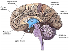
Ø corpus callosum
Ø anterior commissure Ø posterior commissure Ø hippocampal commissure Ø habenularcommissure Ø massaintermedia Ø collicularcommisures |
|
|
Describe the visual pathway form the source of light to the visual cortex. |
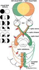
•The left half of each retinareceives light from right visual field (RVF) & connects to the lefthemisphere (LH).
•The right half of each retinareceives light from the left visual field (LVF) connects to the righthemisphere (RH). |
|
|
Describe the auditory pathway from the source of sound to auditory cortex. |
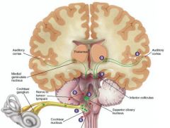
1. cochlear nucleus 2. ipsilaterally to the superior olivary and contralerally to the inferior colliculus. 3. thalamus (MGN) 4. auditory nucleus. 5. some sound sent out by nerve to sensory tympani. |
|
|
Describe the four methods to studying lateralization. |
1. Tachistoscope: a visual method. The only way to present information to one hemisphere. Flash something to one hemisphere (VF) for 200 ms. Peopleare faster when the words are presented in the right visual field because langauge is in the left hemisphere. Justmake take a few seconds longer. 2. Dichotic Listening Task: auditory method. Hear words coming into both ears simultaneously and the ipsilateral connections get cancelled out. Or hear diff words in diff ears. Right ear or left hemisphere has an advantage for lights. left ear or right hemisphere has advantage for non language sounds or emotions. 3. Dichaptic Presentation: the tactile method. Place something in the persons unseen hands and ask to identify. right hand will be better with words as it goes to the left hemisphere. 4. Wada Test: uses Sodium Amobarbital injected into the carotid artery to one hemisphere (right to right). Puts that hemisphere to sleep for 3-5 minutes before crossing over to other side. have to do many tests during this time to test which thing are lateralized. |
|
|
What happens when the corpus collosum is cut? |
Interfers with the communication between hemispheres. |
|
|
What is epilepsy? |
A condition characterized byrepeated episodes of excessive synchronized neural activity.
Seizures start at a specific point in the brain and fan out, and in doing so, stimulate those areas. |
|
|
How can epilepsy be fixed? |
Neurosurgeons may cut the corpuscallosum to prevent the seizures from spreading to the opposite side of thebody.
Can still function because of the smaller commissures that allow information to pass to either hemisphere. Can also locally laser the certain areas. |
|
|
What usually happens in split brain patients? |
- Have normal intelligence and motivation. - Usehands independently in a way others cannot. having trouble using them together. but adapt eventually. - Responddifferently to stimuli presented to only one side of the body. sometimes info doesnt cross over. |
|
|
How did Sperry reveal subtle behavioural differences for split-brain people? |
Because the left side of thebrain is dominant for language in most people, most split brain people:
–Havedifficulty naming objects briefly viewed in the left visual field. •A small amount of information canstill be transferred via several smaller commissures. |
|
|
Explain the behaviour of a split brain patient if the word "Hatband" is flash in front of them. |
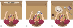
(a) When the word hatband is flashed on a screen,
(b) a woman witha split brain can report only what her left hemisphere saw, “band.” (c)However, with her left hand, she can point to a hat, which is what the righthemisphere saw. |
|
|
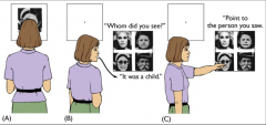
Describe the Chimeric Faces Test. |
|
|
|
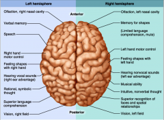
Describe the lateralizations of different functions for each hemisphere of the brain. |
|
|
|
Describe, in terms of emotion, what happens when the right hemisphere is damaged. |
- Right hemisphere is better atperceiving emotions. - Damage to parts of the righthemisphere causes difficulty perceiving other’s emotions, failure to understandhumor and sarcasm, and a monotone voice (lack of prosody). - The right hemisphere is alsobetter at comprehending spatial relationships.
|