![]()
![]()
![]()
Use LEFT and RIGHT arrow keys to navigate between flashcards;
Use UP and DOWN arrow keys to flip the card;
H to show hint;
A reads text to speech;
39 Cards in this Set
- Front
- Back
|
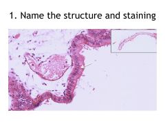
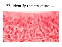
Simple columnar epithelium (Gallbladder, H&E) – Slide 3 |
|
|
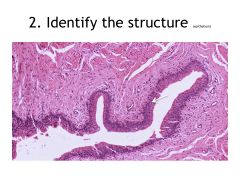
Transitional Epithelium (urinary bladder, H&E) – Slide 8
|
|
|
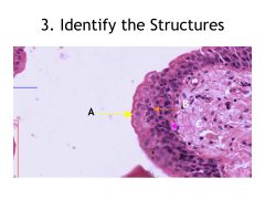
Transitional Epithelium (urinary bladder, H&E) – Slide 8
A) Umbrella Cell B) Pear-shaped cell C) Basal layer
|
|
|
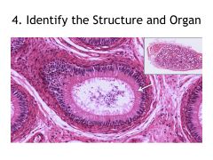
Pseudostratified columnar epithelium (epididymis, H&E) – Slide 4
|
|
|
Mesenchymal tissue (Wharton’s Jelly) (Umbilical cord, H&E) – Slide 19
|
|
|
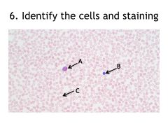
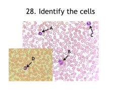
Blood cells (blood smear, May-Grünwald-Giemsa) - Slide 52
A) Eosinophil B) Lymphocyte C) Erythrocyte (red blood cell) |
|
|
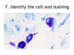
Mast cell (peritoneum, toluidine blue) – Slide 21 |
|
|
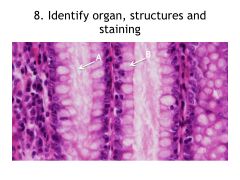
Goblet cells (large intestine, H&E) – Slide 10
A) Crypt of Lieberkuhn B) Goblet cell |
|
|
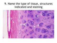
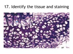
Hyalin cartilage (rib, H&E) – Slide 23
A) Chondron B) Interterritorial matrix C) Chondrocyte D) Perichondrium |
|
|
Elastic fibers (large artery, resorcin fuchsin) – Slide 15 |
|
|
Apocrine secretion (prostate, H&E) – Slide 12
A) Glandular epithelium – cuboidal to columnar, depending on level of activity B) Acinus of the gland C) Prostatic concretions/prostatic stones/corpora amylacea |
|
|
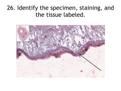
Simple squamous epithelium (endothelium, elastic artery, H&E) – Slide 50
|
|
|
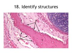
Endochondral ossification – Slide 28
A) Spicule B) Resting zone 3) Zone of proliferation 4) Zone of degeneration 5) Zone of mesenchymal invasion |
|
|
Endochondral ossification – Slide 28
A) Osteoblast B) Osteocyte C) Chondroclast D) Primary bone marrow |
|
|
Fibrous cartilage (meniscus, H&E) – Slide 25
|
|
|
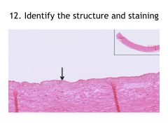
Stratified squamous non-keratinized epithelium (esophagus, H&E) – Slide 5
|
|
|
Elastic cartilage (epiglottis, resorcin fuchsin) – Slide 24
|
|
|
Intramembranous ossification (calvaria, H&E) – Slide 29
1) Spicule 2) Osteoblasts |
|
|
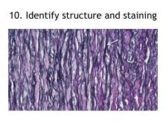
Collagen fibers (tendon, H&E) – Slide 14
tendocyte |
|
|
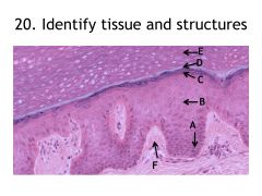
Stratified squamous keratinized epithelium (palmar skin, H&E) – Slide 6 A. Stratum basale B. Stratim spinosum C. Stratum granulosum D. Stratum lucidum (highly refractive; will appear bright pink in your slides) E. Stratum corneum F. Connective tissue papilla |
|
|
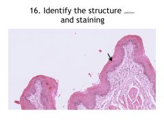
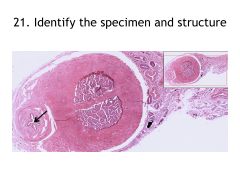
21. Stratified columnar epithelium (penis, H&E) – Slide 7
urethra |
|
|
Stratified columnar epithelium (penis, H&E) – Slide 7 Our specimens are taken from the part of the penis where the urethra shows stratified columnar (e.g. more distal). The epithelium of the penis transitions as it moves distally. |
|
|
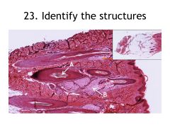
Holocrine secretion (sebaceous gland, hairy skin, H&E) – Slide 11
A. Hair follicle B. Sebaceous gland |
|
|
Reticular fibers (liver, silver impregnation) You should be able to identify reticular fibers. In order to visualize them, we use silver impregnation because they are argyrophilic (so they take up the silver pigment). |
|
|
Bone – cross section (Schmorl)
A. Special lamellae B. Haversian canal C. Osteocyte D. Interstitial lamellae |
|
|
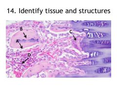
Umbilical cord (H-E), Simple cuboidal epithelium – Slide 19 |
|
|
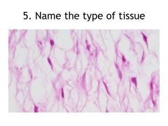
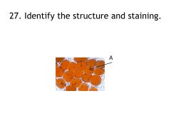
Adipocyte (Sudan stain) – Slide 22
|
|
|
Blood cells (blood smear, May Grünwald-Giemsa) – Slide 52
A. Neutrophil B. Eosinophil C. Neutrophil D. Monocyte |
|
|
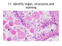
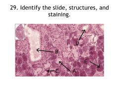
Merocrine secretion (submandibular gland, HE) – Slide 13 A. Mucous acinus B. Excretory duct C. Demilune of Gianuzzi D. Salivary duct E. Serous acinus |
|
|
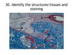
Scalp skin (Azan) – slide 17
A. Hair follicle B. Collagen C. Sebaceous gland |
|
|
Smooth Muscle (Jejunum - H-E) Slide 30 A) Longitudinal B) Cross Section |
|
|
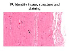
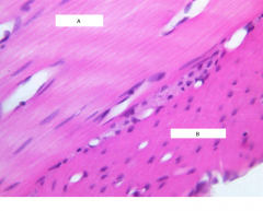
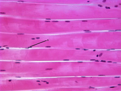
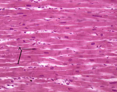
Striated Muscle - Longitudinal Section (H-E) Slide 31 A) Fibrocyte |
|
|
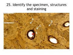
Cardiac Muscle - Longitudinal Section (H-E) Slide 34 A) Cardiac Muscle Cell |
|
|
Cardiac Muscle Cross Section + Purkinje Fibers (H-E) Slide 35 |
|
|
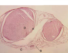
Peripheral Nerve (H-E) Slide 36 A) Perineurium B)Adipose Tissue C) Epineurium D) Endoneurium Note: In Silver Nitrate Impregnation the Myalin sheets are stained |
|
|
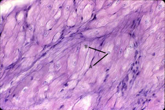
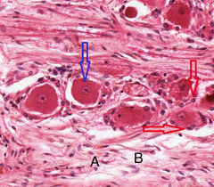
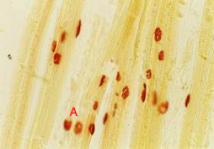
Pseudounipolar Neurones (spinal ganglion, H-E) Slide 37 Blue) Pseudounipolar Neuron Red) Satellite Cells A) Schwan Cells B) Exons w Myalin Sheet |
|
|
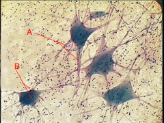
Multipolar Neurones (autonomic ganglion, silver nitrate impregnation) Slide 38 A) Dendrites B) Satellite Cells |
|
|
Motor End Plate (histochemical method for acethylcholinesterase) Slide 43 A) Motor End Plate |
|
|
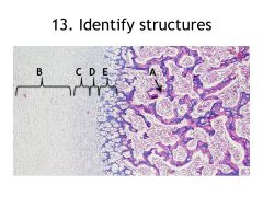
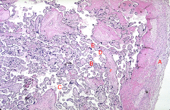
Placenta (H-E) Slide 83 A) Amnion Epithelium B) Chorionic Villi C) Intervillous Spaces D) Fibrinoid E) Anchoring Villus F) Placenta Septa Note: Syncytiotrophoblast around villi |