Reading...
![]()
Play button
![]()
Play button
![]()
Use LEFT and RIGHT arrow keys to navigate between flashcards;
Use UP and DOWN arrow keys to flip the card;
H to show hint;
A reads text to speech;
46 Cards in this Set
- Front
- Back
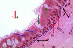
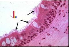
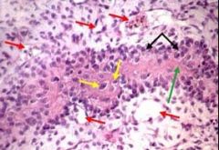
What type of tissue is this? What are the red arrows? Blue arrows Green arrows? Black arrows?
|
Pseudostratified Columnar Epithelium
Red arrows=basal cells Blue arrows=tall columnar cells with cilia Green arrows=goblet cells Black arrows=basal lamina (very pink staining) |
|
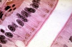
What type of tissue?
|
Simple columnar epithelium
|
|
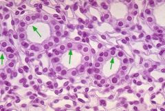
What type of tissue are the green arrows pointing to?
|
Simple cuboidal epithelium
|
|
What are the green arrows pointing to? (tissue type)
|
Simple Squamous Epithelium
|
|
What are the green arrows pointing to?
|
The cytoplasm in simple squamous epithelium
|
|
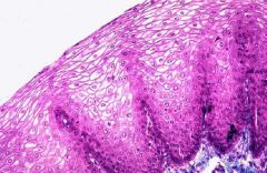
What type of tissue?
|
Stratified squamous epithelium (non-keratonized)
|
|
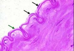
What is the blue arrow? Black arrow?
|
blue arrow: keratonized part of stratified squamous epithelium
black arrow is the entire stratified squamous epithelium tissue (keratonized) |
|

what type of tissue?
|
endothelium (epithelium in blood and lymph vessels)
|
|
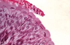
what type of tissue and where is it found?
|
transitional epithelium
found in the urinary bladder |
|
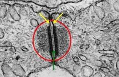
What is in the red circle? What is the green line due to?
|
Desmosome joining two cells
The green line is the INTERMEDIATE LINE, due to an increase in glycalyx also note the increase in cytoplasmic density around the desomosome due to lots of microfilaments present to strengthen the junction. |
|
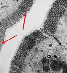
What are the red arrows pointing to?
|
microvilli
|
|
what are the black arrows pointing to?
|
cilia
|
|
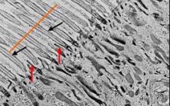
what are the black arrows pointing to?
What are the red arrows pointing to? |
black=cilia
red=microvilli |
|
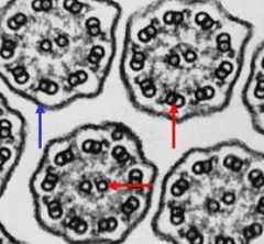
what are these? what is the blue arrow pointing to?
|
cilia! note 9 sets of doublets and 2 central singlets.
blue arrow=plasmalemma, which surrounds each cilia |
|
What type of tissue is this? What makes it distinct?
|
Embryonic connective tissue, MESENCHYME
the DARK nucleouslus is a destinct characterstic |
|
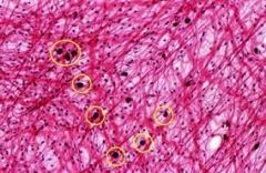
what type of cells are circled in yellow?
|
mast cells
|
|

what are the blue and black arrows pointing to?
|
black=mast cell
blue=collagen fibers |
|
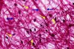
what are the yellow arrows pointing to?
|
elastic fibers
|
|
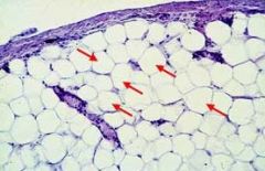
What is the red arrow and what is there in life?
|
adipocyte.
in life it is filled with one fat globule, but the fat is removed during processing for microscopy. |
|
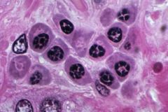
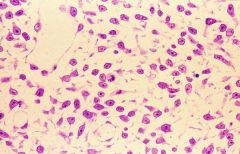
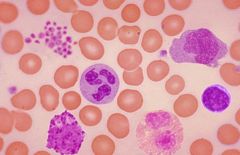
What types of cells are these and how can you tell? What is their function?
|
Plasma Cells
Darkly stained b/c very basophilic b/c lots of rER. chromatin clumps around periphery of nucleus giving it a "cart wheel" appearance. Synthesize and secrete humoral antibodies. |
|
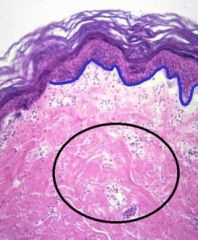
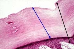
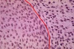
What does the blue line denote?
|
the separation between the dermis and epidermis of the skin.
Epidermis=stratified squamous keratonized epithelium Dermis=dense irregular connective tissue |
|
What is this tissue?
|
Dense connective tissue
|
|
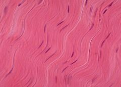
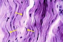
what type of tissue is this? what are it's unique characteristics and what are the main cells?
|
Dense Regular Connective Tissue
-parallel collagen fibers - cells are mostly fibroblasts |
|
What type of cells are present in this slide?
|
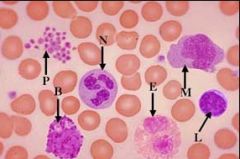
N = neutrophil (light purple, 3-5 nuclei lones)
E = eosinophil (2 nuclie lobes, pink) B = basophil L = lymphocyte (same size as rbcs, dark nucleus) M = monocytes (big, light irregular shape nucleus P = platelets (small fragments of cytoplasm surrounded by plasmalemma) |
|
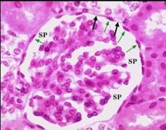
What type of tissue is this? What cells are on the right and on the left?
|
Embryonic Hyaline Cartilage
Left: Chondroblasts (in the process of secreting matrix) Right: Mesenchymal cells |
|
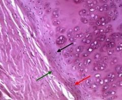
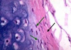
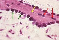
What are the black arrows pointing to?
Red Arrows? Green arrows? |
black is pointing to periphery of hyaline cartilage, the newer, smaller lacunae which contain chondroblasts.
red is pointing to chondrogenic layer, containing chondroblasts that have not yet secreted matrix green is pointing to FIBROUS LAYER, containing collagen fibers, fibroblasts and mesenchymal cells |
|
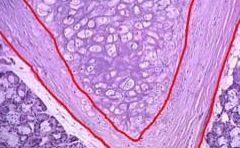
What is the tisue in the center of the V?
What is the red line outlining? |
Elastic Cartilage
Perichondrium |
|
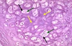
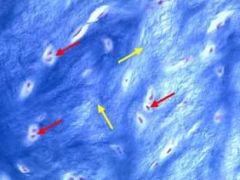
What type of tissue is this?
What are the green arrows? What are the yellow arrows? |
Elastic Cartilage
Green= lacunae containing CHONDROBLASTS yellow=MATRIX (note that you can see a lot of fibers) |
|
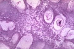
What type of tissue is this?
|
Elastic cartilage
|
|

What is the yellow arrow?
Green arrow? |
Yellow: Elastic cartilage
Green: Perichondrium |
|
What is the green arrow pointing to?
What is the black arrow pointing to? |
Green: Perichondrium
Black: Fibrous portion (contains collagen, fibroblasts and msenchymal cells) |
|
what type of tissue is this?
what is the yellow arrow? what is the red arrow? |
fibrocartilage
yellow: MATRIX red: CHONDROBLASTS |
|
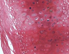
what is the main type of tissue?
|
hyaline cartilage
|
|
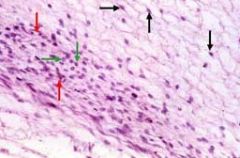
What type of tissue?
What are the green arrows pointing? Black arrows? |
Intramembranous bone (in development phase)
green arrows: osteoblasts black arrows: mesenchymal cells |
|
What type of tissue?
What are the black arrows? Green? Yellow? |
Intramembranous bone
black: OSTEOBLASTS green: OSTEOID yellow: OSTEOCYTES |
|
What type of tissue?
What is the green arrow? yellow? red? |
Intramembranous Bone
green: OSTEOBLASTS yellow: matrix red: OSTEOCYTES |
|
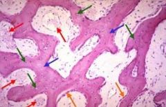
What color arrow are osteocytes?
blood vessels? trabeculae? osteoblasts? |
osteocytes: green
blood vessels: red trabeculae: blue osteoblasts: orange |
|
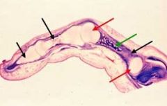
Where is the epiphyses?
Where is the primary center of ossification? |
epiphyses: red arrows
primary ossification center: green arrow |
|
|
What is the metaphysis?
|
the area of epiphysis where the hyaline cartilage starts to be replaced by bone
|
|
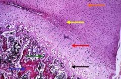
Which arrows represent the following zones:
- proliferation - calcification - ossification - retrogression - hypertrophy |
proliferation: blue
hypertrophy: green calcification: black retrogression: yellow ossification: orange |
|
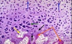
label the zones
|
resting zone - orange arrow
zone of proliferation - yellow zone of hypertrophy - red zone of calcification - black zone of retrogression - green zone of ossification - blue |
|
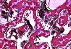
identify the black arrows and the blue arrows
|
black: osteoid - colored red
blue arrows: remnants of hyaline cartilage (colored lavender) |
|

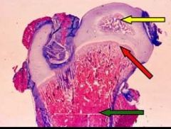
What is the yellow arrow pointing to?
What is it surrounded by? |
secondary ossification center
it is surrounded b hyaline cartilage of the original cartilage model |
|

What is the red arrow pointing to?
|
metaphysis
|
|

Where is the epiphyseal disc?
|
The epiphyseal disc appears as a pale band between the epiphysis and the shaft
|
|
Where is the periosteal collar?
|
it is the part stained dark blue
|