![]()
![]()
![]()
Use LEFT and RIGHT arrow keys to navigate between flashcards;
Use UP and DOWN arrow keys to flip the card;
H to show hint;
A reads text to speech;
210 Cards in this Set
- Front
- Back
|
what is the primary nerve that provides motor innervation to the abdominal wall muscles? where can this nerve be seen on the abdominal wall? |
lateral cutaneous femoral nerve this nerve courses on the top of transversus abdominus |
|
|
what is the origin of the lateral cutaneous femoral nerve? |
ventral branch of the 4th lumbar spinal nerve |
|
|
what is the cutaneous testing area for the lateral cutaneous femoral nerve? |
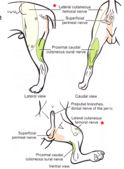
lateral and cranial surfaces of the thigh lateral surfaces of the hip and stifle |
|
|
what is the origin and insertion of external abdominal oblique? |
origin costal part: lateral surface ribs 4-12 origin lumbar part: 13th rib & thoracolumbar fascia insertion by 2 tendons abdominal tendon: linea alba & on pecten ossis pubis pelvic tendon: inserts on pecten ossis pubis |
|
|
what separates the abdominal and superficial tendons of the external abdominal oblique m? |
superficial inguinal ring |
|
|
what is the innervation of external abdominal oblique? |
last 8-9 intercostal nn ventral branches of T13, L1, L2, L3 spinal nerves |
|
|
what is the action of external abdominal oblique? |
compress abdominal viscera (abdominal press) aids in expiration, urination, defecation, partuition flexion of vertebral column (unilateral) bending of ventral comumn (lateral) |
|
|
what is the origin and insertion of internal abdominal oblique? |
O: tuber coxae, thoracolumbar fascia, inguinal ligament I: costal arch, rectus abdominus, linea alba, prepubic tendon by wide aponeurosis |
|
|
what is the innervation of the internal abdominal oblique? |
last few intercostal nn. ventral branches of T13, L1, L2, and L3 spinal nerves |
|
|
what is the action of the internal abdominal oblique? |
compression and support of the abdominal viscera |
|
|
what is the origin and insertion of transversus abdominis? |
O: medial surfaces, last 4-5 ribs transverse processes of lumbar vertebrae via thoracolumbar facia I: linea alba |
|
|
what is the innervation of transversus abdominis? |
last few intercostal nn. ventral branches of T13, L1, L2, L3 spinal nerves |
|
|
what is the action of transversus abdominis? |
compression and support of abdominal viscera |
|
|
what is the origin and insertion of rectus abdominis? |
courses btwn 1st costal cartilage and sternum to pecten ossis pubis by way of prepubic tendon |
|
|
what is the innervation of rectus abdominis? |
intercostal nn. ventral branches of T13, L1, L2, L3 spinal nerves |
|
|
what is the action of rectus abdominis? |
compression and support of abdominal viscera bring pelvis forward flexion of back |
|
|
what is the rectus sheath? what structures is it composed of? |
envelope for rectus abdominis formed by aponeuroses of: external abdominal oblique m. internal abdominal oblique m. transversus abdominis m. |
|
|
what are the layers of the rectus sheath cranial to the umbilicus? |
ext. abdominal oblique ap. int. abdominal oblique ap. rectus abdominis m. int. abdominal oblique ap. transversus abdominis ap. |
|
|
what are the layers of the rectus sheath just caudal to the umbilicus? |
ext. abdominal oblique ap. int. abdominal oblique ap. rectus abdominis m. transversus abdominis ap.
|
|
|
what are the layers of the rectus sheath cranial to the pubis? |
ext. abdominal oblique ap. int. abdominal oblique ap. transversus abdominis ap. rectus abdominis m. |
|
|
what is the linea alba and where does it course? |
strip of collagenous tissue extends from xiphoid process to cranial edge of pelvic symphysis |
|
|
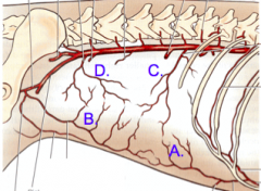
what 4 arteries provide the main blood supply to the abdominal wall? what is the origin of each of these arteries? |
cranial (deep) epigastric (from internal thoracic) caudal (deep epigastric (from pudendoepigastric trunk) cranial abdominal (from phenicoabdominal a. off aorta) deep circumflex iliac (from aorta) |
|
|
A. Cranial (deep) epigastric artery B. Caudal (deep) epigastric artery C. Cranial abdominal artery D. Deep circumflex iliac artery |
|
|
what arteries supply blood to the ventral abdominal wall? |
caudal superficial epigastric artery cranial superficial epigastric artery |
|
|
where are the superficial inguinal lymph nodes located, and what area do they drain? |
located just cranial to pecten ossis pubis drain ventral 1/2 of abdominal wall |
|
|
how are the superficial inguinal lymph nodes drained? |
through 1 or 2 trunks that drain into the medial iliac lymph nodes |
|
|
what structures pass through the inguinal canal? |
external pudendal artery external pudendal vein genitofemoral nerve lymphatics (draining superficial inguinal ln.) vaginal tunic (male)/vaginal process (female) spermatic cord (male) |
|
|
what are the 2 boundaries of the inguinal canal? what forms these boundaries? |
superficial inguinal ring and deep inguinal ring formed by slit in aponeurosis of external abdominal oblique |
|
|
what structures form the 3 boundaries of the deep inguinal ring? |
medial: lateral edge of rectus abdominis m. cranial: caudal edge of internal abdominal oblique m. caudal: inguinal ligament |
|
|
where does the inguinal ligament attach? |
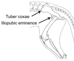
courses between tuber coxae and iliopubic eminence of the pelvis |
|
|
the inguinal ligament separates what two structures? |
the vascular lacuna and the inguinal canal |
|
|
what is the purpose of the vascular lacuna? |
opening in body wall for vessels coursing to pelvic limb |
|
|
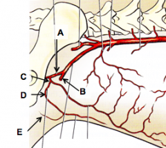
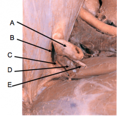
A. deep femoral a. B. femoral a. C. pudendoepigastric trunk D. external pudendal a. E. ventral labial/ventral (cranial) scrotal a. |
|
|
What is the origin of the external pudendal artery (from the descending aorta)? |
descending aorta external iliac a. femoral a. deep femoral a. pudendoepigastric trunk external pudendal |
|
|
what is the origin of the genitofemoral nerve? what are this nerve's sensory and motor functions? |
ventral branches of L3 and L4 spinal nerves motor to cremaster muscle sensory innervation from skin in inguinal region and proximal medial thigh |
|
|
what is the vaginal process? which gender is it seen in (in adults)? |
in females parietal peritoneal "excavation" that passes through the inguinal canal forms a vaginal ring through which the round ligament of the uterus passes |
|
|
vaginal process superficial inguinal ring external pudendal a. external pudendal v. genitofemoral n. |
|
|
what is the vaginal tunic? which gender is it seen in? |
in males vaginal process that in development wrapped around the testis and spermatic cord. forms double-walled structure |
|
|
what are the two anatomical layers of the vaginal tunic in males? what is between these two layers? |
visceral vaginal tunic: inner visceral layer. adhered to testis, epidiymis, and components of spermatic cord parietal vaginal tunic: surrounds components of spermatic cord between layers: vaginal cavity |
|
|
what is the vaginal ring (in males)? what important structure passes through the vaginal ring? |
peritoneal fold at the entrance to the vaginal cavity spermatic cord passes through vaginal ring |
|
|
what layer is included in the clinical vaginal tunic but is not considered part of the anatomical vaginal tunic? |
internal spermatic fascia |
|
|
where does the spermatic cord form? |
at the vaginal ring? |
|
|
what are the components of the spermatic cord? |
testicular artery testicular vein & pampiniform plexus testicular nerve testicular lymphatics ductus deferens deferent a. and deferent v. internal cremaster mesofuniculum |
|
|
what is the origin of the R and L testicular arteries? |
branch off the aorta |
|
|
where do the R and L testicular vein drain? |
R testicular vein: caudal vena cava L testicular vein: L renal vein |
|
|
what is the pampiniform plexus? |
venous complex that wraps around the testicular artery: cooling mechanism |
|
|
what type of nerve is the testicular nerve? |
comprised of both autonomic and sensory fibers autonomic fibers from postganglionic sympathetic fibers |
|
|
what does the testicular lymphatic drain into? |
medial iliac ln and lumbar aortic lnn. |
|
|
what is the course of the ductus deferens? |
from the epididymis to the pelvic urethra |
|
|
what are the three layers of mesentery found in the spermatic cord? what are their functions? |
mesorchium: supports testicular nerves and vessels mesoductus deferens: supports ductus deferens and deferent a. & v. mesofuniculum: anchors other 2 layers to parietal vaginal tunic |
|
|
what is the attachment of the cremaster muscle? |
originates from caudal free border of internal abdominal oblique courses w/i fascia of vaginal tunic (NOT part of spermatic cord) inserts of spermatic fascia and parietal vaginal tunic |
|
|
what is the action and innervation of the cremaster muscle? |
pulls the testis closer to the body in response to cold innervated by genitofemoral n. |
|
|
what is the difference between scrotum position in the dog and the cat? |
dog: inguinal position, has little hair cat: perineal position, covered with hair |
|
|
what two structures are surrounded by the visceral vaginal tunic? |
testis and epididymis |
|
|
what layers make up the scrotum and testes? |
skin tunica dartos external spermatic fascia (clinical) vaginal tunic vaginal cavity visceral vaginal tunic tunica albuginea testicular parenchyma |
|
|
what layers make up the clinical vaginal tunic? |
internal spermatic fascia: dense irregular CT parietal vaginal tunic: serous membrane |
|
|
what structure is responsible for pulling the testes into the scrotum during development? |
the gubernaculum |
|
|
what two remnant structures are left over from the gubernaculum in the male? |
proper ligament of testis (btwn tail of epididymis and testis) ligament of the tail of the epididymis (between tail of epididymus and vaginal tunic) |
|
|
what is the remnant structure left over from the gubernaculum in the female? |
round ligament of the uterus (btwn cranial tip of uterine horn to vaginal process) |
|
|
what structures form the boundaries of the abdominal cavity? |
lateral & ventral: external abdominal oblique, internal abdominal oblique, transversus abdominis, costal arch (lateral) dorsal: vertebrae, hypaxial mm. cranial: diaphragm caudal: pelvic inlet |
|
|
what is the name for the serous membrane in the abdominal cavity? what two layers is this membrane composed of? |
peritoneum parietal peritoneum visceral peritoneum |
|
|
what is the parietal peritoneum attached to? |
transversalis fascia |
|
|
what are three examples of connecting peritoneum? |
mesentery ligament omentum |
|
|
the primitive mesentery is divided into what two portions? |
ventral and dorsal |
|
|
what structures develop from the ventral primitive mesentery? |
lesser omentum falciform ligament median ligament of the bladder |
|
|
what structures develop from the dorsal primitive mesentery? |
greater omentum mesoduodenum mesojejunum mesoileum mesocolon mesorectum |
|
|
what structures are contained within the superficial and deep leaves of the greater omentum? |
superficial leaf: spleen deep leaf: left limb of pancreas |
|
|
what is the space between the superficial and deep leaves of the greater omentum called? |
omental bursa |
|
|
what forms the entrance to the omental bursa? where can this structure be located? |
epiploic foramen dorsal border: caudal vena cava ventral border: hepatic portal vein caudal border: hepatic artery cranial border: liver |
|
|
what two ligaments are formed by parts of the lesser omentum? |
hepatogastric ligament hepatoduodenal ligament |
|
|
what structure can be found ventral to the lesser omentum? |
papillary process of the caudate lobe of the liver |
|
|
what structures form the center for the abdominal quadrant system? |
root of the mesentery (mesojejunum), surrounding the cranial mesenteric artery |
|
|
what are the four abdominal quadrants? |
right cranial quadrant right caudal quadrant left cranial quadrant left caudal quadrant |
|
|
what structure demarcates the division between the L and R quadrate lobes of the liver, and courses from the umbilicus to the diaphragm? |
falciform ligament |
|
|
what structure is enclosed in the falciform ligament in the fetus? in the adult? |
fetus: umbilical vein adult: round ligament of the liver (remnant of umbilical vein) |
|
|
the falciform ligament is a remnant of what structure? |
ventral mesentery |
|
|
what ventral mesentery remnant structure courses along the ventral body wall caudal to the umbilicus? |
median ligament of the bladder |
|
|
what structure contains the remnant of the urachus? |
median ligament of the bladder |
|
|
what are the three openings in the diaphragm called? |
aortic hiatus esophageal hiatus caval foramen |
|
|
what structures course through the aortic hiatus? |
aorta azygos vein throacic duct |
|
|
what structures course through the esophageal hiatus? |
esophagus dorsal and ventral vagal nerve trunks esophageal vessels |
|
|
what structures course through the caval foramen? |
caudal vena cava |
|
|
how is the diaphragm supported dorsally? |
R and L crura |
|
|
how are the R and L lumbocostal arches formed? |
lateral to the crura, between the crus and the body wall |
|
|
what courses through the lumbcostal arches? |
sympathetic trunk major splanchnic nerve |
|
|
what abdominal quadrant is the liver located in? |
R and L cranial abdominal quadrants most cranial abdominal organ |
|
|
what are the 4 lobes of the liver? |
left (L medial and L lateral) quadrate right (R medial and R lateral) caudate (caudate and papillary processes) |
|
|
where is the gall bladder located? |
between the R and quadrate lobes of the liver |
|
|
what structure is found between the L lobe and the quadrate lobe? |
falciform ligament and round ligament of the liver |
|
|
the parietal surface of the liver contacts which structure? |
the diaphragm |
|
|
the visceral surface of the liver contacts which structures |
stomach duodenum pancreas R kidney greater omentum small intestine +/- spleen |
|
|
what ligaments hold the liver in place? |
triangular ligaments (R and L, from the crus to the liver) coronary ligament (from the diaphragm and liver around caudal vena cava and hepatic veins) |
|
|
what vessels provide blood flow to the liver? |
hepatic artery (oxygen rich blood) portal vein (nutrient rich blood from GI tract) |
|
|
what vein drains blood from the liver? where does this vein drain? |
hepatic veins, drain into the caudal vena cava |
|
|
where is the gall bladder located? |
between the quadrate and R medial lobes of the liver R cranial abdominal quadrant |
|
|
What are the three important ducts associated with the gall bladder? |
cystic duct: between gall bladder and bile duct hepatic duct: between bile duct and liver (common) bile duct: union of cystic and hepatic ducts, carries bile to duodenum |
|
|
what is the structure where the bile duct drains into the duodenum? what other duct drains into the duodenum at this location? |
major duodenal papilla pancreatic duct |
|
|
what is the function of the spleen? |
RBC storage RBC destruction immune system component blood filtration |
|
|
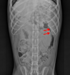
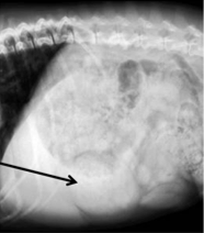
where is the spleen located? |
|
|
|
what are the two surfaces of the spleen and where do they face? |
parietal surface: faces diaphragm and lateral abdominal wall visceral surface: faces greater curvature of stomach and L kidney |
|
|
what is the hilus of the spleen and where is it located? |
on visceral surface where blood vessels enter and leave spleen |
|
|
what structure anchors the spleen to the stomach? |
gastrosplenic ligament |
|
|
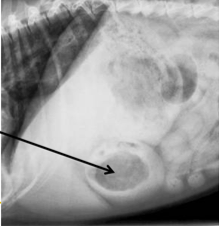
spleen |
|
|
spleen |
|
|
how is identifying the spleen in a cat different from a dog? |
smaller in the cat than in the dog NOT VISIBLE on lateral abdominal view in cats |
|
|
list the path that ingesta takes, starting with the stomach and ending with the rectum |
stomach small intestine
large intestine
|
|
|
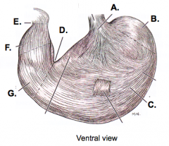
what are the four main parts of the stomach? |
cardia fundus body pyloric part |
|
|
what are the three divisions of the pyloric part of the stomach? |
pyloric antrum pyloric canal pylorus (pyloric sphincter) |
|
|
A. cardia B. fundus C. body D. angular incisure E. pylorus F. pyloric canal G. pyloric antrum |
|
|
what attaches to the greater and lesser curvature of the stomach? |
greater curvature: greater omentum lesser curvature: lesser omentum |
|
|
the stomach is composed of three layers of smooth muscle. what are these layers called? |
tunica muscularis (all 3 layers together) outer longitudinal layer middle circular layer inner oblique layer |
|
|
what structure on the inside of the stomach allows for expansion of the stomach? |
rugae (gastric folds) |
|
|
what two regions of glands can be found in the stomach? |
(fundic) gastric glands: secret acid pyloric glands: secrete mucus |
|
|
what abdominal quadrants are the parts of the stomach located in? |
cardia: L cranial fundus: L cranial body: L cranial pyloric antrum: R cranial (dog), L cranial (cat) pylorus: R cranial (dog), on or slightly L of midline (cat) |
|
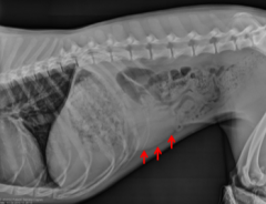
organ? view? |
stomach R lateral recumbency |
|
organ? view? |
stomach L lateral recumbency |
|
|
during development, the intestine rotates around what structure? |
the root of the mesentery |
|
|
what are the 4 parts of the duodenum? |
cranial duodenal flexure descending duodenum caudal duodenal flexure ascending duodenum |
|
|
what is the name for the junction of the duodenum and the jejunum? |
duodenojejunal flexure |
|
|
what is the name for the connecting mesentery between the ascending duodenum and the descending colon? |
duodenocolic fold |
|
|
what abdominal quadrants are the 4 parts of the small intestine located in? the duodenojejunal flexure? |
cranial duodenal flexure: R cranial descending part: R cranial and R caudal caudal duodenal flexure: R caudal and L caudal ascending part: L caudal duodenojejunal flexure: L caudal |
|
|
what abdominal quadrant is the jejunum located in? |
all 4 quadrants! |
|
|
what abdominal quadrant is the ileum located in? |
L caudal and R caudal |
|
|
the ilium is defined by what two structures? |
antimesenteric ileal artery ileocecal fold (more prominent in some species than others) |
|
|
in the dog and cat, ingesta courses from the ileum to the colon through what structure? |
ileocolic orifice |
|
|
what abdominal quadrant is the cecum located in? |
R caudal (in dog and cat) |
|
|
the cecum connects to the ascending colon through what structure? |
cecocolic orifice |
|
|
If ingesta travels into the cecum, what is the full path it would travel from the ileum to the colon (name structures and orifices)? |
ileum ileocolic orifice colon cecocolic orifice cecum cecocolic orifice colon |
|
|
what are the 5 parts of the colon, and what abdominal quadrant are they located in? |
ascending colon: R caudal & R cranial right colic flexure: R cranial transverse colon: R cranial and L cranial left colic flexure: L cranial descending colon: L cranial and L caudal |
|
|
what is the relative location of the cranial mesenteric artery to the caudal duodenal flexure and the transverse colon? |
caudal duodenal flexure: caudal to artery transverse colon: cranial to artery |
|
|
what mesenteric fold connects the ileum and the cecum? |
ileocecal fold (not well developed in cat and dog) |
|
|
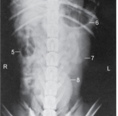
5: cecum 6: fundus part of stomach 7: left kidney 8: bladder |
|
|
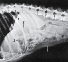
1: liver 2: pyloric part of stomach 2': descending duodenum 3: spleen |
|
|
what is the function of the pancreas? |
exocrine portion: produces digestive enzymes endocrine portion: produces insulin, glucagon, somatostatin, gastrin, etc. |
|
|
what are the regions of the pancreas, and what abdominal quadrants are they located in? |
body: R cranial R lobe (limb): R cranial and R caudal L lobe (limb): R cranial and L cranial |
|
|
what part of the pancreas is located within the mesoduodenum? |
R lobe (limb) |
|
|
what part of the pancreas is located within the deep leaf of the greater omentum? |
L lobe (limb) |
|
|
where does the pancreatic duct empty? |
at the major duodenal papilla (with the bile duct), in the initial part of the duodenum |
|
|
where does the minor pancreatic duct empty? |
minor duodenal papilla, distal to major duodenal papilla in the duodenum |
|
|
what is the function of the kidneys? |
remove toxins from bloodstream produce urine conserve salts, glucose, proteins, water regulate blood pressure |
|
|
how are the kidneys positioned within the abdominal cavity? |
|
|
|
what abdominal quadrant is each kidney located in? |
right kidney: R cranial and R caudal left kidney: L cranial and L caudal |
|
|
what vessels supply blood and drain blood from the kidneys? where does the artery arise? where does the vein drain? |
renal arteries (arise from aorta) renal veins (drain to caudal vena cava) |
|
|
where are the renal lymph nodes located? |
at the hilus of the kidney |
|
|
what is the renal hilus? |
entrance/exit for renal artery, renal vein, ureter, and lymphatics. on the external surface of the kidney |
|
|
what is the renal sinus? |
cavity of the kidney contains renal pelvis, fat, vessels, lymphatics, nerves |
|
|
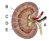
A. renal hilus B. cortex C. renal pelvis D. medula E. renal crest |
|
|
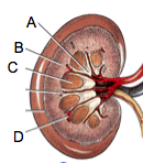
A. pelvic recess B. cortex C. renal pyramid D. arcuate artery and vein |
|
|
what are the 3 main differences between the kidney of the dog and the cat? |
feline kidneys more easily palpated feline kidneys have subscapular veins renal crest is different: more longitudinal in dog, more cone-shaped in cat |
|
|
what is the pathway that urine takes from the kidney to outside the body? |
kidney ureter urinary bladder urethra vestibule (female), tip of penis (male) |
|
|
where are the adrenal glands located? |
cranial to kidneys retroperitoneal |
|
|
the R adrenal gland is closely associated with what structure? |
caudal vena cava |
|
|
what artery and vein course along the surface of the adrenals? |
phrenicoabdominal artery (along dorsal surface) phrenicoabdominal vein (along ventral surface) |
|
|
what are the structures of the female reproductive tract, from internal to external? |
ovary uterine tube uterus (horn, body, cervix) vagina vestibule vulva (with clitoris) |
|
|
the broad ligament of the uterus includes what three structures? |
mesometrium; supports uterus mesovarium: supports ovary mesosalpinx: supports uterine tube |
|
|
where are the ovaries located within the abdominal cavity? |
dorally at the caudal pole of the kidneys R ovary is usually more cranial than the L ovary |
|
|
what structure courses between the ovary and the uterine horn? |
proper ligament of the ovary |
|
|
where is the suspensory ligament of the ovary located? |
cranial border of the mesovarium between ovary and transversalis fascia of the body wall |
|
|
what is the ovarian bursa? how is it formed? |
peritoneal sac that encloses the ovary formed by mesovarium and mesosalpinx |
|
|
what are the three regions of the uterine tube? |
infundibulum ampulla isthmus |
|
|
what kind of uterus do cats and dogs have? |
bicornuate uterus, small body |
|
|
where is the round ligament of the uterus found? what is its developmental origin? |
from the tip of the uterine horn to the vaginal process (dog) or vaginal ring (cat) located within a fold of mesometrium remnant of the gubernaculum |
|
|
where are the internal uterine ostium and external uterine ostium located? |
internal uterine ostium: dorsally located (opening of cervix into uterus) external uterine ostium: ventrally located (opening of cervix into vagina) |
|
|
what is the tissue composition of the vagina? |
circular muscle fibers longitudinal mucosal folds |
|
|
what is the vaginal fornix? |
extension of vagina cranial cervix ventrally located |
|
|
what is the dorsal median post cervical fold? |
narrowing of vagina just caudal to cervix |
|
|
what structure is located between the vagina and the vulva? |
vestibule |
|
|
where does the urethra drain? |
into the vestibule at the urethral tubercle by way of the external urethral orifice |
|
|
where is the clitoris located? |
flood of the vestibule near the vulva glans clitoris sits in the fossa clitoridis |
|
|
what are the parts of the clitoris? |
paired crura short body glans clitoridis (erectile tissue structure) |
|
|
what is the skeletal muscle in the wall of the vestibule called? |
constrictor vestibuli mm. |
|
|
what gland is present in the vestibule to produce mucous secretion during estrus? |
major vestibular gland |
|
|
what arteries provide blood supply to the ovaries and the uterus? what is the origin of these vessels? |
ovarian artery (from aorta) uterine artery (from vaginal artery, which arises from internal iliac artery)
|
|
|
what veins drain blood from the ovaries and the uterus? where do these vessels drain? |
ovarian vein (R -> caudal vena cava, L -> left renal vein) uterine vein (vaginal vein to internal iliac vein) |
|
|
what is the difference between an open or a closed castration? |
open castration: open clinical vaginal tunic: internal spermatic fascia parietal vaginal tunic closed castration: ligate entire vaginal tunic |
|
|
what are the three categories of nerves found in the abdominal viscera? |
parasympathetic neurons (from the vagus) sympathetic neurons (from lumbar sympathetic trunk, splanchnic nerves, and ganglia) visceral sensory neurons |
|
|
what nerves provide parasympathetic innervation to the abdominal cavity? |
dorsal and ventral vagal trunks |
|
|
what nerves provide sympathetic innervation to the abdominal cavity? |
lumbar sympathetic trunk splanchnic nn. lumbar splanchnic nn. major splanchnic n. minor splanchnic nn.
|
|
|
what structure does the lumbar sympathetic trunk course through to get to the abdomen? what other nerve courses through this opening? |
lumbocostal arch in the diaphragm major splanchnic nerve |
|
|
what are plexuses? |
|
|
|
where is the celiacomesenteric plexus located? what ganglia can be found in the celiacomesenteric plexus? |
between the celiac artery and the cranial mesenteric artery contains 3 ganglia: celiac ganglia (R and L) cranial mesenteric ganglia |
|
|
where is the caudal mesenteric ganglion located? |
along the caudal mesenteric a., in the caudal mesenteric plexus |
|
|
what is the name of the plexus between the celiacomesenteric and caudal mesenteric plexuses? |
intermesenteric plexus |
|
|
what nerves supply sympathetic innervation to the pelvic viscera? |
hypogastric nn. (R and L) |
|
|
top: celiac ganglia (R and L) middle: cranial mesenteric ganglion bottom: caudal mesenteric ganglion |
|
|
what are the 5 terminal branches of the aorta? |
external iliac arteries (R &L) internal iliac arteries (R &L) median sacral artery |
|
|
what three branches off of the aorta supply blood to the stomach and intestines? |
celiac a. cranial mesenteric a. caudal mesenteric a. |
|
|
what are the 3 branches off of the celiac artery? |
hepatic artery left gastric artery splenic artery |
|
|
what are the 3 branches off of the hepatic artery? Which of these branch? |
|
|
|
what are the four branches off of the cranial mesenteric artery? |
|
|
|
what is the path that blood takes from the aorta to the antimesenteric ileal branch? |
aorta cranial mesenteric artery common colic artery ileocolic artery cecal artery antimesenteric ileal branch |
|
|
what are the two branches of the caudal mesenteric artery? |
left colic artery cranial rectal artery |
|
|
how is blood supplied to the esophagus in the cervical, thoracic, and terminal regions? |
cervical region
thoracic region
terminal portion
|
|
|
what arteries supply blood to the lesser curvature of the stomach and the lesser omentum? |
left gastric a. (from celiac a.) |
|
|
what arteries supply blood to the greater curvature of the stomach and the greater omentum? |
short gastric aa. (from splenic a. from celiac a.) L gastroepiploic a. (from splenic a. from celiac a.) R gastroepiploic a. (from gastroduodenal a. from hepatic a. from celiac a.) |
|
|
what arteries supply blood to the initial and distal duodenum? |
initial duodenum: cranial pancreaticoduodenal a. (from gastroduodenal a. from hepatic a. from celiac a.) distal duodenum: caudal pancreaticoduodenal a. (from cranial mesenteric a.) |
|
|
what arteries supply blood to the jejunum? |
jejunal arteries (from cranial mesenteric a.) |
|
|
what arteries supply blood to the ileum? |
ileal aa. (from cranial mesenteric a.) mesenteric ileal br. (from ileocolic a. from common colic a. from cranial mesenteric a.) antimesenteric ileal br. (from cecal a. from ileocolic a. from common colic a. from cranial mesenteric a.) |
|
|
what arteries supply blood to the colon? |
cranial mesenteric
caudal mesenteric a.
|
|
|
what arteries supply blood to the liver? |
hepatic brs. (from hepatic a. from celiac a.) |
|
|
what arteries supply blood to the gall bladder? |
cystic brs. (from helpatic a. from celiac a.) |
|
|
what arteries supply blood to the spleen? |
splenic brs. (from splenic a. from celiac a.) |
|
|
what arteries supply blood to the pancreas? |
R lobe: pancreatic brs. (from cranial pancreaticoduodenal a. from gastroduodenal a. from hepatic a. from celiac a.) L lobe: pancreatic brs. (from splenic a. from celiac a.) |
|
|
what veins drain into the caudal vena cava? |
lumbar vv. hepatic vv. phrenicoabdominal vv. renal vv. R testicular and ovarian vv. (L drain into L renal) deep circumflex iliac vv common iliac vv |
|
|
what veins drain into the portal vein? |
cranial & caudal mesenteric veins join to form portal v. gastroduodenal and splenic vv. drain into portal v. |
|
|
what are the 4 lymphocenters in the abdomen? |
lumbar lymphocenter celiac lymphocenter cranial mesenteric lymphocenter caudal mesenteric lymphocenter |
|
|
what lymph nodes are found in the lumbar lymphocenter? |
lumbar aortic lnn. renal lnn. |
|
|
what lymph nodes are found in the celiac lymphocenter? |
hepatic lnn. splenic lnn. gastric lnn. pancreaticoduodenal lnn. |
|
|
what lymph nodes are found in the cranial mesenteric lymphocenter? |
jejunal lnn. colic lnn. |
|
|
what lymph nodes are found in the caudal mesenteric lymphocenter? |
caudal mesenteric lnn. |