Reading...
![]()
Play button
![]()
Play button
![]()
Use LEFT and RIGHT arrow keys to navigate between flashcards;
Use UP and DOWN arrow keys to flip the card;
H to show hint;
A reads text to speech;
93 Cards in this Set
- Front
- Back
|
Because of close proximity opening / exit the lesser curvature forms a ______.
|
deep angular notch
~it is flexed |
|
|
musculoaponeurotic boundary of the external oblique m. may be evident, especially in horses suffering from _________?
What vein might be visible on? |
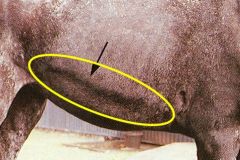
heaves ---heave line (expiratory difficulty)
superficial thoracic (spur) vein |
|
|
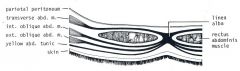
transverse abdominal m. passes over (dorsal to) the rectus abdominis m. to insert on _____ which is _____.
|
transverse abdominal m. passes over (dorsal to) the rectus abdominis m. to insert on LINEA ALBA.
~which is strong and supportive, and relatively avascular. |
|
|
parts of greater omentum
|
greater omentum:
1. gastrophrenic ligament: from greater curvature to crura of diaphragm 2. gastrosplenic ligament: from stomach to spleen 3. lienorenal (renosplenic) ligament - attaches the left kidney to the spleen. |
|
|
Name thick fibroelastic sheet of tissue overlying the aponeurosis of the external oblique muscle, attached to rib cage and tuber coxae
|
Deep fascia becomes yellow abdominal tunic
|
|
|
how many pyloric sphincters?
|
2 (cranial/caudal) are also well
developed, especially that (caudal) guarding the narrow pyloric exit. |
|
|
margo plicatus
|
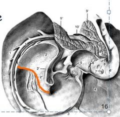
margo plicatus:
- divides distal glandular mucosa from proximal non- glandular, abrasive mucosa - Gastrophilus intestinalis larvae |
|
|
Oesophagus enters ___ curvature quite _____ at cardiac sphincter
|
Oesophagus enters LESSER curvature quite <obliquely> at the cardiac sphincter, which is thick
|
|
|
Contents of the inguinal canal include:
|
* vaginal process, which is diverticulum of peritoneum go thr. vaginal ring
* Spermatic cord within cavity of vaginal process (vaginal tunic), * external pudendal artery and vein, * inguinal lymph vessels and nerves (present in both sexes) |
|
|
• Local anesthesia of flank by blocking ______ branches
|
Local anesthesia of flank by blocking T13-L2 branches
|
|
|
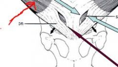
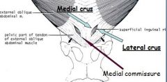
Identify parts of superficial (external) inguinal ring:
(be specific) |
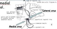
mere slit in aponeurosis of external oblique m. (thus dividing it into the lateral and medial crura of the aponeurosis)
|
|
|
What direction do external abdominal obliques go in?
Border of the internal oblique m. forms a _______? |
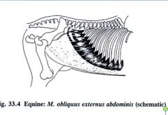
caudo-ventrally (hands in pockets)
caudoventral ridge. |
|
|
The blind sac of stomach, located above cardiac sphincter/ esophagus, is known as the ____
|
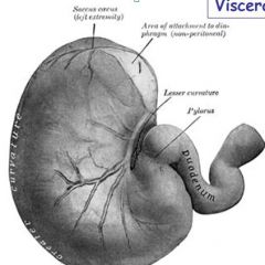
saccus cecus (or blind sack)
Latin cecus means "blind" |
|
|
parts OMENTA:
what do these parts form or enclose? |
Greater and lesser; both along with the visceral surface of stomach, enclose the OMENTAL BURSA.
(entrance to bursa known as epiploic foramen) |
|
|
Where in abdominal cavity is stomach located?
|
LEFT, dorsal of cranial part of abdominal cavity
|
|
|
Duodenum is fixed by mesoduodenum to ______.
|
Duodenum is fixed by mesoduodenum to DORSAL WALL, liver, right dorsal colon and base of the cecum.
(desc. on right side of abd. cavity, but asc. duodenum on left side) |
|
|
Name parts of duodenum and identifiable flexures.
|
(1). Cranial part (sigmoid flexure), (2). descending, (3). caudal flexure (transverse duodenum) and (4). ascending parts]
|
|
|
bile and pancreatic ducts open into what?
|
The bile and pancreatic ducts both open into second curve of the SIGMOID part, opposite each other.
(openings form major and minor duodenal papillae) |
|
|
lateral, segmental sacculations of horse colon known as ?
|
haustra
|
|
|
longitudinal bands on horse colon
|
taeni colli
|
|
|
Another name for large colon?
|
ascending colon
|
|
|
Demarcates the beginning of large colon? location? direction
|
CAECO-COLIC junction; at base of last rib, runs ventrally and cranially on right body wall and abdominal floor to the xiphoid area.
|
|
|
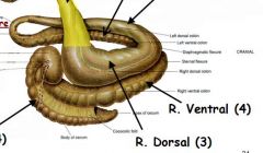
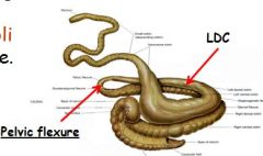
Name parts of ascending (large) colon starting from origin:
|
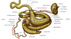
Right Ventral colon-sternal flexure - Left Ventral colon - pelvic flexure - Left Dorsal colon - diaphragmatic flexure - Right Dorsal colon.
|
|
|
Right ventral colon starts where?
Runs in what direction? |
RVC starts at base of last rib, runs ventrally and cranially on right body wall and abdominal floor to xiphoid area.
|
|
|
descending duodenum is related to the ___ and ___ passes around it.
|
descending duodenum is related to the RIGHT kidney and the base of the CECUM (passes around it)
|
|
|
What direction is duod. traveling before transverse duodenum? After transverse duodenum?
|
*Desc..duodenum: travels caudally on right side
*Transv.duodenum (caudal flexure) makes U-turn. *Ascending duodenum: travels cranially on left. |
|
|
where does duodenum end?
|
Reaches the region of the left kidney, and it becomes the jejunum (at duodenojejunal flexure).
|
|
|
Name flexures of duodenum, begins at origin with stomach.
|
* Sigmoid flexure
* caudal flexure * duodenojejunal flexure |
|
|
Jejunoileum is located where?
|
mostly on left dorsal part of the abdominal cavity,
intermingled with small colon |
|
|
loops of small intestine (jejunoileum) are palpable in _______ of abdominal cavity
|
jejunoileum palpable in dorso-caudal aspect of abdom. cavity
|
|
|
epiploic foramen : Between ____ lobe of liver and _____ . And between ____ and _____vein.
|
epiploic foramen : Between right lobe of liver and descending duodenum, and between caudal vena cava and portal vein.
fyi: epip.foramen is RIGHT side; found by lifting liver and placing fingers btw caudal v/c and portal v. |
|
|
____ can get strangulated at epiploic foramen.
|
Loops of jejunum can get strangulated at epiploic foramen.
|
|
|
Which lobe of liver atrophies with age? What happens as a consequence of this?
|
As RIGHT lobe of liver atrophies, epiploic foramen enlarges with age.
~due to pressure on liver from right colon |
|
|
what shape is cecum? volume?
what connective structure extends from body cecum? |
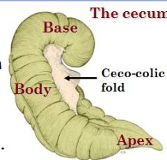
like a coma; holds up to 30 liters
cecocolic fold |
|
|
cecum extends from?
how many taeni colli? |

base extends from pelvic inlet to apex at diaphragmatic area (xiphoid region).
4 |
|
|
Does ventral surface of liver touch ventral abdominal cavity?
|
No
|
|
|
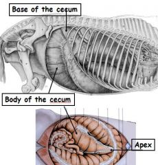
Base of cecum attached to :
|

attached to right sublumbar region and to the right kidney
|
|
|
Body of cecum located where:
|

body runs ventrally on right flank , curves cranio-medially
|
|
|
Which part of cecum is palpable via the rectum?
|
base
|
|
|
How many taeni colli R.ventral colon have? L. ventral colon?
(remember cecum has 4) left dorsal? right dorsal? transverse? small colon? |
RVC = 4
LVC = 4 LDC=1 RDC=3 Trans=2 SC=2 |
|
|
diaphragmatic flexure is considered part of?
|
end of left dorsal colon
|
|
|
which parts of colon are palpable from rectum?
|
left ventral, left dorsal, descending and
|
|
|
widest part of colon?
|
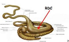
RIGHT DORSAL colon
Mostly within the thoracic cage. Shortest but WIDEST. |
|
|
transverse colon:
attached to? length? shape? |
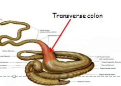
Funnel-shaped
Cranial to root of mesentery; very short Attached to roof of body cavity. Crosses median plane, hence the name. |
|
|
location of desc.colon?
|

Descending colon (also the small colon)
from left kidney to left dorsal part of abdominal cavity to pelvic inlet region; |
|
|
Duodenocecostomy
|
(side to side anastomosis of descending duodenum to base of the cecum in gastro – duodeno – jejunitis).
|
|
|
loops of intestine
|
volvulus
|
|
|
What parts of colon is free-floating?
What does this mean for the horse? |
parts of the ascending colon are free floating
permits intestinal extension during surgery, but also causes twisting of loops of intestine (volvulus), |
|
|
ileal problem? cause?
|
ileal impaction; reasons are not well understood, but thick-walled, and quite frequent
|
|
|
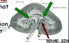
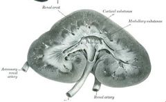
Describe renal crest of equine kidney:
|
Unipyramidal ridge (resulting from fused medullary pyramids) - a common renal crest
|
|
|
Well developed renal crest fuse to form very well developed renal _____, which collects urine from _____.
|
renal pelvis; which collects urine from two polar terminal recesses
|
|
|
Evidence of kidney lobation shown by
|
shown by blood vessels (interlobar arteries) only, but no external evidence
|
|
|
Renal hilus
|
opening into renal sinus (indentation) where ureter and renal vessels enter kidney
~basically it's part coming out |
|
|
Why is urine of equine urine turbid and slimy?
|
Because of glands in renal pelvis and ureters which produce mucus - - so called *physiological albumin* (not pathological)
|
|
|
URETERS on leaving kidneys, travel ____ toward pelvis, where situated on lateral part of the ____ (mare) or ___(stallion).
Travel toward bladder and ____ to the ducti deferentia in male |
Ureters travel along abdominal roof
*in pelvis situated on lateral part of the broad ligament (mare) or genital fold (stallion). * Descend toward bladder VENTRAL to ducti deferens in male |
|
|
Renal artery is branch of?
Where do branches renal arteries enter kidney? |

Arises directly from aorta.
may break into a few branches which penetrate the ventral surface of the organ rather than the hilus |
|
|
Describe location of adrenal glands in comparison to kidneys:
|
ADRENAL glands are on the medial side of the cranial poles of the kidneys.
|
|
|
* rectum is a continuation of the ___.
*shape of rectum? |
Rectum is continuation of small colon, at pelvic inlet
* Sacculated initially but smooth, enlarged sac (ampulla), terminally. Ampulla is retroperitoneal. |
|
|
medical condition in which part of intestine has invaginated into another section of intestine, similar to way in which parts of collapsible telescope slide into one another.[
|
Intussusception
|
|
|
what can happen at Ileo-cecal opening?
|
Can have blockage.
Abnormally high peristalsis -> ileum telescopes into cecum (intussusception). Surgical remedy. |
|
|
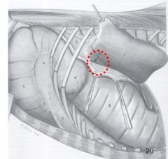
Trocarization necessary when?
|
useful to decompress distended abdomen
e.g. Ceco-colic opening : Enlarged cranial part of base falls over, cranioventrally -> blockage of cecocolic orifice. |
|
|
Aside from ceco-colic opening, which of following most likely to get blockage:
- sternal flexure - pelvic flexure - transverse colon - diaphragmatic flexures |
- pelvic flexure
- transverse colon - sternal & diaphragmatic flexures, also possible, but to a much lesser extent |
|
|
where spleen located?
how does it attach to stomach? |

LEFT side, lies on greater curvature of stomach,
* cranial tip ~6th or 7th rib * only caudo-dorsal projects a bit out of rib cage * cannot be palpated from rear end |
|
|
shape of spleen
What's on cranial border? Caudal border? |

* Cranial border where meets stomach is concave
* caudal border is convex; borders desc.duodenum and loops of small intestine |
|
|
Ligaments from spleen?
|
* attached to stomach via gastrosplenic ligatment (part of greater omentum)
* attached left kidney via lienorenal (renosplenic) ligament – dorsal border of ligament may entrap loops of colon (remember spleen is on left side, so is stomach!) |
|
|
Location of liver:
|
* Mostly RIGHT of median plane, and quite asymmetrical in shape
Cranial border (6th-7th rib): diaphragm Caudal border (16th-17th rib): ventral to right rib |
|
|
which lobe atrophies?
|
RIGHT lobe atrophies with age ( pressure from right dorsal colon)
|
|
|
Parts of cecum? general locations?
|

base - RIGHT, dorsal
body apex |
|
|
Describe gall bladder in horse:
|
●No gall bladder.
● bile or hepatic duct has wide lumen; opens w/major pancreatic duct into hepaticopancreatic ampulla [major duodenal papilla] of the distal and convex part of the duodenal sigmoid flexure. Oblique passage of duct through wall acts as valve. |
|
|
What are the four lobes of the liver?
|
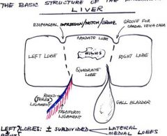
Left lobe (thinnest part)
Quadrate lobe Right lobe Caudate lobe * L&R lobes subdivided in medial/lateral lobes |
|
|
_____ ligament of liver is strong and well formed
|
round (teres) ligament of liver is strong and well formed.
|
|
|
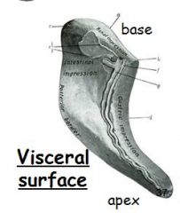
Impressions on liver?
what passs through? |
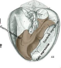
Visceral surface - stomach, duodenal and colic impressions
Renal impression on dorsal part of caudate process! ~Related to pancreas dorsally. Oesophageal impression on dorsal part of left lobe. * caudal vena cava goes though |
|
|
location of pancreas?
parts or recognizable features? |
* right; sublumbar
* right and left lobes and a body (held in place in sigmoid flexure of duodenum). Related to saccus cecus of stomach (left lobe), right kidney (right lobe), aorta, caudal vena cava and sublumbar muscles, dorsally. |
|
|
Supplies the stomach, liver and spleen. The duodenum also, via the _____:
|
- celiac artery; supplies duodenum also via the gastro-duodenal a.
|
|
|
Supplies the small and large intestine (cecum, colon).
|
cranial mesenteric a.
|
|
|
supplies mainly the descending colon and the rectum?
|
caudal mesenteric a.
|
|
|
Lymph Nodes of organs supplied by
|
* celiac artery form a celiac trunk, which empties into the cisterna chyli.
cisterna chyli -> aortic hiatus -> thoracic duct (runs left of aorta); opens into a large vein at thoracic inlet. |
|
|
L.nodes of small intestine (situated at the root of the mesentery) ?
|
intestinal trunk
|
|
|
L.nodes of small colon rectum and anus send efferents to the:
|
lumbar trunk
|
|
|
Which kidney is more cranial?
Give locations: |
Right (left is left behind)
Left Kidney: Ventral to T17 – L2 |
|
|
Give locations of left kidney:
|
Left Kidney: Ventral to T17 – L2
* Related to spleen and stomach cranially, to aorta medially, and small colon and coils of small intestine, ventrally. |
|
|
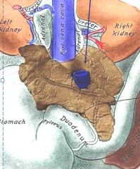
Give location of right kidney:
(in relation to liver? d.duodenum? pancreas? cecum? small....?) |
Ventral to last 2 or 3 ribs and 1st lumbar transverse process (T16 – L1) (mostly covered by ribs)
* cranial pole makes a renal impression on caudate process of liver. - Relations: liver is cranial to it; ventrally is descending duodenum, pancreas and base of cecum; coils of small colon and small intestine; medially, to right adrenal gland and the aorta. |
|
|
Which kidney palpable through rear end?
|
If you're lucky, caudal pole of left kidney may just be within reach in rectal palpation. But it's a crap shoot.
|
|
|
Shape of kidneys:
|
left: bean-shaped.
right: heart shaped |
|
|
coming out of kidney, hilus runs where?
|
hilus is ventromedial
|
|
|
veins are ____ of arteries
|
veins are satellites of the arteries
|
|
|
large arteries (celiac, cranial and caudal mesenteric arteries and their major branches) are the sites of ___ caused by ______.
|
Large arteries (celiac, cranial and caudal mesenteric arteries and major branches) are sites of LESIONS caused by Migrating Larvae! e.g. nematode
~aneurysms may develop |
|
|
"collateral" circulation
|
* advantageous to the animal in event of blockage
* blockage of large arteries is less serious than that of smaller vessels where anastomoses incomplete |
|
|
How accessible is stomach to palpation, etc.?
|
stomach is inaccessible by rectal palpation or through flank incision.
|
|
identify:
|
|
|
|
femoral lamina
|
strands of connective tissue present only in horse, connect lateral crus of superficial inguinal opening to medial thigh fascia
|
|
|
inguinal hernia occurs because ____.
|

When hip joint is maximally extended, there is considerable pull on lateral crus of this opening.
(e.g. problem can arise during mating) |
|
|
• Paralumbar fossa
|
o Concavity immediately caudal to last rib
o Able to be anesthetized during surgery (expand on this) o Palpable structures? |