Reading...
![]()
Play button
![]()
Play button
![]()
Use LEFT and RIGHT arrow keys to navigate between flashcards;
Use UP and DOWN arrow keys to flip the card;
H to show hint;
A reads text to speech;
43 Cards in this Set
- Front
- Back
|
What are the lateral and ventral, dorsal, cranial, and caudal boundaries of the abdominal cavity.
|
Lateral and ventral - The external and internal abdominal oblique mm., the transversus abdominis m., and the costal arch.
Dorsal - vertebrae and hypaxial mm. Cranial - diaphragm Caudal - pelvic inlet |
|
|
What type of membrane is the peritoneum and what two membranes make it up?
|
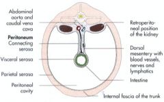
The peritoneum is made of a serous membrane.
The parietal and visceral peritoneum make up the peritoneum and line the peritoneal cavity. |
|
|
What does the visceral peritoneum cover?
|
All of the organs in the abdominal cavity.
|
|
|
Which primitive mesentery, the dorsal or ventral, becomes the abdominal mesentery and greater omentum?
|
The dorsal mesentery becomes the greater omentum, mesoduodenum, mesojejunum, mesoileum, mesocolon, and mesorectum.
|
|
|
What are the six mesenteries that come from the primitive dorsal mesentery?
Where do the mesenteries all course from? |
The greater omentum - or mesogastrium.
Mesojejunum Mesoduodenum Mesoileum Mesocolon Mesorectum Here's an incredibly inappropriate mnemonic for you: Great Jewish dudes have ill conceived rectums. They all course from the dorsal body wall to their respective organs. |
|
|
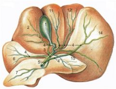
What do the superficial and deep leaves of the greater omentum contain?
|
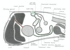
The greater omentum contains fat and vessels (the green line).
The superficial (the more ventral part) leaf contains the spleen. The deep leaf contains the left limb of the pancreas. |
|
|
What is the omental bursa?
What is its clinical signifigance? |
The omental bursa is the space between the superficial and deep leaves of the greater omentum.
It is clinically important to know that the omental bursa only has one opening to the peritoneal cavity through the epiploic foramen - the site of communication between the two. |
|
|
What are the dorsal, ventral, caudal, and cranial boundaries of the epiploic foramen?
|
Dorsal - caudal vena cava
Ventral - portal v. Caudal - hepatic a. Cranial - liver |
|
|
What does the primitive ventral mesentery arise from? Where does it course between? What ligaments (2) are remnants of the primitive structure?
|
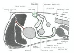
The primitive ventral mesentery becomes the lesser omentum (in orange). It courses from the lesser curvature of the stomach and initial portion of the duodenum to the liver and diaphragm. It's defined by the hepatoduodenal ligament and hepatogastric ligament.
The remnants are the: Falciform ligament and Median ligament of the bladder |
|
|
The falciform ligament both is a remnant of something and contains the remnant of something - what?
What does the falciform ligament demarcate the division between? |
The falciform ligament is itself a remnant of the ventral mesentery.
It contains the round ligament of the liver that is a remnant of the umbilical vein. It demarcates the division between the left and quadrate lobes of the liver. |
|
|
What is the median ligament of the bladder a remnant of? What remnant does it contain?
Is it just me, or is the abdominal cavity like one of those Russian matryoshka dolls - remnants inside remnants ad nauseum. It's like a goddamn grab bag of fun in there. |
The median ligament of the bladder is a remnant of the ventral mesentery.
It contains the remnant of the urachus. |
|
|
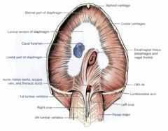
That damn diaphragm - so lazy. Always on a freaking hiatus. Name the three that you might be tested on and what passes through them.
|
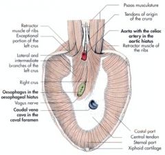
The aortic hiatus - through which the aorta, azygous v. and thoracic duct pass.
The esophageal hiatus - through which the esophagus, dorsal and ventral vagal nerve trunks pass and esophageal vessels pass. The caval foramen - in a fierce marketing campaign, the vena cava decided that "hiatus" had a bad connotation and adopted the name "foramen" that we know it as today. The caudal vena cava goes through that hiatus. |
|
|
To which vertebrae are the roots of the diaphragm connected?
What passes between the crura? What is lateral to each crus connecting it to the body wall? |
The crura are attached to L3 and L4 vertebrae.
The aortic arch passes between them. The lumbocostal arch connects the crura laterally to the body wall. The sympathetic trunk and major splanchnic n. courses through it. |
|
|
What are the functions of the liver? (5)
|
Metabolize carbs, protein and fat.
Produce bile. Store and filter blood. Excrete bilirubin. Detoxify foreign substances. |
|
|
What are the four lobes of the liver and their parts?
|
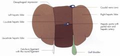
The right medial and right lateral, left medial and left lateral, quadrate, and caudate with the caudate process and papillary process
|
|
|
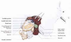
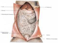
The liver has two surfaces - what are they and what do they make contact with? (that was also a hint as to what they are)
|
The parietal (or diaphragmatic) surface contacts the diaphragm.
The visceral surface contacts the viscera - the stomach, duodenum, pancreas, right kidney, spleen (sometimes), greater omentum and small intestine. |
|
|
Three ligaments function to hold the liver in place - what are they?
|
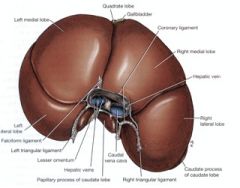
There are two triangular ligaments - the right and left - they course between their respective crus (right or left) and their respective lateral lobe (right or left) of the liver.
The coronary ligament courses between the diaphragm and liver circling the caudal vena cava and hepatic veins. The perspective in the picture is the parietal surface. |
|
|
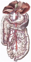
What vessels carry blood to and from the liver?
|
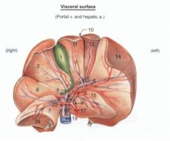
To - the hepatic artery (17) with O2-rich blood from the heart, and the portal vein (18) with nutrient-rich blood from the digestive tract.
From - the hepatic veins (19) going to the caudal vena cava. |
|
|
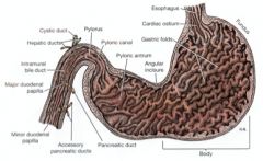
The biliary system consists of one storage organ and three ducts that we need to know - what are they?
|
The gall bladder (1) stores the bile and contracts to eject it into the small intestine.
The cystic duct (3) courses between the gall bladder and bile duct. The hepatic ducts (4) carry bile from the liver. The common bile duct (5) is formed when the cystic and hepatic ducts meet. It carres the bile to the duodenum. |
|
|
Where the common bile duct empties into the duodenum.
|
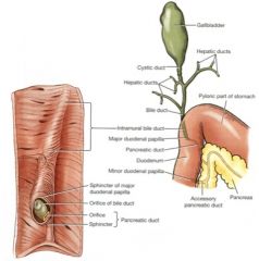
The major duodenal papilla.
|
|
|
What are the functions of the spleen? (4)
Where can you be sure to locate the spleen? What will the spleen's parietal and visceral surface face? |
RBC storage
RBC destruction Immune system functions Blood filtration It's located within the greater omentum and, though shifty, sure to be on the left side of the body. It's parietal surface faces the diaphragm and abdominal wall. It's visceral surface faces the greater curvature of the stomach and left kidney. |
|
|
Why did the barbituates in euthanasia cause the spleen's in our specimens to enlarge?
On which splenic surface does the hilus of the spleen reside? What is the name of the ligament securing the spleen to the stomach? |
Barbituates cause the smooth muscle within the splenic capsule to relax and the spleen to enlarge.
The hilues, or root, of the spleen is on the visceral surface. The vessels of the spleen travel through the hilus. The gastrosplenic ligament anchors the head of the spleen to the stomach. |
|
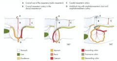
The cranial mesenteric artery is in the root of the mesentery. The root was formed in development by the mesentery being twisted. In which direction does the small intestine wrap around the cranial mesenteric artery? What about the large intestine?
|

The small intestine wraps caudally around the root - the large intestine wraps cranially around it.
|
|
|
It's essential that we know where the root of the mesentery is located as the quadrant system of the abdomen is defined off of it, and if we know what quadrant the regions of the digestive tract should be in, we can know what's gone wrong. So where should the root of the mesentery be located?
|
It's located at L2, surrounding the cranial mesenteric artery. It is the origin of the great mesentery - the mesojejunoileum.
|
|
|
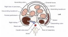
What quadrant(s) is the liver located in?
What about the gall bladder? Spleen? |
The liver spans the left and right cranial quadrant.
The gall bladder is in the right cranial quadrant. The spleen is always on the left side - it is always in the left cranial and caudal quadrants. When in right lateral recumbency, it will also be seen in the right cranial quadrant. |
|
|
What quadrants does the spleen exist in always? What quadrant varies by recumbency?
|
The spleen is always on the left side - it is always in the left cranial and caudal quadrants.
When in right lateral recumbency, it will also be seen in the right cranial quadrant. |
|
|
You team up with microman and get yourself swallowed. Describe your journey through the gastrointestinal tract.
|
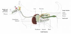
Stomach to small intestine: duodenum, jejunum, and ileum to large intestine: cecum, colon: ascending, transverse, and descending parts, and finally out the pooper - or rectum.
|
|
|
What attaches to the greater and lesser curvatures of the stomach? Which curvature has the least concentration of vasculature?
|
The greater omentum attaches to the greater curvature and the lesser to the lesser.
The greater curvature has less concentration of vasculature and will typically be where stomach incisions are made in surgery for this reason. |
|
|
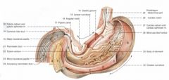
What are the four areas of the stomach? What are the mucosal and submucosal folds called? What might they be mistaken for in radiographs?
|
Cardia, fundus, body, pylorus (with the antrum, canal, and sphincter)
The rugae are the folds permitting stomach expansion - they could be confused for washcloths in the stomach. |
|
|
In what quadrants of the stomach do the areas of the stomach exist in?
|
The cardia, fundus and body are the left cranial.
The pyloric antrum and pylorus are in the right cranial. |
|
|
The areas of the stomach have glands named after them - what are they? What do they secrete? What is the function of this secretion?
|
There are cardiac, pyloric and fundic glands. These secrete mucus, the function of which is to protect the stomach's nonglandular tissue from the digesting acids.
|
|

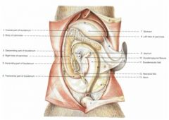
Define where the duodenum (the flexures and de/ascending parts), jejunum, and ileum run in this image.
What quadrants should they be located in? |
Duodenum (A → E) - all quadrants:
Cranial duodenal flexure (A → B) - RCr Descending part (B → C) – RCr, RCa Caudal duodenal flexure (C → D) – RCa, LCa (it actually curves around the cranial mesenteric artery, which isn't depicted accurately here) Ascending part (D → E) – LCa Duodenojejunal flexure (E) - LCa Jejunum (E → F) – all 4 quadrants Ileum (F → G) – terminal part of small intestine – L and R caudal quads |
|
|
In dogs and cats, does food travel through the cecum?
|
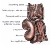
It doesn't have to - it does pass through the ileocolic orifice.
|
|
|
Which quadrant is the cecum located in?
|
The right caudal quadrant in the dog and cat.
|
|

What quadrants do the components of the large intestine reside in? Where do they course between in this picture?
|

Cecum (H) – Rt caudal
Colon (I → L) Ascending colon (I → J) - RCa, RCr Right colic flexure (J) Transverse colon (J → K) – RCr, LCr Left colic flexure (K) Descending colon (K →L) – LCr, LCa Rectum (L → M) |
|
|
What are the functions of the pancreas?
|
The exocrine portion puts digestive proenzymes into the duodenum.
The endocrine portion makes insulin, glucagon, somatostatin, gastrin, etc |
|
|
What are the regions of the pancreas? Where are they located?
(she will ask us about this) |
Find the duodenum, find the pancreas. Didn't they base a tv show on that concept? The duodenum - the cheerleader of the abdomen.
The body is located near the pylorus of the stomach. The right lobe is located within the mesoduodenum The left lobe is located within the deep leaf of the greater omentum. |
|
|
Where do the pancreatic and accessory pancreatic ducts drain? Compare and contrast these ducts in the dog and cat.
|
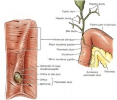
The pancreatic duct empties into the major duodenal papilla.
The accessory pancreatic duct empties into the minor duodenal papilla. In the cat, the pancreatic duct is always present. In the dog its smaller than the accessory pancreatic duct and might be absent. The accessory pancreatic duct is only present in about 20% of cats. |
|
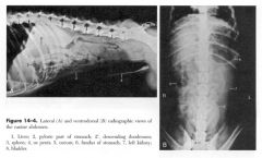
Cover up the key....do it....
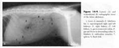
Identify 1, 2, 2', 3, 5, 6, 7, and 8. And for you dirty birdies, go ahead and identify 4. |

Ok, now look.
|
|

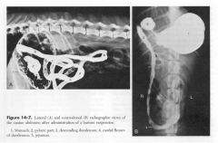
This is a cat.
What are 1, 3', 3", and 7? |
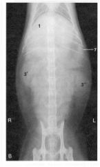
1. Liver
3'. R kidney 3". L kidney 7. spleen |
|

Go to town.
|
|
|

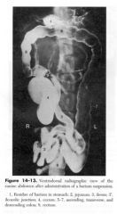
Once more with contrast...name the tagged structures.
|
|
|

After letting the contrast get good and settle in...name the tagged things here.
|
|