Reading...
![]()
Play button
![]()
Play button
![]()
Use LEFT and RIGHT arrow keys to navigate between flashcards;
Use UP and DOWN arrow keys to flip the card;
H to show hint;
A reads text to speech;
91 Cards in this Set
- Front
- Back
- 3rd side (hint)
|
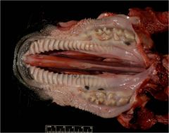
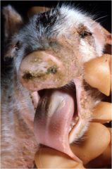
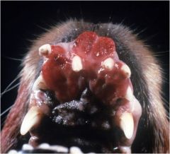
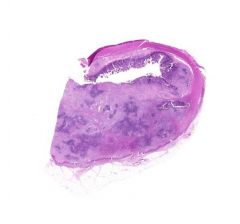
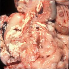
Cleft palate
Causes: Hereditary, prenatal drug or toxin exposure. |
|
|
|
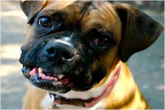
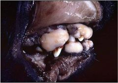
Brachygnathia superior-shortness of maxilla; most common in short nosed dogs.
or Prognathia-long mandible; most common in sheep |
|
|
|
Targeted destruction of ameloblasts during tooth formation.
Dogs: prenatal Canine distemper virus Cattle: prenatal BVD, fleurosis, Ca deficiency |
|
|
|
Inflammatory destruction of the gingiva, periodontal ligament, alveolar bone, and root cementum.
|
|
|
|
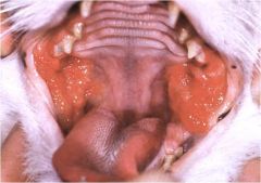
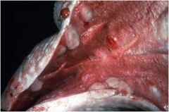
Plasmacytic-Lymphoplasmacytic Stomatitis
Occurs in cats Proliferative lesions, most common at the glossopharyngeal arches |
|
|
|
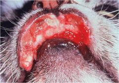
Eosinophilic granuloma complex-rodent ulcer
Cats Cause unknown, may be linked to food hypersensitivity, or feline leukemia virus |
|
|
|
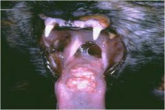
Eosinophilic granuloma complex-oral eosinophilic granuloma
Cats Cause unknown, may be linked to food hypersensitivity, or feline leukemia virus |
|
|
|
Thrush-Candida albicans
Self-limiting, opportunistic fungal infection of young animals. Yellow-white pseudomembranous hyperkeratosis |
|
|
|
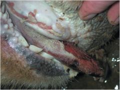
Wooden Tongue
Caused by actinobacillus lignieresii (gram neg). Typically enters the tissue via penetrating wound. |
|
|
|
Oral ulcers
Cattle: vesicular stomatitis, bovine papular stomatitis, BVD, Foot and mouth, Malignant Catarrhal Fever Cats: Calicivirus, herpes virus Pigs: Calicivirus Horses: Herpes virus All: Uremia, trauma/foreign body, ingesting caustic substances. |
|
|
|
Uremic ulcers
Tend to occur on the ventral tongue margin |
|
|
|
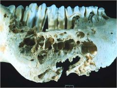
Lumpy jaw
Caused by Actinomyces bovis (gram pos) Enters by penetrating wounds in the oral cavity Granulomatous-suppurative lesions with boney degeneration. |
|
|
|
Proliferative fibrogingival hyperplasia
Benign lesion-secondary to chronic gingivitis Should be smooth Usually multifocal, but can be focal. |
|
|
|
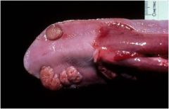
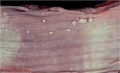
Viral papilloma (papillomavirus)
Species and tissue specific Occur in young puppies and cattle Benign, multifocal, and tend to spontaneously regress. Can be removed if interfering with eating. |
|
|
|
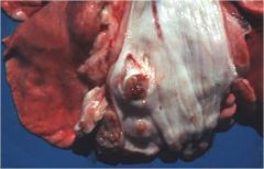
Epulides (sing-epulus)
Benign lesion originating from the periodontal ligament. Can be fibrous or ossifying. Does not invade the bone! |
|
|
|
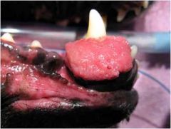
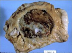
Acanthomatous ameloblastoma
Originates from the odontogenic epithelium. Locally invasive (often into alveolar bone), but does not metastasize. |
|
|
|
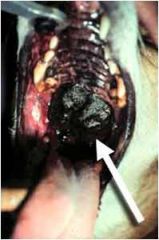
Oral melanosarcoma
Malignant-invasive and metastatic Most common oral neoplasia in dogs. |
|
|
|
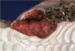
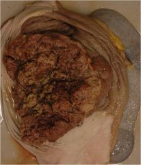
Oral squamous cell carcinoma
Malignant-infiltrative, boney destruction, metastatic (late). Most common malignant oral tumor of cats, second most common in dogs. |
|
|
|
Oral fibrosarcoma
Malignant-infiltrative, low metastatic rate. Second most common oral neoplasia in cats. |
|
|
|
Esophageal submucosal gland dilation
Incidental finding in the distal esophagus of dogs. |
|
|
|
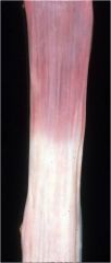
Distal esophageal muscular hypertrophy
Incidental finding in horses |
|
|
|
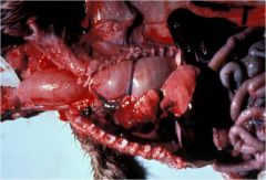
Bloat line
Well demarcated line at or cranial to the thoracic inlet. Indicates that the cow actually died of bloat, vs bloating during decomposition. |
|
|
|
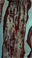
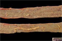
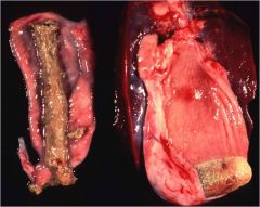
Esophageal ulceration
Causes: Trauma, foreign body, caustic substances, medication lodged in esophagus (esp. tetracycline and doxycycline), BVD infection in cattle. |
|
|
|
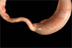
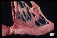
Megaesophagus
Causes: PRAA, myesthenia gravis, stricture, idiopathic |
|
|
|
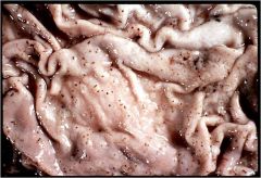
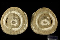
Gongylonema pulchrum
Nematode embedded in the mucosa Typically not clinically significant |
|
|
|
Esophageal viral papillomas
Occur in cattle, caused by papillomavirus |
|
|
|
Spirocera lupi
Spirurid nematode Causes granulomatous and eosinophilic esophagitis May be associated with esophageal osteosarcoma and fibrosarcoma |
|
|
|
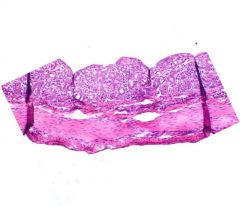
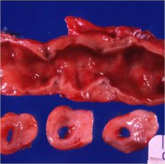
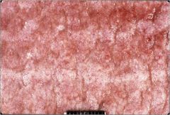
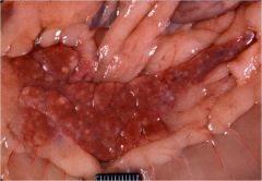
Rumenitis/Rumen ulcers
Common causes are grain overload or fungal ruminitis. |
|
|
|
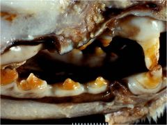
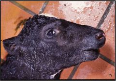
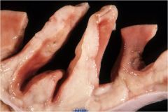
Brachygnathia inferior-short mandible
Common in calves and long-nosed dogs |
|
|
|
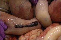
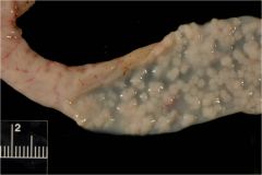
Pigs: Happens most commonly at the pars esophagea.
Cattle: Abomasal ulcers; caused by acidosis, fungal infection, stress, or LSA. Canine: Common causes are NSAIDs, mast cell tumors, and gastrinomas Feline: Uremia, and similar causes to dogs. |
|
|
|
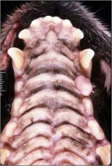
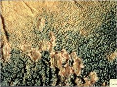
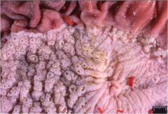
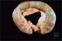
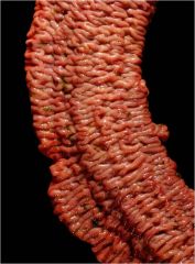
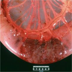
Ostertagia (cattle) or Teladorsagia (small ruminants).
Characteristics "cobblestone" appearance. |
|
|
|
Equine Gasterophilus
Incidental finding-no clinical signs |
|
|
|
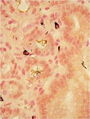
Helicobacter
Must use special stain to see. Probably an incidental finding, hard to verify as the cause of clinical disease. |
|
|
|
Proventricular dilatation disease
Bornavirus. GI primarily, also heart, CNS, and adrenals |
|
|
|
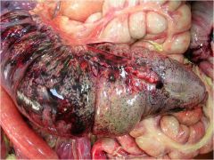
Gastric rupture
Most common in horses Muscle wall tears along the greater curvature, mucosa tears last. |
|
|
|
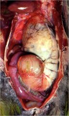
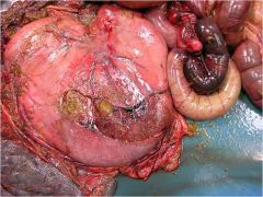
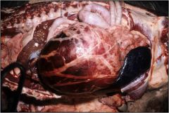
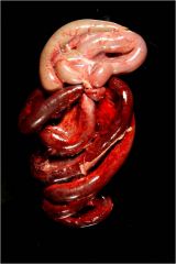
Gastric dilatation volvulus
Dogs Pulls mesentery over the stomach. Causes organ ischemia. |
|
|
|
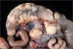
GI lymphosarcoma
Common in cattle and cats. Cattle: Caused by bovine leukemia virus; abomasum is most common GI location. Cats: stomach and ileum are most commonly affected. |
|
|
|
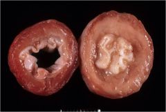
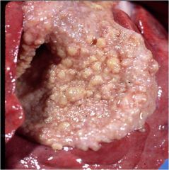
Gastric squamous cell carcinoma
Most common in horses Cauliflower like mass in the squamous portion of the stomach. Can metastasize. |
|
|
|
Hyperplastic GALT
Incidental finding |
|
|
|
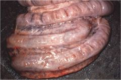
Hemomelasma ilei
Incidental finding in horses |
|
|
|
Lymphoplasmacytic enteritis
Type of inflammatory bowel disease |
|
|
|
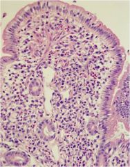
Eosinophilic enteritis
Lots of pink! Type of inflammatory bowel disease Causes: Parasites, fungal or allergy |
|
|
|
Feline infectious peritonitis
Wet or dry form |
|
|
|
Peyers patch necrosis
Cattle: Caused by BVD Dogs: Caused by Parvovirus (may also cause intestinal hemorrhage). |
|
|
|
Fibrinous enteritis
Raccoons: Parvovirus |
|
|
|
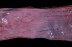
Rotavirus/coronavirus
Intestinal wall-flaccid, thin, transparent. Due to villous atrophy and loss |
|
|
|
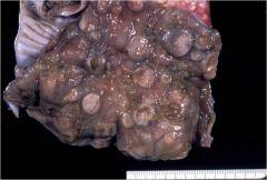
Johne's disease
Mycobacterium avium ssp. paratuberculosis Cattle Corrogated appearance in the ileum and large intestines (looks similar to Lawsonia intracellularis in pigs) |
|
|
|
Lawsonia intracellularis
"Proliferative ileitis" Occurs in pigs and horses May also cause necrosis and hemorrhage in pigs (Porcine proliferative enteropathy) Appears similar to Johne's disease |
|
|
|
Clostridial enteritis
Occurs in dogs, pigs, and horses Causes hemorrhage |
|
|
|
E. coli, edema disease
Most common in pigs less than 5 days old. Watery diarrhea Shiga-toxin causes edema in the intestinal tissues. |
|
|
|
Salmonella colitis
Horses: Bloody diarrhea and ulceration Pigs: Button ulcers, associated with strictures |
|
|
|
Most common in horses, does occur in ruminants
Ulcerative enterocolitis |
|
|
|
Colonic histoplasmosis
Dogs Thickened large intestines |
|
|
|
Pythium insidiosum
Invades the muscular layer Causes a palpable abdominal mass |
|
|
|
Endarteritis-strongyle infarction
Migration irritates vasculature, causes non-strangulating infarct. |
|
|
|
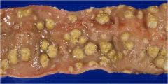
Coccidiosis
Fibrinohemorrhagic or fibrinonecrotic enteritis with white nodules in the mucosal surface Chickens and Pigs |
|
|
|
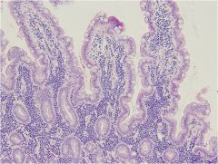
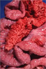
Lymphangectasia
"shag carpet"-prominent white, thickened villi, may also see granulomatous lesions along the mesenteric lymphatics Occurs in dogs and horses Leads to PLE |
|
|
|
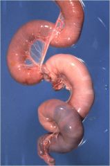
Intussusception
Caused by anything altering motility-eg. parvo, parasites, diarrhea |
|
|
|
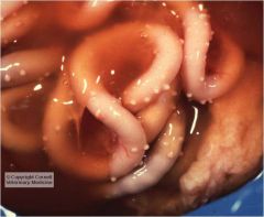
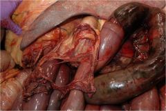
Intestinal strangulation
Common in horses-pedunculated lipomas |
|
|
|
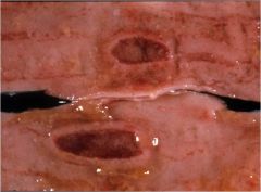
Intestinal obstructive entrapment
Intestine gets trapped in a mesenteric rent, note well demarcated line |
|
|
|
Intestinal torsion/volvulus
Common in the large colon of horses, and pigs |
|
|
|
Enterolith
Common in horses Intestinal obstruction |
|
|
|
Intestinal lymphosarcoma
White-grey, bulging thickened wall |
|
|
|
Intestinal carcinoma
Causes stenotic thickening with dilation proximal to the tumor site. Metastasizes regionally, distantly, peritoneally. |
|
|
|
Pancreatitis with fat necrosis (saponification)
|
|
|
|
Pancreatic acinar atrophy
Leads to exocrine pancreatic insufficiency Most common in German shepherds and rough-coated collies |
|
|
|
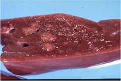
Nodular pancreatic hyperplasia
|
Benign
Variable sized white nodules |
|
|
Which hepatic diseases exhibit centrilobular/midzonal patterns?
|
Lipidosis, glycogen storage disease, and necrosis
|
|
|
|
Which hepatic diseases exhibit periportal patterns?
|
Infiltrates, lymphoma, inflammation, and necrosis
|
|
|
|
Which hepatic diseases exhibit centrilobular (only) patterns?
|
Congestion and necrosis
|
|
|
|
Which hepatic diseases exhibit random patterns?
|
Bacterial and viral infections.
|
|
|
|
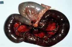
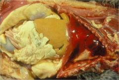
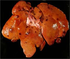
"nutmeg liver"
Chronic passive congestion associated with right heart failure Liver will have rounded edges with fibrin on the capsule. |
|
|
|
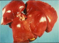
Steroid hepatopathy
Pale tan, enlarged, rounded, friable liver Dogs |
|
|
|
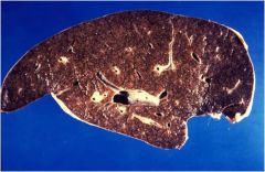
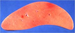
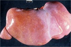
Hepatic lipidosis
Pale tan to yellow, enlarged, may float Cats: Starvation->fat mobilization->overwhelms liver Ruminants: Overconditioned and pregnant->increased nutritional demand with decreased food intake->fat mobilization->liver overwhelmed |
|
|
|
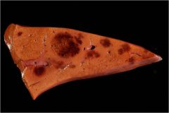
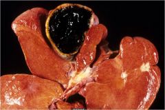
Amyloidosis
Pale tan, WAXY, friable to firm Most common in Abyssinian cats and Sharpei dogs |
|
|
|
Incidental finding in cats and cattle.
Proliferation of small blood vessels within the liver. Looks the same as hemangiosarcoma in the dog, but telangiectasia does not occur in dogs. |
|
|
|
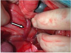
Congenital portosystemic shunt
Liver will also be small. Causes portal hypoperfusion, which can only be differentiated from primary portal vein hypoplasia by the presence of a shunt. |
|
|
|
Describe feline inflammatory liver disease.
|
Two types: lymphocytic portal hepatitis and cholangiohepatitis.
Causes pale, tan-white liver with reticular pattern. |
|
|
|
Describe chronic hepatitis in dogs.
|
Small firm liver with histologic fibrosis.
|
|
|
|
Hepatic necrosis
|
|
|
|
Hepatic abscess-caseous lymphadenitis
Corynebacterium pseudotuberculosis Green/white abscesses with "onion skin" appearance in a variety of organ tissues |
|
|

|
Cirrhosis
Non-specific end stage lesion of liver disease. Commonly associated with toxic insult (eg. aflatoxin) |
|
|
|
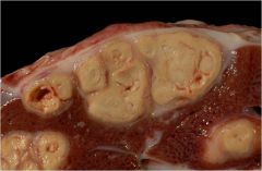
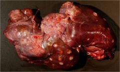
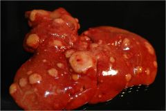
Hepatic nodular hyperplasia
Benign, older animals |
|
|
|
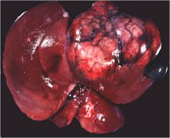
Cholangiocarcinoma
Multifocal, with 1-2 large masses Arises from biliary epithelium |
|
|
|
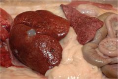
Hepatocellular carcinoma
Typically one large mass on a single lobe. Good prognosis if entire lobe is removed. |
|
|
|
Hepatocellular lymphosarcoma
Nodular to diffuse, white-tan, bulges |
|
|
|
Feline visceral mast cell tumor
Big, pale tan liver Spleen is typically also affected. |
|
|
|
Hemangiosarcoma
Dogs Looks similar to telangectasia in cats and cattle. |
|
|
|
Gallbladder-cystic mucinous hyperplasia
Thickened and bumpy mucosa |
|
|
|
Gallbladder mucocele
"kiwi" appearance on ultrasound Causes obstruction, mural necrosis, and may rupture the gallbladder leading to peritonitis |
|
|
|
Gallbladder fibrinous casts
Associated with salmonella infection in cattle |
|