Reading...
![]()
Play button
![]()
Play button
![]()
Use LEFT and RIGHT arrow keys to navigate between flashcards;
Use UP and DOWN arrow keys to flip the card;
H to show hint;
A reads text to speech;
47 Cards in this Set
- Front
- Back
|
astigmatism
|
abnormal curvature of cornea leading to refractive errors and blurred images
|
|
|
what is in the outer layer of the eye (2)?
|
1) sclera
2) cornea |
|
|
what is the sclera?
|
white covering where eye muscles attach
|
|
|
What is the corena
|
main refractive surface of eye
sensitive to touch |
|
|
what is in the middle layer of the eye?
|
1) choroid
2) ciliary body 3) lens 4) iris 5) pupil 6) vitrous cavity |
|
|
Whatis the choroid?
|
blood vessels and CT between sclera and retina
|
|
|
What is the ciliary body?
|
ciliary muscles + ciliary processes resides at junction between cornea and sclera
|
|
|
What is the purpose of the ciliary process?
|
secrete aqeous human to anterior and posterior chamber
|
|
|
where is anterior chamber
|
between cornea and iris
|
|
|
Where is posterior chamber?
|
between iris and lens
|
|
|
What is glaucoma?
|
increased fluid pressure in eye due to reduction or blockage of aqeous humor from anterior to posterior chamber
|
|
|
How does accomodation occur?
|
contraction of ciliary muscle --> reduce tension on zonule fibers --> lens relax
|
|
|
What are the two layers of the iris?
|
stroma - connects to spinchter pupllae and dilater pupillae
epithelial pigmented layer - blocks light from passing through iris into retina |
|
|
What is the vitreous cavity?
|
behind lens filled with vitreous human
|
|
|
What are the problems with vitreous cavity?
|
1)floaters - blood debris gets in
2) vitreous detachment - vitreous pulls away from retina |
|
|
What are the ten layers of the retina?
|
1)pigment epithelium
2) outer inner sigment of rods/cones 3) outer limiting membrane between glia and inner photo receptors 4) outer nuclear layer - cell bodies of rods and cones 5)outer plexiform layer - photoreceptorsmake contact with bipolar and horizontal cells 6)inner nuclear zone-cell bodies of bipolar,horizontal and amarine cell 7) inner plexiform layer - thick synaptic zone where bipolar cells make contact with retinal ganglion cells 8)ganglion cell layer - contains cell bodies of ganglion cells 9) nerve fiber layer - retinal ganglion cell axons 10) inner limiting membrane, basal lamina between vitreous and proximal glia |
|
|
An mneumonic for remember the ten layers of retina?
|
POOOIIGNI
POOO I, I, get nervous instantly |
|
|
What is the vertical chain in neuron? (3)
|
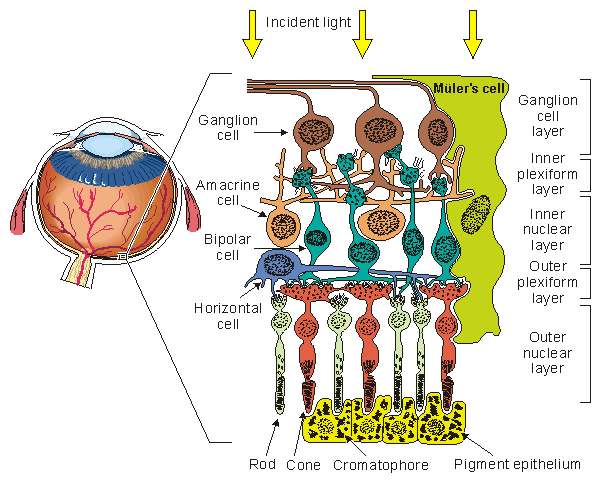
1)photoreceptor synapse bipolar and horizontal cell
2)bipolar cell terminate on ganglion and amacrine cell 3) ganglion cell -terminal out put </img> |
|
|
What is the vertical chain in the eye with the off shoots?
|
</img>1)photo receptors terminate on bipolar and horizontal cells
2)horizontal cells can mediate lateral interactions in outer plexiform layer 3)bipolar cells terminate on ganglion and amacrine cells 4)amacrine cells can mediate lateral interactions in inner plexiform layer 5)ganglion cells are output cells of retina |
|
|
What are cGMP levels in light vs dark?
|
dark - cGMP increases, Na influx, K efflux --> depol
light - cGMP decreases, reduced Na influx, K efflux,hyperpol |
|
|
Is glutamate released in dark ?
|
yes
|
|
|
What is a lower frequency?
|
400-near infared, red
700- near blue, ultravoilet |
|
|
Why do you use red to adapt humans to max rod sensitivity?
|
red is not absorbed by rods to any significant exptent
|
|
|
What is a trichromat?
|
has all 3 cone types
|
|
|
What is protanopia
|
lack red
|
|
|
What is deuteranopia
|
lack green
|
|
|
What is tritanopia?
|
lack bule cone pigment
|
|
|
Which is more sensitive to low light- rods/cones?
|
rods
|
|
|
Which is better for day/night vision?
|
cones - day vision
rods - night vision |
|
|
Which is more sensitive for sunlight and moon light?
|
moon light-mesopic - rods and cones
photopic - sunlight - cones scotopic - starlight - rods |
|
|
Which has more photo pigment?
|
rods, each photo pigment has signle photo sensitivity
|
|
|
Which has a easier dark adaptation time?
|
cones
|
|
|
Which is more sensitivity to a different wavelengths of light and which one only has a signle type of pigment?
|
cones - different wavelengths
rods - rodopsin |
|
|
Which has high acquity?
|
cones
|
|
|
Which is concentrated in fovea?
|
cones
|
|
|
Which is not present in fovea?
|
rods
|
|
|
Where are rods usually located?
|
periphery of retina
|
|
|
What is visual acuity and how is it measured?
|
spatial resolution andmeasured by snellen chart
|
|
|
Wha tis the fovea centralis?
|
pit of cones for fine stimulus detail
|
|
|
What is papilledema?
|
swelling of optic nerve head associated with increased intercranial pressure
|
|
|
What is macular degeneration?
|
macula and fovea become compromised because pigment epithelium degenerates and forms drusen and allows leaking behind fovea
|
|
|
What is glaucoma?
|
pressure in anterior chamber increases because fluid cant get out axons of ganglion cellsand optic vessels compromised
|
|
|
What is retinitis pigmentosa/
|
rods of peripheral retina begin to degenerate causing night blindness and eventual tunnel vision
|
|
|
What is emmetropia?
|
normal eye shape
|
|
|
What is myopia?
|
near sightedness
eyeball is elongated |
|
|
What is hyperopia?
|
eyeball flattened and only distant objects are in focuse
|
|
|
What is presbyopia?
|
loss of lens elasticity with age
|