![]()
![]()
![]()
Use LEFT and RIGHT arrow keys to navigate between flashcards;
Use UP and DOWN arrow keys to flip the card;
H to show hint;
A reads text to speech;
70 Cards in this Set
- Front
- Back
What are coat brushes good for identifying? |
Identifying mites like Chorioptes in horses feathers. |
|
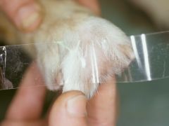
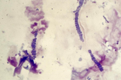
What are hair plucks good for identifying? How long does it take a fungal culture to come back? |
Broken hair, crusts and scales to look for fungal spores. Fungal culture for ringworm. Fungal cultures take 14-21 days to come back. |
|
What diagnostic techniques can we use in our approach to a horse with skin disease? |
Coat brushings-->mites. Hair plucks--> crusts, scales, broken hairs--> fungal Skin scrapes. Swabs--> moist lesion. Fungal culture. Aspirates--> masses and abscesses. Impression smears. Biopsy/punch--> diffuse skin disease like trusting. Allergy testing--> intradermal or serum. Photographs for sarcoids. |
|
|
If we are taking a biopsy in the case of diffuse skin disease what must we not do to the skin? |
We must not prepare it. |
|
What are causes of pruritic disease in horses? |
1) Pediculosis (Lice) biting or sucking. 2) Chorioptes equi. 3) Trombiculosis- T. autumnalis. 4) Oxyuris equi 5) Onchocercal dermatitis. 6) Sweet itch. 7) Atopy. 8) Urticaria/Hives. 9) Pemphigus foliaceus. 10) Multisystemic eosinophilic epitheliotrophic enteritis 11) Strongyloidosis- S. westeri. |
|
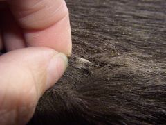
Signs of pediculosis in horses?Diagnosis? |
Pruritis, scaling and alopecia. Wintered or poorly managed horses. Cushings horse. Diagnose: demonstration of lice and nits. |
|
|
Treatment for pediculosis? |
Insecticidal shampoos, sprays or powders with pyrethrins or pyrethroids. Two applications at 14 day intervals. |
|
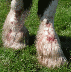
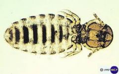
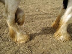
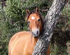
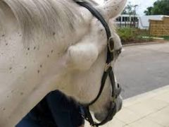
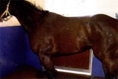
Where on horse is usually infected with Choriopte equi?Signs? |
Distal limb of heavily feathered horses. Horses stamp and bite limbs. Scaling and exudation with secondary bacterial infection. |
|
How is chorioptes equi spread? |
Asymptomatic carriers, bedding, brushes, contact. |
|
|
How do we treat chorioptes in horses? |
Removal of hair and scabs compined with application of a insecticidal like fipronil or permethrins. Systemic ivermectins and avermectins. Seleen shampoo to reduce itch. |
|
Name the two types of louse that affect horses? |
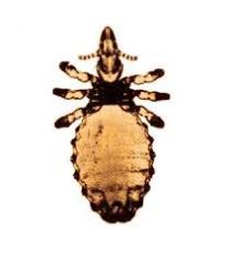
Damalinia equi- biting louse. Haematopinus asini- sucking louse. |
|
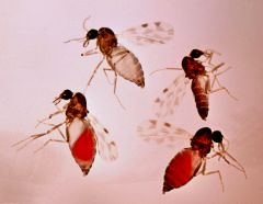
When do horses usually get infestation with trombiculosis in terms of season? |
Late summer and early autumn. Nymphs lead to pruritus of the head and limbs. |
|
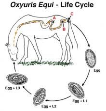
Why does Oxyuris lead to itching what is the problem with this disease in terms of treatment? Treatment? |
Oxyuris equi migrates from small colon rectum to lay eggs on the perianal skin causing intense pruritius and tail rubbing. NEED TO TREAT THE ENVIRONMENT ALSO WHERE THE HORSE RUBS!!!!!!! Treat with an ivermectin. |
|
How do we diagnose oxyuris equi? |
Skin tape on the perianal region!
May see worms coming out. |
|
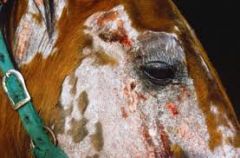
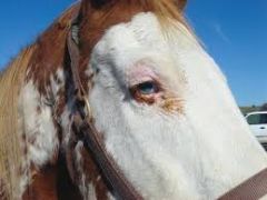
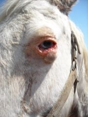
What is the causative agent of Onchocercal dermatitis? How is it spread? Signs? |
Dermatitis caused by onchocerca cervicalis microfilaria transmitted by culocoides and other biting insects. Larvae lodge in capillaries and signs are associated with type 1 and type 3 hypersensitivity reactions. Puritius, allopecia, scaling of head, neck, ventral midline, withers and chest. Focal dermatitis and exudation. |
|
What is the pathogenesis of sweetitch? |
Hypersensitivity reaction to the bite of culicoides midges. |
|
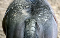
Where does sweet itch commonly effect on the body? What are signs? |
Tail and mane. Season signs of pruritis that is worse at dusk and dawn, aggressive rubbing causes self inflicted trauma. |
|
|
Treatment options for sweetitch? |
1) Management of skin lesions. 2) Corticosteroids. 3) Immunotherapy/ desensitisation treatment. 4) Fly repellants. |
|
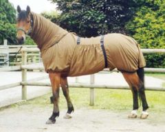
How do we prevent midge bites? |
1) Oil based liquids like benzyl benzoate. 2) Stabling at dawn and dusk. 3) Rugs/hoods e.g. boett rug 4) Insect proof stables with meshes. 5) Fans/wind prevents midges landing. 6) Avoid rivers/water/woodland. |
|
What type of hypersensitivity reaction is atopy? Signs? DIagnosis? Treatment? |
Type 1 hypersensitivity to allergens. Intense pruritis with secondary self inflicted trauma. Diagnosis: intradermal skin testing, skin biopsies, serum ELISA or RAST tests. Treat: avoid the allergen, immunotherapy or corticosteroids. |
|
Pathogenesis of uticaria/hives? |
Immune mediated/ allergy arises due to an injected drug like quinidine, ingestion of chemicals or feeds, inhaled pollens, moulds, chemicals. Direct skin contact or mucous membranes. |
|
Clinical signs of Urticaria? |
Wheals that pig on pressure. Pruritus from mild to severe. Exudation sometimes from wheals. Angio-oedema. |
|
|
Diagnosis and treatment plan for urticaria? |
Diagnosis based on signs, history, skin tests, ELISA or RAST, food elimination tests. Treatment: Treat with short course of corticosteroids. Single dose of I/V or I/M dexamethasone or oral prednisolone. |
|
What is the pathogensis of pemphigus foliaceus? |
Autoimmune disease characterized by exfoliative dermatitis leads to a type 2 hypersensitivity reaction due to auto-antibodies directed against cell membrane of epidermal cells. |
|
Signs of pemphigus folicaeus? |
Vesicles, pustules, erythematous scaling, crusting begins on the face and limbs, inflammation of coronary band, ergot and chestnut. Painful/pruritic lesions. Systemic signs of weight loss, pyrexia, inappetence, lethargy, ventral oedema. |
|
|
How do we diagnose and treat pemphigus? Prognosis ? |
Diagnose on biopsy. Direct immunfluroscene shows immunoglobulin in epidermis. Treat: High doses of corticosteroids. Prognosis guarded. |
|
Signs of multisystemic eosinophilic epitheliotrophic enteritis? |
GIT and skin signs. Ulcerative coronitis, alopecia, hyperkeratosis and exudation. Intensely pruritic lesions. Signs of generalized disease: pyrexia, weight loss, diarrhoea, lymphadenopathy,ventral oedema and dullness. |
|
Diagnosis and treatment of MEED? |
Skin biopsy also rectum and intestines.
Hypoprotaenmia, hypoalbumnaemia, hyperfibrogenaemia. Treat: steroids. |
|
|
DDX list for alopecia in horses? |
Rain scald. Dermatophytosis. Sarcoids, Linear keratosis. Selenium toxicity. |
|
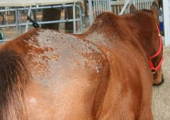
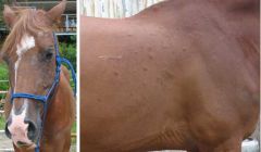
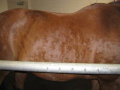
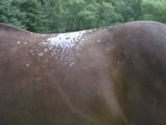
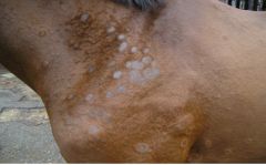
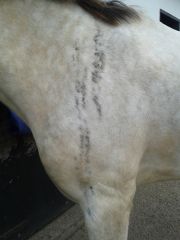
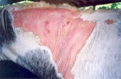
Cause of rain scald and signs? |
Cause is dermatophilus congolensis. Signs: exudation, matted hairs, scabs in areas wetted by rain- back and quarters. Paint brush lesions. |
|
Treatment for rain scald? |
Antibiotics. Clipping and soaking of skin with chlorohexidine to remove exudates and scabs. Topical AB. |
|
|
Dermatophilus congolensis. |
|
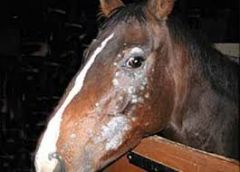
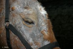
What species commonly causes ringworm in horse? Less commonly? Signs? |
Tricophytan commonly. Less common microsporum sp. Signs are circular erect lesions of hair, alopecia and scaling. |
|
|
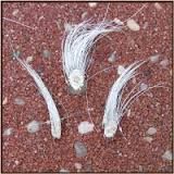
How do we diagnose ringworm? |
Hair plucks and scrapes. Woods lamp- trichophytan don't fluoresce, microsporum canis and equinum some fluoresce |
|
How do we treat ringworm? |
Topical antifungal like enilconazole, natamycin. Treat enviroment with sporocidal natamycin or virkon. Griseofulvin C/I in pregnant mares as teratogenic. |
|
What is alopecia areata? Signs? treatment? |
Cell mediated autoimmune skin disease characterized by thinning of the main and tail and reasonably circumcised areas of alopecia. No effective treatment. |
|
Talk to me about linear keratosis and what horses it is mainly seen in? |
Idiopathic inherited dermatoisis mainly seen in quarter horses. Unilateral, linear, vertical bands of hyperkeratosis and hairloss on neck, thorax and upper hind limb |
|

Name the most common skin tumour of horses? |
SARCOIDS. |
|
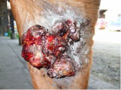
Name the 6 types of sarcoid? |
Occult--> flat plaque with hairloss. Verrucose--> cauliflower. Nodular--> complete s/c or intradermal or burst through skin. Mixed Fibroblastic--> ulcerative. Malevolent--> invasive. |
|
|
Treatment of sarcoids? |
Benign neglect. Surgery-laser,cryo, sharp excision. Ligation. BCG injection. Liverpool cream. |
|
|
Believed cause of sarcoids? |
Bovine papillomavirus plays a role. |
|
|
DDX list for nodules, tumours and swellings? |
Insect bites. Papillomas- viral and congenital and aural. Sarcoids. Melanoma. Lymphosarcoma. Eosinophilic collagen necrosis/granuloma. Axillary nodular necrosis. Cysts- dermoid, dentigerous, epidermoid. |
|
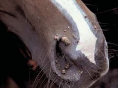
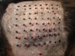
What age of horses is equine papilloma virus seen? Signs and treatment? |
Under 4 years old. Signs lesions as small white-grey papules in huge numbers on muzzle, lips, eyelid, external genitals, limbs. No need to treat as spontaneous resolve. |
|
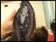
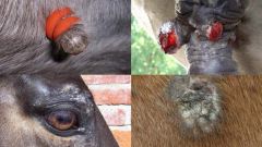
What is shown in this image? Treatment? |
Aural papillomas. No need to treat. |
|
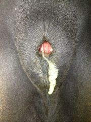
Most common type of melanocyte tumor in horse? What colour of horse is predisposed? Where are they usually found?
|
Melanomas around the perineum, sheath, parotid region and less commonly guttural pouch, eyelids, lips and iris. |
|
|
How do we diagnose/treat melanomas/melanosarcomas? |
Biopsy, FNAC- differentiation between two. Treat by surgical excision, cryosurgery. Cytotoxic drugs like cisplatin. Cimetidine orally as histamine may be involved in tumour growth. |
|
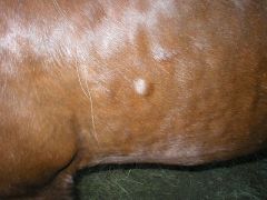
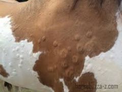
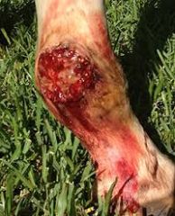
What is shown here and what are signs? Treatment? |
Eosinophillic collagen necrosis/ granuloma. Firm, painless skin nodules usually along back and side of chest. Usually do not need to treat. Intralesional steroid or surgical excision. |
|
Where are epidermoid cysts most common? |
False nostril. |
|
DDX list for ulcerations and erosions in horses? |
SCC. Habronemiasis. |
|
Aetiology of SCC in horses? Diagnosis and treatment? |
UV lights. Smegma. Diagnosis- Biopsy or impression smears for the ocular form. Treatment options: Surgical- excision like penile amputation, cryosurgery. Cytotoxic/antimitotic drugs- intralesional cisplatin. Radiation- most effective |
|
Cause of habronemiasis and signs? |
Granulating nodules/ wounds causes by larvae of habronema muscae, habronema majus and drachia megastoma. |
|
DDX for papules, pustules, vesicles? |
Staphylococcal dermatitis- s. aureus, s. intermedius, s. hyicus. |
|
|
Signs associated with staphylococcal dermatitis? Treatment? |
Painful skin lesions with folliculitis and furunculosis. Treat with antibiotics based on C&S like penicillin and topical chlorohexidine. REMEMBER CHECK RUGS AND REMOVE DAILY! |
|
|
DDX scaling and crustings?
|
Pemphigus folliaceous- rare immune mediated disease- type II hypersensitivity to epidermal cells. Actinic dermatoses. |
|
|
What must we differentiate sunburn from? |
Photosensitisation- Primary due to st johns wort or secondary due to liver failure. |
|
Reasons for actinic dermatoses? |
Sunburn. Photosensitisation. |
|
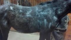
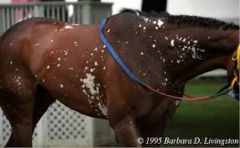
Main ddx for abnormal coat? |
CUSHINGS- PPID Caused by excessive POMC secretion leading to dysregulated release of ACTH and cortisoL! |
|
What is vitiligo? |
Leucoderma loss of pigment of the skin. |
|
DDX abnormal coat pigment? |
Leucotrichia- white hairs. Leucoderma- vitiligo. |
|
|
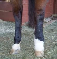
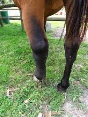
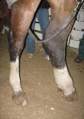
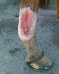
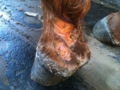
DDX for pastern dermatitis? |
Chorioptes equi- feathered horses. Secondary photosensitisation- accumulation of phylloerythrin due to failure by liver to exrete. Mud fever- dermatophilus congolensis. Pastern/cannon leucocytoclastic vasculitis. Coronary band dystrophy/coronitis. MEED. Chronic progressive lymphoedema. |
|
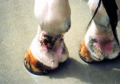
Signs of mud fever? Diagnosis and treatment? |
Exudation, scabs, matting hair on distal limbs. Diagnose with a smear. Treat- A/B, anti inflammatories, soaking skin with chlorohexidine to remove scabs and exudate, clip hair, dry skin and apply topical A/B ointment. |
|
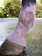
What is pastern/cannon leucocytoclastic vasculitis? Cause? Signs?Diagnosis? Treat? |
Skin condition of unpigmented distal limb. Caused by UV light? Signs: erythema, oozing and crusting of white distal limbs, similar to mud fever. DIagnosis: Biopsy reveals a leucocytoclastic vasculitis, vessel wall necrosis and thrombosis. Treat: clipping hair and removal exudate and crusts with chlorohexidine. Topic antiseptic, antibiotics and steroids. Sunblocks. Leg wraps when out in sun. |
|
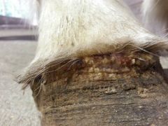
Cause of coronary band dystrophy and how do we treat it? Signs? |
Idiopathic/ immune mediated defect in cornification of coronary band. Signs crack, fizzures, bleed and ooze. Treat: symptomatic care of lesions, keep dry, and use steroids. |
|
|
Sequalae to mite infestation leading to dermatitis of the pastern? |
Chronic progressive lymphoedema. |
|
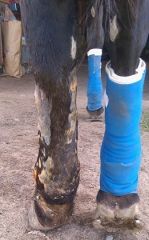
DDX list for limb swelling? |
Lymphangitis. Cellulitis. Vasculitis. Purpura haemorrhagica. |
|
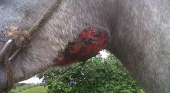
Aetiology of lymphangitis? What limbs usually affected? Signs? |
Inflammation of cutaneous lymphatics secondary to a bacterial infection obtained via a small cut/abrasion. Hindlimb more than forelimb. Signs: swelling, exudation, lame |
|
Treatment of lymphangitis of the limb? |
A/B based on culture and sensitivity. NSAIDs/corticosteroids. Diuretics and potassium iodide. Hydrotherapy, cleaning with antimicrobials, leg wraps, gentle exercise. |
|
What is the aetiology of cellulitis? Signs and treatment? |
Wound infection that spreads through a tissue plane. Affects limbs causing acute swelling, inappetence and pyrexia. Treat: A/B, NSAID, hydrotherapy, cleaning with antimicrobials, gentle exercise. |
|
Aetiology of vasculitis? Causes? Signs? Treat? |
Inflammatory reaction in blood vessel walls usually type 1 or type 3 hypersensitivity. Various infections or drugs may induce. Signs oedema, necrosis and ulceration. Treat: steroids. |
|
What is purpura haemorrhagica associated with in the limbs? |
Strangles infection. Immune mediated vasculitis. |