![]()
![]()
![]()
Use LEFT and RIGHT arrow keys to navigate between flashcards;
Use UP and DOWN arrow keys to flip the card;
H to show hint;
A reads text to speech;
160 Cards in this Set
- Front
- Back
|
Normal QRS |
Normal transition across Precordium |
|
|
QRS abnormalities |
Abnormalities in height - high/low voltage Abnormalities in width - WPW - BBB - IVCD Hemiblocks could be with axis determination |
|
|
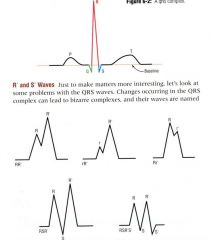
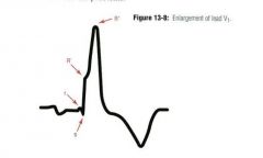
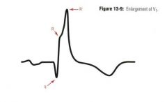
Abnormal QRS EKG complex components |
|
|
|
If the first wave of QRS is positive, then it is the ________ wave |
R wave |
|
|
If the first wave of the QRS is negative, then it is the ______ wave |
Q wave |
|
|
Any negative wave after a preceding R wave is the _______ wave |
S wave |
|
|
QRS is due to __________ ___________ |
ventricular depolarization |
|
|
QRS complex normally lasts |
0.6 to 0.11 seconds |
|
|
Axis of QRS is between ________ and ________ |
-30 and +105, downward and to the left |
|
|
The QRS is the __________ of the vectors |
summation |
|
|
Parts of the QRS can be ___________ in some leads |
Isoelectric |
|
|
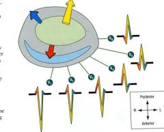
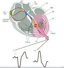
QRS depolarization sequence |
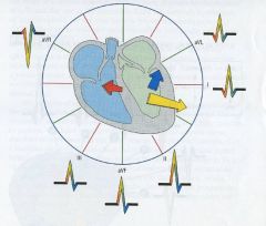
1) Septum depolarizes L to R 2) Main vector is then posterior and inferior 3) Then finally, main vector is posterior-superior |
|
|
LV contains ________% of cardiac muscle in most people |
70% - most electrical activity points towards the LV NO matter what lead you're reading |
|
|
Example of LV predominance on EKG |
A lead over right heart will amplify the right heart voltage a little because it is sitting over the RV BUT the major voltage is going away from that lead toward the dominant left ventricle |
|
|
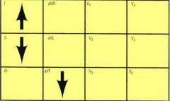
QRS changes across the pericordium |
|
|
|
QRS complex in V1 is _________ |
Negative |
|
|
QRS complex in V2 is _________ |
Negative |
|
|
QRS complex in V3 is ____________ |
Negative but leaning more towards isoelectric |
|
|
QRS complex in V4 is _________ |
positive |
|
|
QRS complex in V5 is ___________ with prominent but equal _________ and ________ waves |
QRS in V5 is positive Prominent negative Q and S waves |
|
|
QRS complex in V6 is ___________ with larger ______ wave than _______ wave |
Positive Larger negative Q wave than S wave |
|
|
Areas of change in the precordial leads where the QRS switches from mostly negative deflection to mostly positive deflection |
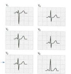
Transition zone |
|
|
The normal transition zone is between __________ and _______ |
V3 and V4 |
|
|
Q waves are significant if they have what characteristics? |
Greater than 0.03 seconds Depth is equal to or greater than 1/3 of R wave |
|
|
Q waves in MI are _________ and not large enough with most only occurring in 1 lead |
Benign |
|
|
Q waves are more indicative of _________ __________ |
PRIOR MI |
|
|
An MI of significant size must have Q waves in at least __________ contiguous leads |
2 continguous leads |
|
|
What do you need to diagnose an Acute MI? |
ST-T wave changes |
|
|
Insignificant Q waves can be seen in __________ ___________ |
septal depolarization |
|
|
Significant Q's can be seen in ________ muscle, ______ in more than _______ lead |
DEAD muscle, MI, in more than one lead |
|
|
QS waves can occur with no ___________ inbetween |
No R wave |
|
|
Pseudo Q waves can appear in _________ |
WPW |
|
|
Septal Q waves are seen in leads _________ and _______ |
I and aVL |
|
|
QS waves are seen only in _____________ |
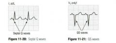
VI |
|
|
Significant Q graphical representation |
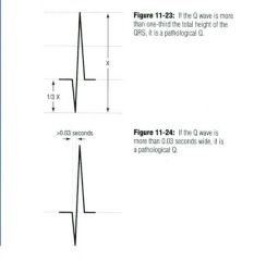
Q > 1/3 of R height Q > 0.03 s wide |
|
|
Which leads have the Q waves? This defines the site of infarction |
Leads I, II, aVR, aVF, V4-6 |
|
|
Small notch is often seen at the ________ of QRS |
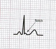
End |
|
|
QRS notching is insignificant unless the person is a ______________ |
Curly haired Italian Male |
|
|
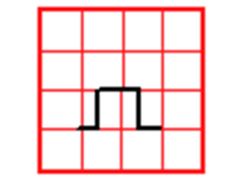
Osborn Wave is a _______ seen in _________ |
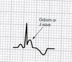
J wave HYPOTHERMIA |
|
|
The colder the temp, the ____________ the J wave |
Higher the J wave |
|
|
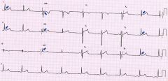
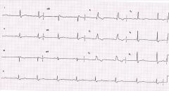
J waves on EKG (blue arrows) |
|
|
|
Another example of J wave EKG |
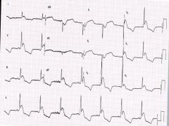
Pronounced positive deflection on QRS complex due to hypothermia |
|
|
LVH would cause _______ voltages on EKG |
Increased |
|
|
MI and concurrent loss of LV muscle would cause _________ voltage on EKG |
Decreased - Yellow scar, wall thinning - Decrease in voltage - Increase in Q waves |
|
|
Pericardial effusion would cause _________ voltage |
Decreased |
|
|
Men and youth have _______ voltages leading to _______ amplitudes |
higher voltages, greater amplitudes |
|
|
Precordial leads are __________ voltage than limb leads because the precordial leads are _________ to the heart |
Higher voltage Closer to the heart |
|
|
Pathological increases in amplitude caused by |
LVH/RVH |
|
|
Low voltage is _____ common than high voltage |
LESS common |
|
|
Causes of Lower voltages on EKG |
MI's - scar tissue is electrically inert Marked increases in body fat Huge left pleural effusion Pericardial effusion Recording at half standard |
|
|
What do you have to make sure to look at when recording EKG's? |
Make sure you're not at half standard, and if you are, adjust the reading accordingly |
|
|
Scar tissue is electrically __________ |
inert and causes decreased voltages |
|
|
In pericardial effusions, the limb leads will record less than ________ mm voltage |
5 mm voltage |
|
|
In pericardial efusions, the precordial leads will record less than _________ mm voltage |
10 mm voltage |
|
|
The larger the effusion, the ________ the QRS |
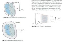
Smaller - Affects all components, not just QRS |
|
|
Half Standard |
Rectangle height is only 1 big square - Need to double to normalize |
|
|
Standard speed is ____________ |
25 mm/sec |
|
|
Each little box equates to _______ |
.04 seconds |
|
|
Each big box equates to ___________ |
.2 seconds (5 little boxes) |
|
|
Whole EKG strips spans __________ seconds |
6 |
|
|
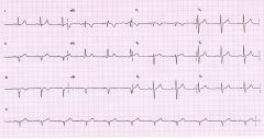
Pericardial effusion EKG |
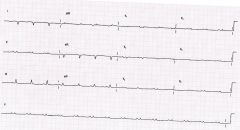
Low voltage all waves |
|
|
LVH leads to ___________ voltages |
Increased |
|
|
LVH causes |
Outflow obstruction - Aortic stenosis/HTN Less likely with volume overload problems - Mitral Regurg/Aortic Regurg |
|
|
Increased muscle mass causes ______ AP, _____ vector with resultant increased EKG _________ |
Increased action potential Increased vector Increased EKG amplitude |
|
|
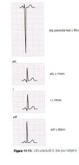
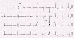
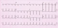
EKG Criteria for LVH |
S in V1/V2 + R of V5/V6 > 34mm Any precordial lead > 45mm R wave in aVL > 10 mm R wave in lead I > 11mm R in AVF > 19mm |
|
|
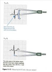
In LVH, the ____ wave in leads V1/2 are added to the ______ wave of V5/V6 with a sum greater than _______ |
S wave of V1/2 R wave of V5/6 Greater than 34 mm |
|
|
In LVH, precordial lead voltage has to be greater than ___________ |
45 mm |
|
|
In LVH, The R wave in aVL has to be greater than ________ |
10mm |
|
|
In LVH, the R wave in lead I has to be greater than ____________ |
11mm |
|
|
In LVH, the R wave in AVF has to be greater than __________ |
19mm |
|
|
Can't have LVH if _______________ is present |
Left bundle branch is present |
|
|
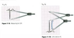
You must use calipers to calculate LVH |
|
|
|
LVH EKG changes |
|
|
|
LVH EKG |
|
|
|
RVH is ________ common than LVH |
Less common |
|
|
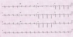
An additional vector produced by the enlarged RV causes increased voltages in leads ______ and _________ |
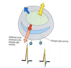
V1 and V2 |
|
|
Right Ventricle is located right underneath the _________ |
sternum |
|
|
RVH vector is directed to the ______ and ________ |
anterior and right |
|
|
In RVH, you have the summation of the ___________ forces and ________ RV forces |
Septal forces and enlarged RV forces |
|
|
RVH measurement via calipers |
|
|
|
RVH causes |
Secondary to pathology of RV or distal to RV
- lungs - left heart failure |
|
|
Mimicry of RVH (can't make diagnosis) includes |
Right BBB Very young children WPW Posterior wall MI |
|
|
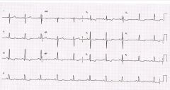
RVH EKG |
|
|
|
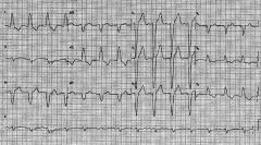
Abnormal QRS duration is greater than |
0.12 seconds |
|
|
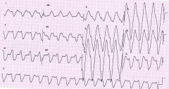
If tachycardia with a wide QRS think, _______________ until proven otherwise |
V tach |
|
|
Etiology of wide QRS complexes |
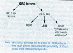
LBBB/RBBB Intraventricular conduction delays WPW |
|
|
Bundle Branch Block EKG |
|
|
|
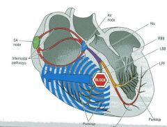
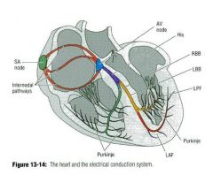
How many bundles are there? |
Two, Right and Left |
|
|
Left bundle divides left _________ and ________ fascicles |
anterior and posterior fascicles |
|
|
Blocked bundle affects both ______ and _______ |
conduction and axis via cell to cell transmission |
|
|
Bundle branch blocks have slow, _______, and _________ QRS complexes |
slow, wide, and bizarre |
|
|
Bundle branch blocks have ___________ conduction until the block |
Normal conduction until the block |
|
|
If there's a wide QRS consider |
LBBB RBBB IVCD - HYPERkalemia Ventricular tachycardia - sinus tachycardia with a bundle branch block |
|
|
Any lead in which the QRS is greater than 0.12 seconds or greater means that _____________ lead is of that duration |
EVERY lead is at least 0.12 second |
|
|
When checking for QRS width, look at the ________ QRS in any lead |
widest QRS in any lead |
|
|
During RBBB, there is ______ impulse to the left bundle |
Normal impulse to left bundle |
|
|
In RBBB, part of the ______ and right ______ are delayed secondary to cell to cell spread on the Right |
Septum and Right Ventricle |
|
|
Delayed cell to cell spread slows depolarization time leading to ___________ QRS |
prolonged R' is the additional slow vector, LATE |
|
|
RBBB has a QRS greater than ________ |
0.12 seconds |
|
|
In RBBB, there is an _________ pattern with _______ > _______ in lead V1 |
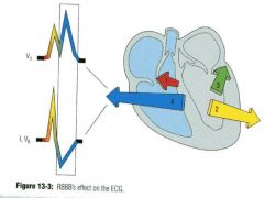
RSR' pattern R' > R |
|
|
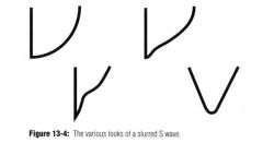
Slurred ____ wave in leads 1 and V6 during RBBB |
Slurred S wave - Lead overlies L ventricle, looking at slowed electrical activity going away from you toward R ventricle |
|
|
In RBBB, the ST wave is in the _________ direction of QRS |
opposite direction of QRS |
|
|
RBBB EKG |
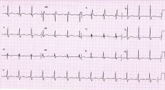
Look for slurred S wave in leads I and V6 RSR' pattern in lead VI |
|
|
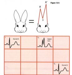
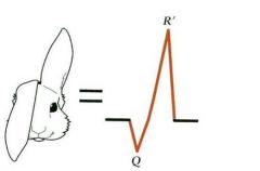
RSR' Bunny Ears picture |
|
|
|
Slurred S waves picture |
|
|
|
RBBB EKG |
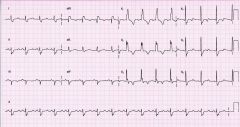
Look for Rabbit Ears RSR' |
|
|
In RBBB, __________ has a small _________ wave at the beginning aka Mutant Rabbit |
V1 has a small r wave |
|
|
Floppy Eared Bunny = _____________ with a negative Q wave and Positive R' |
MI
|
|
|
Floppy Ear (MI) in VI |
|
|
|
Floppy Ear Example with Q, R, and R' on VI |
|
|
|
LBBB is called an ___________ by Heibel |
Unglygram |
|
|
If both parts of the left side are involved, the doctor should be ___________ |
more concerned |
|
|
LBBB is a ____________ wave |
Monomorphic wave |
|
|
In LBBB, there are discordant _______ waves that occur in ____________ direction |
discordant T waves opposite directions |
|
|
In LBBB, the RV is ________ with the left being ________ via cell to cell |
RV is OK, left is slow cell to cell |
|
|
LBBB criteria |
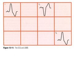
Duration of atleast 0.12 seconds Broad, monomorphic R waves in I and V6 with no Q's Broad monomorphic S in V1, small R wave possible |
|
|
LBBB must last for at least _____________ |
0.12 seconds |
|
|
LBBB have broad __________ R waves in _____ and ________ with no Q's |
Monomorphic I and V6 |
|
|
In LBBB, there is broad monomorphic ______ in V1 with a small ______ wave |
Monomorphic S wave in V1 Small R wave possible |
|
|
The R waves in V1 - variation of LBBB |

|
|
|
In LBBB, there may be notching of the R wave in lead ________ |
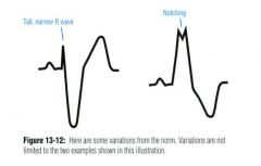
V6 |
|
|
LBBB EKG |
Look for notching in R wave on V6 R waves in V1 Monomorphic S wave in V1 Monomorphic R waves in 1 and V6 |
|
|
Can you diagnose LVH and LBBB on the same EKG? |
No |
|
|
Causes of LBBB |
Hypertension CAD Cardiomyopathy Rheumatic Infiltrative Idiopathic |
|
|
IVCD Criteria |
Intraventricular Conduction Delay - Doesn't have to be 0.12 seconds - Has some but not all features of either RBBB/LBBB - LVH, peri-infarction, Hyperkalemia, Quinidine and Flecanide (1A/C) |
|
|
In IVCD, the QRS is __________ |
wide |
|
|
In IVCD, the S in slurred in ___ and ________ |
S slurred in 1 and V6 |
|
|
In IVCD, there is an ___________ R:S ratio in V1 |
Increased |
|
|
IVCD EKG |
|
|
|
Hemiblocks occur only on the ________ and involves __________ of the ________ bundle |
left, half of left bundle |
|
|
Left anterior hemiblock is called ____________ |
Left anterior fascicular block |
|
|
Left posterior hemiblock is also called _______________ |
left posterior fascicular block |
|
|
Left anterior fascicle is ___________ and innervates the __________ walls of the LV |
Thin, anterolateral walls of LV |
|
|
Left posterior fascicle is _________ dispersed and fans supplying the ___________ and ________ walls with electrical stimulation |
inferior and posterior walls |
|
|
Graphical representation of Hemiblocks |
|
|
|
Left Anterior Hemiblock sequence |
1) Depolarization has to come from the septum, inferior wall and posterior wall toward the anterior and lateral walls 2) Unopposed vector arises that points superior and leftward 3) Expect a qR complex or Large R in lead I and an rS complex in III 4) cause of small q and r is the abnormal direction of septal depolarization |
|
|
In Left Anterior Hemiblock, expect a qR complex or large R wave in lead ____________ and an rS complex in lead ________ |
qR complex/R wave - lead I rS complex - lead III |
|
|
Left anterior hemiblock graphical representation |
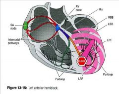
Vector is superior and leftward |
|
|
Left anterior hemiblock criteria |
Left axis deviation with axis -30 to -90 qR or an R wave in Lead I rS complex in III |
|
|
To recognize left Anterior Hemiblock, use leads |
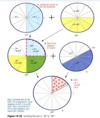
I, AVF, and II |
|
|
In Left anterior hemiblock, lead I will be ___________, lead II will be ____________, and aVF will be ____________ |
I - positive II - negative avF - negative |
|
|
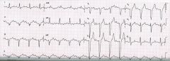
Left anterior hemiblock EKG |
R wave in lead I rS complex in lead III (no Q wave) |
|
|
Left Posterior hemiblock is ______ to diagnose and ________ |
difficult to diagnose and RARE |
|
|
Left posterior hemiblock sequence |
1) Depolarization of inferior and posterior aspects of LV delayed 2) Unopposed vector from the septum and anterior walls is to the right 3) small q in III, and S in lead I |
|
|
Criteria for left posterior hemiblock
|
1) Axis 90 to 180 to the right 2) S wave in lead I 3) q wave in lead III 4) Exclusion of RAE/RVH - can't call LPH unless these two are absent |
|
|
In Left posterior hemiblock, there is an _______ wave in lead I |
S wave |
|
|
In left posterior hemiblock, there is a ______ wave in lead III |
q wave |
|
|
Left posterior hemiblock requires _______________ |
Exclusion - other causes of R axis deviation - exclude RVH, RAE S1, Q3, T3 can indicate PE |
|
|
Most common cause of R axis deviation is ____________ |
RVH |
|
|
Most common cause of RAE is ___________ |
anything distal to the RA |
|
|
What three leads/waves can indicate PE? |
S wave in lead I Q wave in lead III T wave in lead III |
|
|
Left Posterior Hemiblock EKG |
|
|
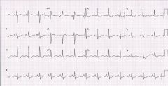
What is the diagnosis of this EKG? |
Not a clue |
|
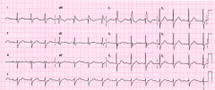
What is the diagnosis of this EKG? |
Not a clue |
|
|
Bifasicular Block must have a __________ component |
RBBB component |
|
|
Bifasicular Block Types |
Must be RBBB+ RBBB + LAH - stable unless acute RBBB + LPH - Unstable |
|
|
RBBB + LAH is __________ unless acute |
stable unless acute |
|
|
RBBB + LAH is ___________ |
UNSTABLE |
|
What is the diagnosis of this EKG? |
No clue |
|
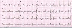
What is the diagnosis of this EKG? |
No clue |