![]()
![]()
![]()
Use LEFT and RIGHT arrow keys to navigate between flashcards;
Use UP and DOWN arrow keys to flip the card;
H to show hint;
A reads text to speech;
21 Cards in this Set
- Front
- Back
|
SA node |
Pacemaker, initiates depolarization |
|
|
AV node |
Passes depolarization to ventricles, brief delay to allow for ventricular filling |
|
|
Purkinje fibers |
Throughout ventricles |
|
|
P wave |
Atrial depolarization |
|
|
QRS complex |
Ventricular depolarization and atrial repolarization |
|
|
T wave |
Ventricular repolarization |
|
|
ST segment depression vs elevation (always an emergency) |
Depression: Myocardial ischemia Elevation: Myocardial infarction |
|
|
U wave |
Repolarization of the purkinje fibers |
|
|
ECG interpretation (look at the following areas in order) |
Rate- P wave (heart rate), Rhythm (intervals)- AV blocks, Axis |
|
|
To measure HR from ECG strip |
30 large boxes = 6 seconds. Then, count the number of R waves in 6 seconds then multiply by 10 |
|
|
1st degree AV block |
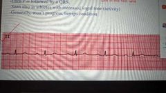
AV nodal disease. PR interval > .2 seconds (beyond one small box) Each P is followed by a QRS Seen in athletes with increased vagal tone (activity) Generally, won't progress, benign condition |
|
|
2nd degree (mobitz) |
Type I (Wenckebach): PR interval gets progressively longer until a QRS is dropped, pattern is predictable Type II is different from I where the PR interval remains consistent but QRS drop is unpredictable) Type I continue exercise and monitor, slow down Type II stop exercise |
|
|
Bruce protocol |
Stress test maximal, pushes person to the limit and stopped until a person can no longer continue. You do not slow down, you also do not stop then resume. All or none |
|
|
3rd degree AV block |
The atrial rate is independent from the ventricular rate (P wave and QRS March out separately) The PR interval is constantly changing, the QRS is usually wide and bizarre When noticed during exercise, stop immediately and activate emergency |
|
|
Supraventricular arrhythmias (never an emergency) |
Premature atrial contractions Atrial tachycardia (100-250) Atrial flutter (250-350) Atrial fibrillation (400-600) |
|
|
Premature atrial contractions |
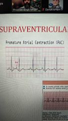
T wave overlap with new P |
|
|
Atrial tachycardia |
Flutter waves between R waves, stop exercise but not an emergency Monitor (slide before question 5) |
|
|
Ventricular fibrillation |
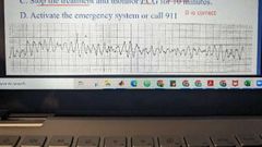
Emergency, stop and call 911 |
|
|
Premature ventricular contractions |
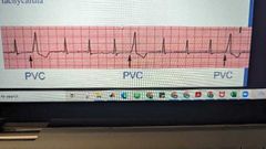
Heartbeat initiated by purkinje fibers |
|
|
PVC |
Bigeminy 1 normal beat followed by 1 pvc Trigeminy 2 normal beats followed by 1 pvc Multifocal more than 1 pvc is present and 2 do not appear similar in configuration (STOP exercise) Couplet 2 consecutive PVC together with no normal beat between them (STOP exercise) |
|
|
Stops vs referral |
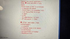
See pic |