Reading...
![]()
Play button
![]()
Play button
![]()
Use LEFT and RIGHT arrow keys to navigate between flashcards;
Use UP and DOWN arrow keys to flip the card;
H to show hint;
A reads text to speech;
23 Cards in this Set
- Front
- Back
|

1, White mark prob dt fixer spilt on film before processing
2. Multiple black finger prints dt developer on the hands of the processor |
|
|

1. Multiple white lines = contrast material spilt onto animals coat (contrast used for cystography here)
Note: The coat should be clean and dry to minimise artefacts |
|
|
1. Tape from xray film box
2. Crimp marks dt rough handling of film 3. Dirt on rollers in automatic processor |
|

|
1. Grey background prob dt faulty processing eg expired chemicals, incorrect temp, insufficient time
RTFM!!! |
|
|

Multiple bright white specks dt dirt on ISs
Clean screens regularly |
|
|

Film too light, could be dt;
underexposure (insufficient kVp or mAs, or, incorrect film-screen combination Underexposure commonly dt incorrect measurement of the patient or incorrect settings on the xray machine |
|
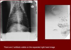
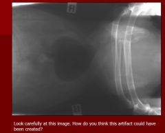
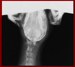
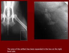
How do you think this artefact was created?
|

The double image occurs when a large xray cassette has been placed under the xray table to utilised the bucky grid but has not been removed for processing.
A second radiograph has been taken of the smaller dog on top of the table as no grid was required. This has created the impression that the small dog is inside the large one! This is the film that was underneath the table and was exposed twice. |
|

|
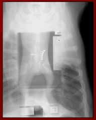
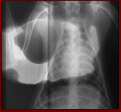
The xray film has been exposed twice - it has been flipped 180 degrees around the long axis.
(this study is a pneumocytogram - air - negative contrast in the urinary bladder) |
|
This image has been manually processed
|

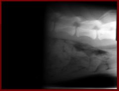
Irregular white marks - film touching the side of the tank and removal of emulsion
** Emulsion is easily damaged especially when wet |
|

|
Black area = film fogging
- dt film exposed to light before or after xray exposure Oft dt film hopper left open, lid left off box, faulty cassettes or opening the dark room door before the film has fully enetered the automatic processor |
|

|
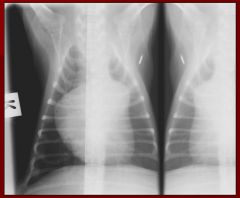
Mirror image dt film being folded onto self
-> dark line at fold -> mirror image either side of fold |
|
|

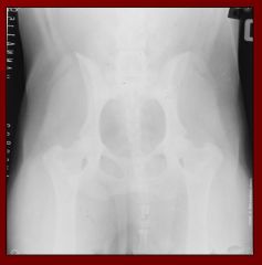
"Grid cut off"
- focussed grid upside down in xray beam -> malalignment b/n diverging xray beam and lead strips in grid ** Imp that beam are grid are correctly alignned and at correct focal spot |
|

|
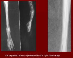
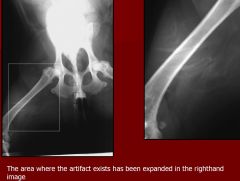
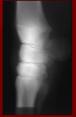
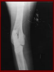
Fine parallel lines dt lead lamellae in grid - oft detected when using a stationary grid
- grid not necessary if object is less than 10cm thick (like this cat limb) |
|
|
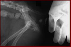
Fingers in image (when animal restrained)
***NEVER place any part of operator in primary beam even if shielded by lead. - sedate or anaesthetise - use sandbags or tape |
|
|

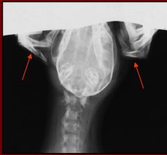
Handlers fingers in primary beam
** Lead gloves and gowns only protect wearer from SCATTER radiation Note in this case the finger bones can be seen indicating that the gloves are old |
|

|
Blur due to motion at time of exposure
- often dt patient movement - can also occur dt movement of the cassette or xray tube |
|
|

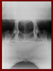
Radiograph too dark -> soft tissues difficult to see
Could be due to overexposure, over development or over measurement of the area being radiographed |
|
|

Static electricity
- prob dt film being removed too quickly from box |
|
What two artefacts can you see?
|

Large white marks dt fixer on fingers of processor before developer
Bright white spots dt dirt in cassette |
|

|
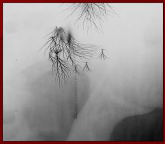
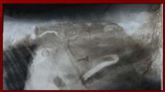
Scratches on film that have removed emulsion prior to processing
|
|

|
Brown/yellow stain dt residual fixer on film caused by insufficient washing (should be 30-40 mins if manual)
|
|
|
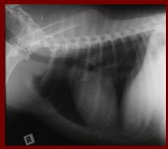
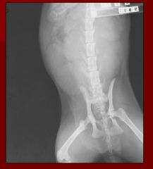
Not well positioned - thorax should be central and only a small portion of the cranial abdomen should be visible - as in this well positioned example above
Achieve this by centering the xray beam just caudal to the scapula and approx the level of the 4th intercostal space. Note that the forelimbs are pulled forward to prevent superimposition of the musculature |
|
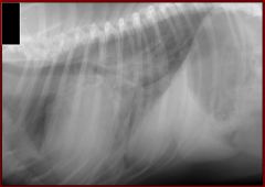
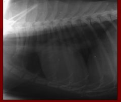
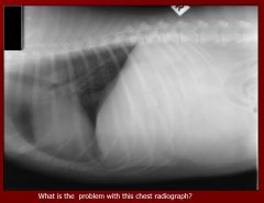
What is the problem with this radiograph?
|

No attempt at collimation
- good radiation safety practice to limit the size of the primary beam and collimate to the area of interest |