![]()
![]()
![]()
Use LEFT and RIGHT arrow keys to navigate between flashcards;
Use UP and DOWN arrow keys to flip the card;
H to show hint;
A reads text to speech;
30 Cards in this Set
- Front
- Back
|
What range of pitch/frequency is normal speech? |
200 - 500 Hz |
|
|
Above what decibel of sound can do permanent damage to hearing and what is the relationship of sound and damage above this level with time? |
75-80 dB. It is a logarithmic relationship with time and damage. Loud noise --> death of outer hair cells in organ of Corti |
|
|
What is the advantage of having an asymmetric ear and two ears? |
This allows localisation of sound Infront or behind someone with the head shadow effect benefit of low frequency sounds bending around the head. |
|
|
What is the function of the external ear? |
To catch the sound and direct it on to the tympanic membrane |
|
|
What are the different types of earwax (worldwide demographic)and what is it's purpose? |
Dry/flaky in Asia Wet in Western and Africa Earwax is for waterproofing |
|
|
What are the functions of the external auditory meatus and how does it carry out these? |
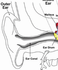
Direct sound on to the tympanic membrane - The ear canal has a resonance frequency of 2-5Khz which is the same as speech = efficient transmission Protection and cleansing through trapping foreign bodies - growth of skin unidirectionally outwards |
|
|
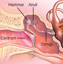
How is sound transmitted in the middle ear? |
Sound hits the tympanic membrane causing vibration in the flaccid portion which vibrates the handle of the malleolus --> incus --> stapes (lever system) --> oval window. Sound is increased by 20:1 / 20 dB as impedance increases due to the tympanic membrane and bones = 50% transmission to inner ear cochlear fluids --> liquid vibration |
|
|
How is pressure balanced in the middle ear |
Pressure is typically atmospheric (low) to increase vibration displacement. Air is exchanged with the mucosa and skin. If abnormal pressure does occur then the eustachian tube can open to balance the pressure. Middle ear gas is the same as mixed venous blood. |
|
|
What is the function of the eustachian tube? |
Equalise air pressure in the middle ear to Atmos. Pressure |
|
|
What is the tegmun tympani |
The boney roof of the middle ear |
|
|
What is the function of tensor tympani and stapedius and neuronal mechanism? |
Regulate loudness of sound vibrations on the oval window. 3/4 neurone reflex arc takes 25ms to tense the muscles --> pushes ossicles together, dampening sound |
|
|
What are the different functions of the round window and oval window? |
Oval window receives sound from the stapes which vibrates perilymph in the scala vestibuli Round window releases sound from perilymph of the scala timpani |
|
|
How is sound transmitted in the inner ear and what impact does the location within the cochlear have on hearing? |
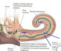
Sound is transmitted up the scala vestibuli to the helicotrema and then back down the scala timpani. Sound heard is lower in frequency as we move up to the helicotrema, each point = 1 resonance frequency = 1 neurone Also decrease in frequency along basilar membrane from base (near limbus) to apex) |
|
|
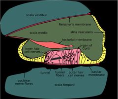
Where is perilymph and the endolymph found and what is the difference of their composition? |
Perilymph is in the scala vestibuli and scala timpani. Endolymph is in the scala media Perilymph: CSF/serum: high Na+ and low K+ Endolymph: intracellular: low Na+ and high K+ |
|
|
Tectorial membrane |
Mediates the cochlear echo reflex --> Receives motor nervous action to focus sound on to the outer hair cells - pressing down on them: fine tuning sound |
|
|
How does sound cause vibration of the basilar membrane? |
Pressure differences between the scala vestibuli and scala timpani perilymph causes the basilar membrane to vibrate. "perilymph compression wave" |
|
|
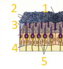
Describe the function of the organ or corti? |
Basilar membrane movement presses up against hair cells pushing them upwards --> deflection of stereocilia of Inner hair cell's --> positive ion selective channels open - potassium and calcium enter the cell, depolarising it |
|

|
1. Scala Vestibuli - Perilymph 2. Reissner's membrane 3. Scala Media - Endolymph 4. Tectorial membrane 5. Organ of Corti - IHC and OHC 6. Basilar membrane 7. Scalat tympani - perilymph 8. Cochlear nerve |
|
|
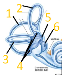
Functions of the vestibular system |
- Balance - Orientation of head in space - Send information to the CNS to rapidly compensate for change in balance/head orientation |
|
+ function |
1. Posterior semi-circular canal for Pitch 2. Anterior semi-circular canal for Roll 3. Lateral semi-circular canal for Yaw 4. Ampullae of semi-circular canal to detect changes in movement of endolymph and convert this into neuronal signal 5. Utricle (posterior) - Detect horizontal acceleration 6. Saccule (anterior) - Detect vertical acceleration |
|
|
Maculae and function(s) |
Comprised of Utricle and Saccule. Utricle: Horizontal acceleration Saccule: Vertical acceleration |
|
|
Which semi-circular canal detects which type of head movement? |
Posterior - Pitch (nodding head) Anterior - Roll (stretching side of neck) Transverse - Yaw (Shaking head in "no" fashion) |
|
|
Otolith organs |
Comprise fixed system to detect linear acceleration: Maculae (Utricle and saccule) |
|
Otolith organ Function and process of signal generation |
1. Striola: comprised of Otoconia 2. Otoconia: CaCo3 crystals which press down on hair cells 3. Otolithic membrane membrane: gelatinous to allow movement to be sensed 4. Nerve fibres and suppporting cells 5. Hair cells: Displacement of hair cells --> receptor output increases - alignment of hair cells --> repeatable signal pattern. |
|
|
Semi-circular canal angular acceleration mechanism of detection |
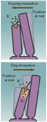
Movement of endolymph in the semi-circular canal due to head movement --> moves cupula in the ampullae of the semi-circular canal --> Alteration of kinocilum and stereocilium interaction mediates signalling. Shorter stereocilium moving towards and hitting longer kinocilium --> depolarisation Stereocilium moving away from Kinocilium --> Hyperpolarisation |
|

|
1. Cupula - displaced by endolymph 2. Ampulla 3. Semi-circular canal with Endolymph 4. Kinocilium 5. Stereocilium Nerve cells underneath |
|
|
Mechanism of signal transduction in semi-circular canals: differential effect in each ear |
Stereocilium moving towards Kinocilium --> Mechanically-gated potassium channels opening intrinically --> Depolarisation of stereocilium--> Glutaminergic transmission to vestibular nerve Stereocilium moving away from Kinocilium --> removal of potassium --> hyperpolarisation of stereocilium--> no Glutamine release. Opposites occur in each ear and the CNS compares this. |
|
|
Vestibulo-ocular reflex - function and mechanism |
Prevent retinal slip (image movement on the surface of the retina) Is modulated by the cerebellum Vestibular nucleus (within medulla) receives +ve or -ve signals from the vestibule --> passes on information to the abducens (pons) and oculomotor (midbrain) nuclei --> mediates movement of eye through eye muscles. |
|
+Function of each |
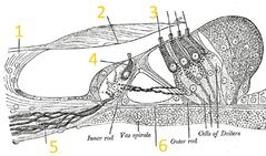
1. Limbus 2. Tectorial membrane - focusing sound vibrations on to organ of corti through the Cochlear-echo reflex 3. Outer hair cells - also attached to tectorial membrane to help tense and focus sound on inner hair cells 4. Inner hair cells - sensory to inner fluid movement through stereocilia that depolarise to transmit signal. 5. Nerve cells 6. Basilar membrane - moved by change in fluid pressure between scala vestibuli and tympani. |
|

|
1. Vestibular nucleus (Medulla) 2. Abducens nucleus (pons) 3. Oculomotor nucleus (Midbrain) 4. Medial longitudinal fasciculus |