![]()
![]()
![]()
Use LEFT and RIGHT arrow keys to navigate between flashcards;
Use UP and DOWN arrow keys to flip the card;
H to show hint;
A reads text to speech;
45 Cards in this Set
- Front
- Back
- 3rd side (hint)
|
Regulatory T-cell markers Memory T-cell markers |
Regulatory = CD25, FOXP3 Memory = CD45RO, CCR7 Both = CD4+ |
|
|
|
Roles of PGE2 |
GI and kidney function Vasodilation Uterine contraction Chemotaxis |
|
|
|
Roles of PGD2 |
Bronchoconstriction Anti-platelet aggregation |
|
|
|
Roles of PGF2 |
Bronchoconstriction Uterine contraction |
|
|
|
Roles of PGI2 |
Vasodilation Anti-platelet aggregation Renal, epithelial and CNS functions |
|
|
|
Role of IL-6 |
Increased cytokine release |
|
|
|
Role of INFy |
Priming agent |
|
|
|
Role of IL-12 |
Activates NK cells |
|
|
|
Role of IL-1B |
PG production (vasodilation + inflammation) Activates lymphocytes Apoptosis |
|
|
|
Roles of leukotrienes |
Bronchoconstriction Vascular permeability Chemotaxis |
|
|
|
Roles of thrombin |
Mobilise P selectins Produces PGs, PAF and NO Produces chemokines Expresses CAM Stimulates COX-2 |
PPP N CCC |
|
|
Where is the sensory component of pain processed? |
Lateral pain system: - S1 + S2 - Lateral thalamic nuclei |
|
|
|
Where are the affective and cognitive-evaluative components of pain processed? |
Medial pain system: - ACC - Medial thalamic nuclei - Insula |
|
|
|
Sartorius (attachment sites) |
ASIS - superomedial tibia |
|
|
|
Rectus femoris (attachments) |
AIIS - tibial tuberosity (via patellar tendon) |
|
|
|
Gracilis (attachments) |
Body and inferior ramus of pubis - superomedial tibia |
|
|
|
Semitendinosus and semimembranosus (attachments) |
Ischial tuberosity - superomedial tibia / medial condyle of tibia (respectively) |
|
|
|
Biceps femoris (attachments) |
L: Ischial tuberosity S: Linea aspera and lateral supracondylar line - head of fibula |
|
|
|
Gastronemius |
L: lateral condyle of femur M: popliteal surface of femur - calcaneus (via calcaneal tendon) |
|
|
|
Actions and innervation of gluteus maximus |
Hip extension + lateral rotation Inferior gluteal n. |
|
|
|
Actions and innervation of gluteus medius and gluteus maximus |
Abduction Superior gluteal n. |
|
|
|
Actions and innervation of TFL |
Hip flexion Superior gluteal n. |
|
|
|
Pain classified by nature |
Nociceptive: somatic + visceral Non-nociceptive: neuropathic + sympathetic |
|
|
|
Actions and innervation of piriformis |
Lateral rotation Branches of anterior rami of S1, S2 |
|
|
|
Actions and innervation of superior gemelli and obturator internus |
Lateral rotation N. to obturator internus |
|
|
|
Actions and innervation of inferior gemelli and quadratus femoris |
Lateral rotation N. to quadratus femoris |
|
|
|
Cause of hip drop |
Damage to superior gluteal n. |
|
|
|
Terminators of inflammation |
Lipoxin Resolvins, protectins Anti-inflammatory cytokines (IL-10, TGF-b) ACh (inhibits TNF production) |
|
|
|
Local effects of macrophages in response to TNFa and IL-1 |
More receptors on endothelium More chemokines More WBC activation Clotting More fibroblasts (more collagen) |
|
|
|
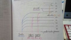
How long does it take for neutrophils to be replaced by macrophages? |
2-4 days |
|
|
|
Innervation of hamstrings |
Semitendinosus, semimembranosus and long head of BF = tibial n.
Short head of BF = common fibular n. |
|
|
|
Blood supply of hamstrings |
Inferior gluteal and perforating branches of profunda femoris a. |
|
|
|
Action, innervation and blood supply of medial thigh |
Adduction (+obturatur externus causes lateral rotation) Obturator a. and n. |
|
|
|
Hip flexors |
Psoas major Iliacus Pectinus Sartorius Rectus femoris |
|
|
|
Knee extensors |
Vastus lateralis Vastus intermedius Vastus medialis |
|
|
|
Innervation and blood supply of anterior thigh |
Femoral n. Femoral a. (+pectineus supplied by anterior branch of obturator a.) |
|
|
|
Innervation and blood supply of posterior leg |
Tibial n. Posterior tibial a. |
|
|
|
Innervation and blood supply of anterior leg |
Deep fibular n. Anterior tibial a. |
|
|
|
Innervation and blood supply of lateral leg |
Superficial fibular n. Fibular a. |
|
|
|
Origin of gluteal and obturator arteries |
Internal iliac |
|
|
|
Branches of profunda femoris |
Medial circumflex femoral a. Lateral circumflex femoral a. Perforating branches |
|
|
|
Venous drainage of lower limb (cutaneous) |
Great saphenous runs medially into femoral v. Small saphenous runs laterally into popliteal v. |
|
|
|
Lymphatic drainage of lower limb |
Popliteal to deep inguinal Superficial inguinal to external iliac (+ some deep inguinal) |
|
|
|
Spinal roots of femoral, obturator, superior/inferior gluteal and sciatic n. |
|
|
|
|
What do PT, APTT and bleeding time measure? |
PT = extrinsic pathway APTT = intrinsic pathway bleeding time = platelet function |
|