![]()
![]()
![]()
Use LEFT and RIGHT arrow keys to navigate between flashcards;
Use UP and DOWN arrow keys to flip the card;
H to show hint;
A reads text to speech;
38 Cards in this Set
- Front
- Back
|
Know the definitions of the Q wave, R wave, S wave, and J point. |
Q Wave-1st negative deflection after P Wave. R wave-1st positive deflection after P wave. S wave-1st negative deflection after either Q or R wave. J point-Exact point at which QRS complex stops and where ST segment starts. |
|
|
Know the 2 criteria for pathological Q waves. |
Wider than 40 milliseconds, depth below isolectric line more than 1/3 of the height of the R wave. |
|
|
Know which leads look at the lateral, inferior, septal, anterior, & posterior walls of the left ventricle & the right ventricle. |
Lateral- I, aVL, V5 & V6 Inferior- II, III, aVF Septal- V1 & V2 Anterior- V3 & V4 Posterior- V8, V9 (15 Lead) R. Ventricle- V4r |
|
|
Know the 3 reasons for running a 15 lead. |
1. Inferior wall MI. 2. Lateral wall MI. 3. ST segment depression in 2 or more anterior septal leads. |
|
|
Know the turn signal method for finding a bundle branch block (all steps). |
1. QRS greater than 120 milliseconds in diameter. 2. You have ruled out a ventricular rhythm. (Atrial Rhythm) 3. Find the J point. 4. J point below baseline = LBBB. 5. J point above baseline = RBBB. |
|
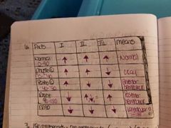
Know how to fill in the QRS directions on an axis chart and what pathological left & right axis each indicate. |
AXIS. I. II. III. MEANS NORMAL. ⬆️ ⬆️ ⬆️. Normal (0-90)
PHYSIO L. ⬆️ ⬆️ ⬇️ Okay 0-Neg 40
PATHO L. ⬆️ ⬇️. ⬇️ Anterior Hemiblock Neg 40-Neg 90
RIGHT. ⬆️. ⬆️⬇️= Posterior Hemiblock 90-180
ERAD. ⬇️. ⬇️. ⬇️. Ventricular? |
|
|
Know the limits for the PR interval. |
120 milliseconds |
|
|
Know the reasons a QRS may be wide and how to determine why a QRS is wide. |
1. BBB. 2. Originated in the ventricles. 3. If it is a BBB it will show evidence of atrial origin. |
|
|
Know the 3 reasons to call a pt unstable and cardiovert. |
1. Altered Mental Status. 2. Pulmonary Edema. 3. Acute Coronary Syndrome (ACS). |
|
|
Know the Bradycardia algorithm from top to bottom and where to start in different situations. |
1. 02, Monitor, IV 2. Atropine .5mg Rapid IV Push every 5 minutes to a max of 3mg total. 3. Pacing 4. Dopamine 2-10mcg/kg/min 5. Epi 2-10mcg/min |
|
|
Know what each of the 3 large coronary arteries perfuses. |
R. Coronary Artery- most of the atria (SA node 50-55%) R. Ventricle- Inferior wall of the L. Ventricle & bottom third of the posterior wall (AV node 90-95%) L. Circumflex- Rest of the atria, top 2/3 posterior wall of lateral wall of L. Ventricle (SA node 45-50%) LAD- Anterior & Septal walls.
|
|
|
Know the 2 most commonly used calcium channel blockers in EMS, to include doses & contraindications. |
1. Cardizem- .25mg/kg given over 5-10 min/ repeat after 20 min at .35mg/kg given over 5-10 min. 2. Verapamil- 5mg given over 5-10 min/ repeat after 20 min at 10mg given over 5-10 min. |
|
|
Know how to treat stable SVT. |
1. 02, Monitor, IV, 12 Lead, Vagal Maneuvers. 2. Adenosine- 6mg Rapid IV push. 3. Adenosine- 12mg Rapid IV push. 4a. Beta Blockers- Labatilol or Metoprolol at 10-20mg 4-5 min IV 4b. Cardizem or Verapamil 4c. Amio 150mg over 10 min. |
|
|
Know what an ECG consistent with pericarditis looks like. |
Global ST elevation or almost global ST elevation (all 12 leads or most of the 12 leads) with no reciprocal changes. |
|
|
Know the different antiarrythmics work (sodium, calcium, or potassium blocker). |
Lidocaine- sodium channel blocker (ventricular rhythms). Verapamil & Cardizem- calcium channel blockers (Atrial rhythms). Amiodarone & Procainamide- both sodium and potassium channel blockers (ventricular & atrial rhythms). |
|
|
Know why NTG can drop pressure so profoundly in a right ventricular infarct. |
Lowers preload. (Vasodilator). |
|
|
Know the 3 ECG changes that occur as COPD progresses. |
1. Right Axis Deviation. 2. Right Atrial Enlargement. 3. Right Ventricular Hypertrophy. |
|
|
Know what synctium is. |
When all of the muscle depolarizes and contracts at one time. (How wide or long QRS duration). |
|
|
Know at what QRS duration you can assume a patient has lost 50% of his contractile force. |
When QRS is wider than 120 milliseconds (7 1/2 blocks). |
|
|
Be able to explain Starlings Law. |
The more you stretch a muscle during diastole phase (the relaxation & filling period) the greater the force during the systole phase (the contraction period). |
|
|
Know what A1, B1, & B2 receptors do. |
A1- Causes periphovasoconstriction (aterioles). B1- Cause increased heartrate and contractile force (1 ♡). B2- Cause bronchioldiolation (2 lungs). |
|
|
Know the dosing regimen of procainamide, to include the end points. |
Loaded at infusion rate 20-50mg/min to one of 4 end points: 1. You resolved the rhythm. 2. Neutral end point of max dose of 17mg/kg. 3. QRS widens by 50%. 4. Hypotension occurs. |
|
|
Know the preferred drug to treat hypotension with pulmonary edema (carcinogenic shock). |
Dobutamine 2-20mcg/kg/min |
|
|
Know the H's & T's. |
H's Hypoxia Hypovolemia Hydrogen ion (acidosis) Hypo/hyper electrolytes Hypo/hyper thermia Hypo/hyper glycemic T's Toxins (overdose) Thrombosis Tension pneumothorax Tamponade Trauma |
|
|
Be able to define intotrope, chronotrope, & dromotrope. |
Inotrope- something that pertains to conctrile force. Chronotrope- something that pertains to heartrate. Dromotrope- something that pertains to the electrical conduction. |
|
|
Know how atropine works. |
Blocks the vagus nerve. |
|
|
Know the firing rates for the SA Node, the AV Node, and a ventricular pacemaker. |
SA Node- 60 to 100 BPM. AV Node- 40 to 60 BPM (Junctional). Ventricular Pacemaker- 20 to 40 BPM (Idoventricular). |
|
|
Be able to list at least 3 vagal maneuvers. |
1. Bare down. 2. Blow through an occluded straw, or a syringe, or pinch nostrils & blow. 3. Ice packs to face or coughing. |
|
|
Know the drug of choice for the management of torsades. |
Magnesium: Torsades w/ pulse: 2-4g infused over 20-40 minutes. Torsades w/o pulse: 2g IV push, repeat in 5 minutes as needed. Max dose of 4g. |
|
|
Know the voltage setting for cardioverting the different tacharrhythmias. |
Torsades: 360j or biphasic equivalent unsynchronized. SVT: 100j synchronized. A-Fib/A-Flutter: 100j synchronized. |
|
|
Know when to use bicarb and calcium early on a cardiac arrest. |
Use bicarb & calcium early on a cardiac arrest when a pt is on dialysis and is due or overdue for dialysis. |
|
|
Know the criteria for a new onset A-Fib and the danger of cardioverting A-Fib. |
New onset of A-Fib less than 48 hours. Danger of cardioverting: may throw a blood clot and cause a pulmonary embolism or a stroke. |
|
|
Know the 2 most common STEMI imposters. |
1. Left Bundle Branch Block. 2. LVH. |
|
|
Know both methods for determining LVH. |
1. Sv1+Rv5 or Rv6=35 or greater. 2. R in I+ S in III=25 or greater. |
|
|
Know the criteria for calling a STEMI. |
ST elevation greater than 1mm in 2 or more contiguous leads. |
|
|
Know the antidote for calcium channel blockers. |
Calcium 1-2g Slow IV Push. (Titrate to effect) |
|
|
Know when the electricity is delivered in synchronized cardioversion. |
Delivered during the absolute refectory period. |
|
|
Know why medications are withheld in hypothermic arrest. |
Receptors do not work until the pt is rewarmed. |