![]()
![]()
![]()
Use LEFT and RIGHT arrow keys to navigate between flashcards;
Use UP and DOWN arrow keys to flip the card;
H to show hint;
A reads text to speech;
99 Cards in this Set
- Front
- Back
|
Two types of cartilage growth |
Interstitial Appositional |
|
|
Chondrocytes in lacunae undergo mitosis, both cells then occupying the same lacuna, the two cells synthesize new matrix and push apart |
Interstitial growth |
|
|
Stem cells at internal edge of perichondrium divide forming new stem cells and chondrobladts New chondrocytes produce new matrix |
Appositional growth |
|
|
Classifications of bones |
Long bones Short Flat Irregular |
|
|
Bone cells, which are stem cells in this case, derived from mesenchyme and found in the periosteum and endosteum |
Osteoprogenitor |
|
|
Calls produced from stem cells which produce new bone |
Osteoblasts |
|
|
What does blast mean |
Build |
|
|
Mature bone cells |
Osteocytes |
|
|
Bone cell that dissolves bone matrix |
Osteoclasts |
|
|
What does class mean |
Dissolve |
|
|
Organic and inorganic material formed by bone cells and lying in between them |
Bone matrix |
|
|
Bone that lies on the outside of individual bone |
Cortical bone |
|
|
Organic substances of bone matrix |
1/3 organic components: collagen fibers, and ground substance |
|
|
Inorganic components of bone matrix |
Calcium phosphate Calcium hydroxide Calcium carbonate Icons of sodium, magnesium, sulfate, and fluoride |
|
|
Type of bone that is latticework within a bone |
Cancelous (spongy) bone |
|
|
Basic structure of cortical bone |
Osteon (haversion system) |
|
|
Two synonyms for bone growth |
Osteogenesis and ossification |
|
|
Type of growth that occurs on flat bones of the skull, som facial bone, mandible, and central part of clavicle. |
Intramembranous ossification |
|
|
Describe intramembranous ossification |
Ossification centers in thick regions of mesenchyme Bone matrix (osteoid) undergoes calcification Woven bone and periosteum form Lamellar bone replaces woven as compact and spongy bone. |
|
|
Type of growth that occurs in bone extremities, vertebrae, and ends of clavicles |
Endochondral ossification |
|
|
Describe endochondral ossification |
Hyaline develops Cartilage calcifies, bone collar forms Primary ossification center forms diaphysis Secondary ossification centers form epiphysis Bone replaced cartilage (except articular, and epiphyseal plate) Epiphyseal plate ossify you form epiphyseal lines |
|
|
Anatomical term for growth plate |
Epiphyseal plate |
|
|
Where does the bone increase in length |
Epiphyseal plate |
|
|
Type of bone growth that increases the length of a bone |
Interstitial growth |
|
|
Type of bone growth that increases the diameter of a bone |
Appositional growth |
|
|
Where does a bone increase in diameter |
Periosteum |
|
|
Opening in bones through which blood vessels carry nutrients |
Nutrient artery |
|
|
Large smooth rounded articulating oval structure |
Condyle |
|
|
Small flat shallow articulating surface |
Facet (means face) |
|
|
Prominent, rounded epiphysis |
Head |
|
|
Smooth grooves pulley like articular process |
Trochlea |
|
|
Deep pit or socket in maxillae or mandible |
Alveolus |
|
|
Flattened or shallow depression |
Fossa |
|
|
Narrow groove |
Sulcus |
|
|
Narrow prominent ridge like projection |
Crest |
|
|
Projection adjacent to a condyle |
Epicondyle |
|
|
Low ridge |
Line |
|
|
Any marked bony prominence |
Process |
|
|
Angular extension of a bone relative to the rest of the structure |
Ramus |
|
|
Pointed slender process |
Spine |
|
|
Massive rough projection only found in the femur |
Trochanter |
|
|
Small round projection |
Tubercle |
|
|
Large rough projection |
Tuberosity |
|
|
Names of openings and spaces in bones |
Canal Fissure Foramen Sinus |
|
|
Passageway through a bone |
Canal (meatus) |
|
|
Narrow, slit line opening through a bone |
Fissure |
|
|
Rounded passageway through a bone (a hole) |
Foramen |
|
|
Cavity or hollow space in a bone |
Sinus |
|
|
How Classification of joints is done |
What’s it’s made of and how it moves |
|
|
How do mobility and flexibility relate to one another |
Inversely: as one increases the other decreases |
|
|
Types of joints based on structure |
Fibrous Cartilaginous Synovial |
|
|
Three types of joints based on function |
Synarthrosis (immovable) Amphiarthrosis (slightly movable) Diarthrosis (freely movable) |
|
|
Characteristics of fibrous joints |
Joined by dense regular connective tissue Immovable or only slightly movable No joint cavity |
|
|
Functional and structural classifications of gomphoses joint, and where they’re located |
Synarthrodial Fibrous Found only in the teeth |
|
|
Structure and functional classifications of sutures, and location |
Fibrous Synarthrodial Only in the skull |
|
|
Fibrous joints in which articulating bones are joined by long strands of dense regular connective tissue, fibrous, and amphiarthrodial |
Syndesmoses |
|
|
Bones joined directly by cartilage. Sometimes hyaline, sometimes fibrocartilage. No cavity |
Cartilaginous joints |
|
|
Locations, and classifications of synchondroses |
Growth plate, rib to costal cartilage, synarthrodial, cartilaginous. |
|
|
Locations, and classifications of symphyses joint |
Intervertebral discs, symphysis pubis Amphiarthrodial Cartilaginous (fibrocartilage) |
|
|
Joints with a fluid filled capsule, bones have articular (hyaline) cartilage to protect against friction. Always diarthrodial |
Synovial joints |
|
|
Structure that surrounds synovial joints and forms it’s cavity |
Articular (joint) capsule |
|
|
Outer layer of joint capsule |
Fibrous (joint) capsule |
|
|
Inner layer of joint capsule, creates synovial fluid |
Synovial membrane |
|
|
Slimy fluid that nourishes, lubricates, and protects a synovial joint |
Synovial fluid |
|
|
Contains synovial fluid around the tendon, reduces friction |
Synovial sheaths |
|
|
Fat pads |
Pads made of fat |
|
|
Fills space in larger joint capsules, guides movement |
Cartilage discs |
|
|
Extra bag of fluid (synovial), disperses pressure |
Bursa |
|
|
One side has a slight oval protrusion, the other an elliptical recess, two planes of motion |
Condyloid joint |
|
|
Flat or mostly flat surfaces gliding across each other, one or two planes of motion |
Gliding joints |
|
|
Protruding condyle fits into a corresponding recess, has one plane of motion |
Hinge joints |
|
|
One bone makes a ball shape, which fits into the other like a socket |
Ball and socket joint |
|
|
One bone convex, the other concave, two planes of motion. |
Saddle joints |
|
|
Structures vary, but always simply rotate along a central axis |
Pivot joints |
|
|
Angular Movements of synovial joints |
Extension Hyper extension Flexion Abduction Adduction Circumduction Rotation (pronation, supination) |
|
|
Lateral carrying away from the midline of the body |
Abduction |
|
|
Medial movement of a body part towards the midline |
Adduction |
|
|
Decreases the angle of a joint |
Flexion |
|
|
Increases the angle of a joint |
Extension |
|
|
Increases the angle beyond the normal anatomical range |
Hyperextension |
|
|
Type of movement when the trunk of the body moves laterally along a coronal plane |
Lateral flexion |
|
|
Distal end of an appendage moves in a circular motion |
Circumduction |
|
|
Pivot motion along an axis |
Rotation |
|
|
Pivoting motion in which a long bone turns on its long axis so that the anterior surface moves laterally |
Lateral rotation |
|
|
Pivot motion that turns the anterior side of a long bone medially |
Medial rotation |
|
|
Inferior movement of a body part |
Depression |
|
|
Superior movement of a body part |
Elevation |
|
|
For sum of the foot or hand moves so that the angle between it and the limb decreases |
Dorsiflexion |
|
|
When the anchor joint moves to point the toes inferiorly |
Plantar flexion |
|
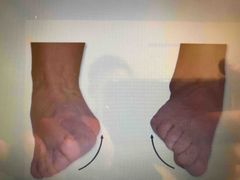
When the intertarsal joints are moved to turn the sole of the foot medially |
Inversion |
|
|
When intertarsal joints move to face the sole of the foot laterally |
Eversion |
|
|
When the palm of the hand is directed posteriorly or inferiorly |
Pronation |
|
|
When the palm is directed anteriorly or superiorly |
Supination |
|
|
When the body is in a face down position |
Prone position |
|
|
When the body is in a face up position |
Supine position |
|
|
When a body part moves anteriorly in a horizontal plane |
Protraction |
|
|
When a body part moves posteriorly along a horizontal plane |
Retraction |
|
|
When the thumb moves towards the Palmer tip of the fingers |
Opposition |
|
|
When the thumb moves away from the Palmer tips of the fingers |
Reposition |