![]()
![]()
![]()
Use LEFT and RIGHT arrow keys to navigate between flashcards;
Use UP and DOWN arrow keys to flip the card;
H to show hint;
A reads text to speech;
23 Cards in this Set
- Front
- Back
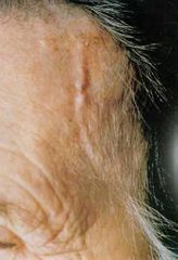
Diagnosis? |
Giant cell (temporal) arteritis. Temporal artery shows thickened, nodular and tender segment. |
|
|
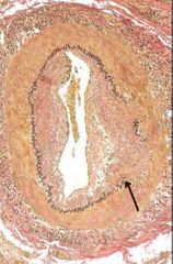
Arrow shows focal destruction of internal elastic membrane and intimal thickening characteristic of long standing or healed arteritis.
Stain is Elastin stain. |
|
|
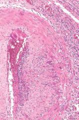
Giant cell arteritis. |
|
|
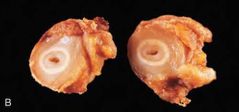
Takayasu arteritis. |
|
|
Takayasu arteritis. Gross appearance of cross section of carotid artery. Shows marked intimal thickening and adventitial fibrosis with minimal residual lumen. |
|
|
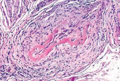
Polyarteritis Nodosa.
Segmental fibrinoid necrosis and thrombotic occlusion of lumen of this small artery. |
|
|
Microscopic Polyangitis (leukocytoclastic vasculitis). Fragmentation of neutrophils in and around blood vessel walls( nuclear dust). |
|
|
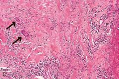
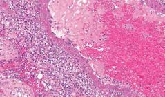
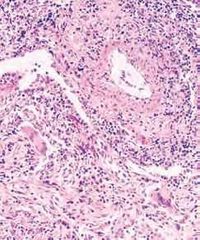
Granulomatosis with Polyangitis. Also Wegner's granulomatosis. Granulomatous inflammation showing giant cells. |
|
|
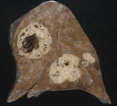
Wegner's granulomatosis. Lung of patient with fatal granulomatosis with Polyangitis demonstrating large, nodular, centrally cavitating lesions. |
|
|
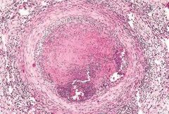
Thromboangiitis obliterans (Buerger disease) Lumen is occluded by thrombus containing microabscesses composed of neutrophils , vessel wall is infiltrated with leukocytes.
|
|
|
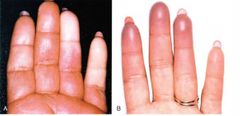
RAYNAUD PHENOMENON A) Sharply demarcated pallor of distal arteries due to closure of distal arteries. B) Cyanosis of the fingertips. |
|
|
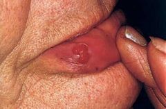
Pyogenic granuloma of lip. These are capillary hemangiomas. |
|

|
Hemangioma of the tongue. |
|
|
Juvenile capillary hemangioma. Strawberry type hemangioma. Rapidly grow for few months but then fade by 1-3 yrs of age and completely regress by age of 7. |
|
|
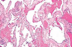
Cavernous Hemangioma. Large cavernous blood filled vascular spaces separated by thick connective tissue stroma. |
|
|
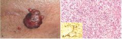
Bacillary angiomatosis. A) characteristic cutaneous lesion. B) acute neutrophilic inflammation with capillary proliferation Warthin-starry stain demonstrates cluster of tangled bacilli(black). |
|
|
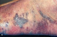
Kaposi Sarcoma. Coalescent red-purple macules and plaques of the skin. |
|
|
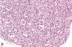
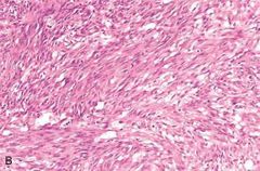
Kaposi Sarcoma. Nodular stage of it. Sheets of proliferating spindle cells. |
|
|
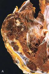
Angiosarcoma.
|
|
|
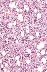
Angiosarcoma. Moderately differentiated angiosarcoma with dense clumps of atypical cells lining distinct vascular lumens. |
|
|
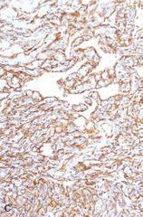
Angiosarcoma. IHC staining for endothelial cell marker CD31 demonstrating endothelial nature of tumor cells. |
|
|
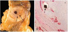
Restenosis after angioplasty and stenting. Movat stain used in B which stains collagen fibres. |
|
|
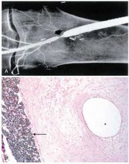
Intimal hyperplasia at distal anastamosis of a synthetic femoral-popliteal graft. A) Angiogram shows constriction. B) arrow shows Gore-Tex graft with prominent intimal proliferation and very small residual lumen"*". |