Reading...
![]()
Play button
![]()
Play button
![]()
Use LEFT and RIGHT arrow keys to navigate between flashcards;
Use UP and DOWN arrow keys to flip the card;
H to show hint;
A reads text to speech;
21 Cards in this Set
- Front
- Back
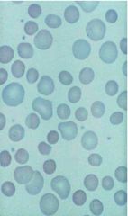
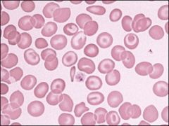
This change seen in what conditions (5)?
|
spherocytes
Seen in: Warm AIHA Old blood Burns Venoms Oxidant damage |
|
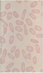
Seen in what conditions? (2)
|
Eliptocytes
Conditions: Hereditary ellipotcytosis Iron deficiency |
|
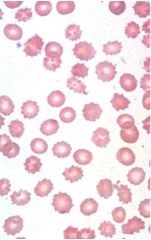
Seen in what conditions? (4)
|
Echinocytes
Conditions: ARF prolonged EDTA Low pH slides Pyruvate kinase deficiency, post splenectomy |
|
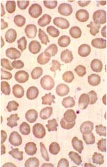
Seen in what conditions? (6)
|
Acanthocytes
Conditions: Liver failure a-beta-liproporteinemia hypothyroid post-splenectomy McLeod (no Kx) MDS |
|
Seen in what conditions? (4)
|
Target cells
Conditions: Liver failure Hemoglobinopathies Asplenia Iron deficiency |
|
What condition? (1)
|
Bite cells
Condition: G6PD deficiency (munching of Heinz body), may be accompanied by blister cells |
|
Causes? (7)
|
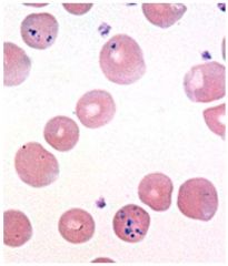
Basophilic stippling (ribosomal RNA)
Causes: Lead poisoning Hemoglobinopathy Pyrimidine 5' nucleotidase deficiency MDS Infection Sideroblastic anemia Porphyria |
|
causes? (3)
|
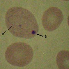
Howell Jolly body (nuclear remnant)
Causes: Post-splenectomy/asplenia Megaloblastic anemia MDS |
|
Causes (2)?
|
Pappenheimer body (mitochondrial remnants)
Causes: post-splenectomy/asplenia Fe overload |
|
Causes (2)?
|
Cabot ring: microtubule, remnants of the mitotic spindle
Causes: Disordered erythropoiesis Severe megalobastic anemia |
|

Crystal violet stain. What condition?
|
Heinz body
Conditions: G6PD deficiency |
|
|
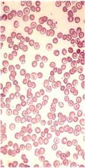
Iron deficiency anemia, post transfusion. The cells are pale with an enlarged central pallor, in sharp contrast to the transfused normochromic cells.
|
|
|
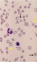
Blood film, megaloblastic anemia. Macrocytosis and a circulating megaloblast with abnormal, binucleated nucleus and open chromatin.
|
|
|
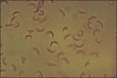
Sickle cell preparation, in sodium metabisulfate; sickle cell anemia. Even in sickle cell trait, all cells eventually sickle.
|
|
|
Blood film, sickle cell anemia. The erythrocytes are far from one another, suggesting a severe anemia. Numerous pointed sickle cells and target cells are present. Close to the nucleated red blood cell, elliptical cells are seen. These cells have a dense center, instead of the usual central pallor, suggesting that they are incipient sickle cells (500?).
|
|

|
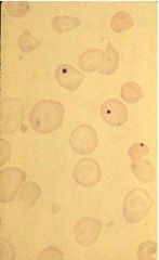
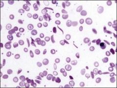
Hemoglobin (Hb) C disease, after splenectomy. Before splenectomy, the only morphologic abnormality was the presence of target cells. After splenectomy, Howell-Jolly bodies and Hb crystals, such as those in the center, were present. Note that almost all of the Hb in this particular cell is in the dark bar, and the membrane is still visible. Some such crystals are distinctly hexagonal (1000?).
|
|
|
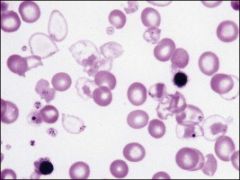
Homozygous β-thalassemia. A few cells contain hardly any hemoglobin (Hb), and the Hb is often precipitated at the membrane. Bizarre target cells, Howell-Jolly bodies, and poorly hemoglobinized nucleated red blood cells are seen.
|
|
|
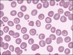
β-Thalassemia trait. Hypochromic, microcytic red blood cells with frequent targeting. Mild anemia.
|
|
|
Hemoglobin H disease. Mild anemia and target cells.
|
|
|
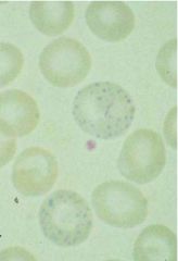
Hemoglobin H preparation. Film made after incubation of blood with brilliant cresyl blue. Several red cells contain multiple small pale blue inclusions.
|
|
|
Heinz bodies. Normal red cells incubated with an oxidant drug and stained while in suspension with methyl violet; finally, an air-dried film was made and stained with Wright's stain. Purple-staining Heinz bodies are precipitates of denatured hemoglobin that tend to attach to the cell membrane.
|