Reading...
![]()
Play button
![]()
Play button
![]()
Use LEFT and RIGHT arrow keys to navigate between flashcards;
Use UP and DOWN arrow keys to flip the card;
H to show hint;
A reads text to speech;
40 Cards in this Set
- Front
- Back
|
What are the main functions of the digestive tract?
|
Mechanical Digestion (mastication, grinding)
Chemical digestion (to break down food) Absorption (of nutrients, water) Elimination (of wastes) Endocrine acivity |
|
|
What is the embryological origin of the epithelial lining of the digestive tract? Of the tract wall?
|
Tract: Endoderm
Wall: Splanchnic mesoderm for the most part |
|
|
Describe the layers of the digestive tract wall.
|
Mucosa: epithelium, underlying CT (lamina propia), thin layer of smooth muscle (muscularis mucosa)
Submucosa: CT layer, includes Meissner's Plexus (ANS) Muscularis Externa: inner circular, outer longitudinal; sandwiches Auerbach's plexus Serosa or Adventitia: outermost part of GI tract |
|
|
In which layer of the digestive tract wall is Auerbach's plexus found? Meissner's plexus?
|
Auerbach's: Muscularis Externa
Meissner's: Submucosa |
|
|
What is serosa? What does it comprise? Where is it found?
|
Serosa: single layer of squamous cells and underlying CT; comprises visceral peritoneum
Found on portions of digestive tract within abdominal cavity |
|
|
Which portions of the digestive tract exhibit an adventitia?
What is the adventitia? |
Parts of tract fixed to adjacent tissues: esophagus, anal canal; retroperitoneal regions
|
|
|
What does the mucosa of the esophagus consist of?
|
Epithelium: Stratified Squamous
Lamina Propria (loose CT) Muscularis Mucosa |
|
|
What does the submucosa of the esophagus consist of?
|
Vascular CT; some glands near stomach jn to lubricate/protect surface
INNERVATION: symp (inhibits motility) and parasymp (promotes motility) |
|
|
What type of muscle is present in the esophagus?
|
Upper 1/3: striated skleteal muscle
Middle 1/3: skeletal and smooth muscle Lower 1/3: Smooth muscle |
|
|
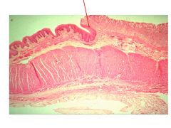
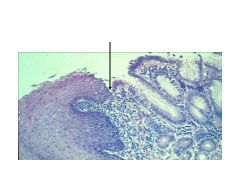
What characterizes the Gastro-Esophageal junction?
|
Abrupt change from stratified squamous of esophagus to simple columnar of stomach
|
|
|
What is the epithelium of the stomach lining? How do glands differ in the cardiac region? Fundus/Body? Pyloris?
|
Simple columnar epithelium
Cardiac regions have cardiac glands: mostly mucous cells, long glands, short pits Fundic/Body glands: pit and gland areas are ~ equal Pyloric glands: short with long pits |
|
|
Where are surface mucous cells located? Mucus neck cells?
|
Surface mucous cells: On surface of stomach (comprise simple columnar epithelium)
Mucus neck cells - in upper regions of gastric glands; secrete mucous into lumen |
|
|
Where are parietal cells found? What is their role?
|
Found in middle part of gastric glands; transport H+ and Cl- into lumen
Secrete gastric intrinsic factor (GIF) - impt for B12 absorption |
|
|
Where are chief cells found? What's their role? What stimulated their activity?
|
Deepest bases of gastric glands
Secrete pepsinogen (precursor to pepsin; gets cleaved by acid) Secretin stimulates release of pepsin |
|
|
Where are enteroendocrine cells found? What's their role?
|
Scattered among epithelium near neck of glands
Synthesize and release gastrin (promotes parietal cell activity)--secrete paracellularly into CT; diffuses into nearby target cells |
|
Identify.
|
Esophagus
|
|
Identify.
|
Esophagus: striated squamous epithelium; mucous cells; muscle layer
|
|
|
Gastro-esophageal junction: Esophagus on left with striated squamous; stomach on right with simple columnar
|
|
|
Gastro-Esophageal Junction: esophagus on left with striated squamous, stomach on right with simple columnar
|
|
|
How do parietal cells appear under the microscope? Under EM, what structure would you look for to identify a parietal cell (with certainty)? What are its functions?
|
Fried egg look; very eosinophilic (no protein secretion function, so very little RNA)
Under EM, would be able to see intracellular canaliculi (invaginations with grooves)--increase surface area (for HCl release?) |
|
|
What are the functions of the small intestine? What are its three sections?
|
Neutralize chyme from stomach
Chemical digestion Absorption of nutrients, water, salts Immune surveillance Duodenum, jejunum, ileum |
|
|
What is the epithelial lining of the small intestine? How does the small intestine appear on gross dissection? Histologic?
|
Simple columnar
Gross: Plicae (pleated folds of mucosa); have villi (projections of mucosa), which have microvilli (brush border) |
|
|
What is the effect of gastrin?
|
Stimulates parietal cells
|
|
|
What is the effect of intrinsic factor? Where is it produced?
|
Intrinsic factor--allows for absorption of B6; produced and secreted by parietal cells
|
|
|
Where are goblet cells found in the GI tract?
|
Some in the small intestine, greater quantities in ileum and large intestine
|
|
|
What cells are contained in the mucosa of the small intestine?
|
Absorptive cells (ENTEROCYTES)
Scattered goblet cells |
|
|
What are lacteals? Where are they found?
|
Lacteals are lymph capillaries; absorb dietary fats; found in lamina propria of small intestine villi
|
|
|
What are Brunner's glands? Where are they found?
|
Submucosal glands that produce alkaline mucous to neutralize chyme; found ONLY IN DUODENUM
|
|
|
How would you distinguish a low magnification view of the jejunum from one of the ileum?
|
Jejunum has longer, leafy villi
Ileum has stubby villi |
|
|
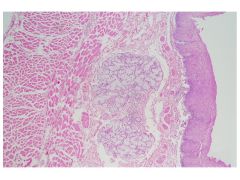
How is immune surveillance accomplished in the small intestine?
|
MALT: Peyer's Patches may be present in mucosa or submucosa; utilize M cells (microfold cells) to present antigens to APC's
|
|
|
What is the role of glycocalyx? Where is it found?
|
Plays role in absorption of nutrients; found associated with microvilli (brush border) at apex of absorptive cells
|
|
|
What are the three main secretions of enterendocrine cells? What are their actions? Where are they secreted?
|
Gastrin: stomach to jejunum; stimulates parietal cells (HCl)
CCK: small intestine; stimulates gall bladder to release bile, stmiulates pancreatic cells to secrete digestive enzymes Secretin: small intestine; stimulates pancreatic duct cells to release bicarbonate (to neutralize acid) |
|
|
What are Argentaffin cells? Where are they found? How are they viewed histologically?
|
Argentaffin = Enteroendocrine cells in epithelium of small intestine; love silver, need silver stain to identify
|
|
|
What are Paneth cells? Where are they found?
|
Present at bases of intestinal glands; secrete lysozyme and antimicrobial molecules into lumens of crypts
|
|
|
What are the distinguishing characteristics of the duodenum? Jejunum? Ileum?
|
Duodenum: Brunner's glands in submucosa
Jejunum: plicae circulares--folds containing core of submucosa; visible with naked eye (most prominant in distal duodenum and jejunum). NO GLANDS IN SUBMUCOSA Ileum: Peyer's patches; less prominent plicae circulares. NO GLANDS IN SUBMUCOSA. lots of goblet cells in terminal ileum. |
|
|
What is the function of the large intestine? What are its key histologic characteristics?
|
Continued absorption
Lots of goblet cells, NO VILLI, has crypts (glands) |
|
|
What is the epithelium of the colon?
|
Simple columnar with absorptive cells and mucous cells; crypts with enteroendocrine cells
|
|
|
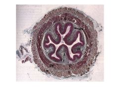
Where is the appendix in relation to the large intestine?
|
Appendix is diverticulum of colon
|
|
|
What is the histological appearance of the colon?
|
Simple columnar until ano-rectal junction; crypts disappear and epithelium becomes stratified squamous. Lamina propria has large veins (can cause hemorrhoids)
|
|
|
Describe the epithelial lining and muscle content of the colon.
|
Simple columnar-->stratified squamous (non-keratinized)-->stratified squamous (keratinized): at skin portion
Colonic smooth muscle-->skeletal muscle at voluntary anal sphincter |