![]()
![]()
![]()
Use LEFT and RIGHT arrow keys to navigate between flashcards;
Use UP and DOWN arrow keys to flip the card;
H to show hint;
A reads text to speech;
25 Cards in this Set
- Front
- Back
|
What is Cell Theory? |
- The cell is the basic component of a living thing - All cells arose from pre - existing cells - Within the cell, chemical reactions can occur eg metabolism |
|
|
What are cells organised into? |
Tissues |
|
|
What are tissues organised into? |
Organs |
|
|
What are organs organised into? |
Organ system |
|
|
Nucleus |
- Contains DNA - codes for protein synthesis - DNA is arranged as chromosomes (long strands). Unless the cell is dividing they appear as granular material called chromatin - Surrounded by a double membrane, called nuclear envelope which has many pores |
|
|
Nucleolus |
- Darkly staining part of nucleus - Synthesises ribosomes which leave though pores and attach to RER |
|
|
Ribosomes |
- In Eukaryotic Cells, ribosomes are 80S - Made from RNA and are the site of protein synthesis - Some are free in cell but most are attached to RER |
|
|
Rough Endoplasmic Reticulum |
- Network of flattened sacs - Found extending from the nucleus outwards into cytoplasm - Proteins synthesised by ribosomes pass into RER for packing - small vesicles form at the end to be transported out of the cell |
|
|
Smooth Endoplasmic Reticulum |
- This is similar to the RER but has no ribosomes - It's function is to synthesise lipids eg steroid hormones, fat and cholestrol |
|
|
Golgi Apparatus |
- Stack of 4 - 8 flattened membrane sacs - It joins substances together - eg proteins & lipids - and package them into vesicles to be secreted from the cell |
|
|
Mitochondria |
- These have a double membrane with many folds to increase surface area - They have a matrix in the middle which contains the enzymes for aerobic respiration - The inner part of mitochondria is infolded to form cristae (singular, crista). |
|
|
Cell Wall |
- This is laid down by the plant cell as it grows. It is formed by overlapping cellulose fibres, giving it strength and elasticity. It is very permeable to water |
|
|
Plasmodesmata |
- These are extensions of the cytoplasm passing through the cell wall, from one cell to the next |
|
|
Vacuole |
- The large fluid filled permanent vacuole is surrounded by a single membrane called the tonoplast - The vacuole stores soluble substances – sugars, minerals, salts, etc |
|
|
Chloroplasts |
- These organelles have a double membrane surrounding a matrix called a stroma, this contains the enzymes used in photosynthesis - There is also a series of membranes called Thylakoids. The light absorbing pigments and carrier molecules embedded in them to turn light energy into chemical energy |
|
|
Prokaryotic Cells (bacteria) |
- These cells don’t have membrane bound organelles, e.g. Mitochondria - They don’t have a nucleus, their DNA exists as a loop - They have a cell wall but it is not made from cellulose, instead it is made from Peptidoglycans (protein and carbohydrates) |
|
|
Eukaryotic Cells (plant and animal cells) |
- They have much larger structure, so need an internal system of membranes - They have mitochondria, where aerobic respiration takes place - The DNA is contained in a nucleus, with a nuclear membrane |
|
|
Magnification Equations |
magnification = size of image / size of object magnification = length of scale line measured / length of scale line given |
|
|
Resolution |
- Resolution is defined as the smallest distance between two particles which allows them to be distinguished from one another - It depends on the wavelength. The shorter the wavelength of light the better the resolution |
|
|
Electron Microscope |
NEGATIVE: - Expensive to buy and run - Living specimens can’t be observed - Images in black and white - Preparing specimens is complex POSITIVE: - High resolution so greater depth of field may be observed - High magnifications |
|
|
Light Microscope |
POSITIVE: - Relatively cheap to buy and run - Living and dead tissues may be observed - Colour images are obtained - Preparing specimens relatively simple NEGATIVE: - Resolution limited by wavelength of light - Depth of field is restricted - Limited magnification |
|
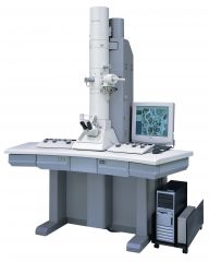
What type of microscope is this? |
Electron microscope |
|
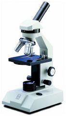
What type of microscope is this? |
Light microscope |
|
|
Gram positive and Gram negative bacteria |
Gram positive: mainly have peptidoglycans which pick up a stain called crystal violet Gram negative: have an outer layer composed of lipids and polysaccharides which resits crystal violet stain but they pick up a red dye called safranin |
|
|
Stages of Gram Staining |
1. bacteria in an air dried smear on a microscope slideappear colourless 2. smear of bacteria is treated with crystal violet (abasic stain). All the cells appear violet when the stain is washed from theslide 3. smear is flooded with Lugol’s iodine (a mordant (fixing)treatment to combine the dye to those bacteria which it will react) 4. the smear is now treated with a decolourising solutionof acetone and alcohol – this removes the violet dye from the cells with whichit has not reacted. Gram – positive bacteria remain purple 5. red dye safranin is briefly added as acounterstain – it is taken up by the colourless bacteria of the treated smear.Gram – negative bacteria now appear red and gram – positive bacteria remainpurple |