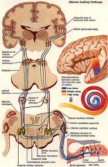
Reading...
![]()
Play button
![]()
Play button
![]()
Use LEFT and RIGHT arrow keys to navigate between flashcards;
Use UP and DOWN arrow keys to flip the card;
H to show hint;
A reads text to speech;
26 Cards in this Set
- Front
- Back
- 3rd side (hint)
|
Describe the role of CNVII and CNVIII in the ear?
|
• CN VIII is always lateral to the CN VII. CN VII is carrying efferent fibers to the muscle of the ear. CN VIII is carrying afferent fibers away from the ear.
|
|
|
|
Describe energy transfer in the ear?
|
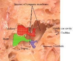
• This is the inside of the tympanic membrane.
• Energy is transferred from air pressure to mechanical pressure at the tympanic membrane. At the oval window the energy is mechanical energy is tranferred to fluid pressure which will be changed to electrical energy in the neurons. |
|
|
|
Compare the organization of these systems:
1. Somatosensory system 2. Visual system 3. Auditory system 4. Taste and smell |
1. The somatosensory system is organized by dermatomes
2. For the visual system, info is organized according to visual space 3. For the auditory system, info is organized by frequency. -Frequencies are organized by position of hair cells on the basilar membrane 4. Taste and smell are the so-called chemical senses. Air-borne molecules come in and are dissolved in a fluid (saliva in mouth for taste and nasal concha for smell) |
|
|
|
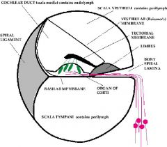
Describe the 1st order relay system of the auditory system?
|

• This is a picture of the fluid filled cavities of the central portion of the auditory system
• The pink nerve cells shown are the 1st order relays of the auditory system • As the interior of the fluid filled ascends, the penetration by the bony spiral membrane gets shallower and the basilar membrane gets broader. |
|
|
|
Compare frequencies at different parts of the basilar membrane and the reason for this?
|
Different places on the membrane will vibrate at different frequencies:
1. At the base of the cochlea, the bony spiral membrane projects far in to the cavity, so the basilar membrane is short and will only vibrate at high frequencies. 2. At the apex of the cochlea the bony spiral membrane does not project into the cavity very far so the basilar membrane will be longer and will vibrate at low frequencies. -The basilar membrane will determine the frequency at which cells in a particular point will vibrate. |
|
|
|
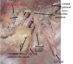
What are the 3 divisions of axons of CN VII as it leaves the CNS at the pontomedullary junction and enters the internal acoustic meatus?
|
1. Sensory axons carrying taste information from the anterior two thirds of the tongue. These axons are the afferent limb of the salivation reflex.
2. The axons of the efferent limb of the salivation reflex are here causing secretion of saliva. 3. The somatic motor axons of the facial nerve spreading out to end on the many muscles of the face, causing them to contract. |
|
|
|
Compare the location of the cell bodies of these and their termination:
1. Axons that are the afferent limb of the salivation reflex 2. Axons that are the efferent limb of the salivation reflex 3. Somatic motor axons of the facial nerve |
1. The cell bodies of axons of afferent limb of salivation are in the geniculate ganglion, and the axon terminals innervate the rostral half of the nucleus of the solitary tract in the medulla.
2. The axons of the efferent limb of this reflex are preganglionic parasympathetic and their Their cell bodies are located in the superior salivatory nucleus of the medulla, and they terminate on the cells of the submandibular ganglion, composed of postganglionic parasympathetic cells, whose axons terminate on the submandibular and sublingual glands. 3. The cell bodies of somatic motor axons of the facial nerve are located in the facial motor nucleus of the pons, and the axons course through the auditory canal, exit via the stylomastoid foramen, and then move anteriorly., coursing through the parotid gland and then to facial muscle. |
|
|
|
What are 7 features of auditory stimuli?
|
1. Frequency modulation (FM)=pitch differences
2. Amplitude modulation (AM)=intensity differences 3. Temporal patterning (change, on/off) = start a word, stop a word 4. Stimulus duration 5. Phase: whether action potential is on ascending side or descending side 6. Localization in space 7. Biological significance (aggression, excitement, food, sexual status) |
F A T Boy's LiPS
|
|
|
What are 4 characteristics of primary auditory neurons?
|
1. Increase in firing rate (excitation) of short
latency 2. Firing rate matched to absolute intensity of sound 3. No adaptation of response 4. High frequency selectivity (tonotopic organization of auditory CN VIII) NOTES: • Primary auditory neurons follow what is happening with the stimulus very truly • The firing rate of the neuron matches the duration, frequency, and intensity of the stimulus • The response does not adapt; as long as the stimulus is occurring, the firing is also occurring |
|
|
|
Describe the dorsal auditory pathway?
|

Dorsal pathway = discrimination functions.
• Some neurons excitable to a narrow frequency range, others inhibited to adjacent frequencies. • This allows for sharpening of sound. It is much like lateral inhibition is the visual system • Others respond to a complex sequence of excitation and inhibition, depending on stimulus frequency and intensity. |
D = D
|
|
|
Illustrate projections of the Dorsal Cochlear Nucleus until it gets to the brachium of the inferior colliculus?
|
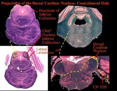
It is Contralateral Only!
CN VIII enters brain at the pontomeduallry junction and synapes on dorsal cochlear nucleus. It then crosses the midline (contralateral) to lateral lemniscus and ascends rostrally and synapses in the chief sensory nucleus of the inferior colliculus (tonotpoically organized) and synapses in the inferior colliculus and leaves thru the brachium of the inferior colliculus going to the medial geniculate nucleus |
|
|
|
Illustrate projections of the Dorsal Cochlear Nucleus from the brachium of the inferior colliculus until its final destination?
|

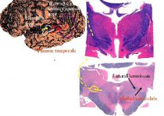
From the brachium of the inferior colliculus, the axons go to the medial geniculate nucleus and the axons synapse at medial geniculate nucleus. Then the medial geniculate projects thru internal capsule of the thalamus to the temporal lobe. In the temporal lobe, the axons go to Heschl’s transvere gyrus (primary auditory cortex on the dorsal surface of the superior temporal gyrus ). Then the axons project further to the planum temporale and then go to Wernicke’s sensory speech area (in order to understand the sounds you hear as words)
|
|
|
|
What is the significance of wernicke’s speech area and • Planum temporale?
|
• For most people Wernicke’s speech area is only on the left side
• Each language has a different interpretation of different tones, so the interpretation of sounds in Wernicke’s area is a learned process. • Planum temporale is an auditory association cortex and a relay between primary auditory cortex and wernicke’s speech area. • A lesion in planum temporale on the left side will not cause deafness (deafness only from CN VIII) but will cause a “word deafness”. You are not deaf; you can hear tones but you cannot comprehend speech because the relay to Wernicke’s area is damaged. |
|
|
|
Describe the ventral pathway?
|

Ventral pathway (ventral cochlear nucleus-trapezoid body-superior olive)
o spatial localization and reflex functions. • Localization of a sound source in the horizontal plane only. • Same functional features as first order relay. • Retains temporal information (spike discharge rate) • Many cells time-locked to the phase of the auditory stimulus • The pathway of axons originating in the ventral cochlear nuclei have many of the same connections as the dorsal nucleus projections, but are bilateral, as shown in the next panel. |
|
|
|
What is the interaural line?
|
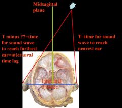
• The interaural line is the line between the two ears
• An auditory stimulus occurs at some point off the midline. It will reach one ear before it reaches the other. • In this picture, the stimulus will reach the right ear before it reaches the left because the right ear is closer to the stimulus. |
|
|
|
What does the head do in relation to stimulus and why?
|

The head rotates to the stimulus so that the sound reaches both ears at the same time and to fix the eyes on the source of the sound.
|
|
|
|
Describe the Projections of the Ventral Cochlear Nucleus until it gets to the superior colliculus?
|
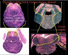
This is Bilateral!
-CN VIII enters at the pontomedullary junction and goes to the ventral cochlear nucleus -The axons from the ventral cochlear nucleus send bilateral projections into the superior olives of the pons -The axons from the superior olives send axons up to the inferior colliculus. -Some of the axons from the inferior colliculus go to the deep layers of the superior colliculus and some go to the medial geniculate nucleus |
|
|
|
Describe the Projections of the Ventral Cochlear Nucleus from the superior colliculus until it gets to its final destination?
|

Some of the axons from the inferior colliculus go to the deep layers of the superior colliculus and some go to the medial geniculate nucleus
From the medial geniculate, the axons go thru the internal capsule to the Heshel’s gyrus on both sides |
|
|
|
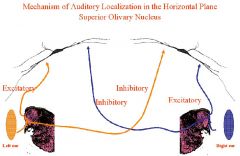
Describe the mechanism of Auditory Localization in the Horizontal Plane of
Superior Olivary Nucleus? |

• There is a reflex of turning the head towards a sudden loud sound. There is also an emotional (fear) response. It a preparation to flee (fight or flight—autonomic response).
• It all begins with CN VIII • The cells at the top of the slide are stained with a Golgi stain: the cell bodies have dendrites that stick out left and right and the are found in the superior olives of the pons • If there is an auditrory stimulus 10 degrees off the midline to the right: the sound will reach the right ear first. The 1st order neurons of the right ear will send projections to the ventral cochlear nucleus. • The ventral cochlear nucleus will send out bilateral projections to the superior olives of the pons. • The ipsilateral projection will go to the lateral dendrites of the superior olive • The contralateral projection will synapse will medial dendrites of the superior olive on the other side. • The contralateral projections are inhibtory. • The ipsilateral projections are excitatory. • These excitatory and inhibitory fibers go to the head turning muscles to make the head turn and localize the sound. • This happens only in the horizontal plane. • The interaural timelag will be longer the further off the midline the auditory stimulus occurs |
|
|
|
1. What will happen to a sudden sound occurring, for example, 4 degrees to the right of the midline?
2. What is the physical basis of the information by which the nervous system computes the angle of the sound from the midsagittal plane? |

1. It will arrive at the two ears at different times and different intensities due to the distance that the head separates the two ears.
2. The interaural time lag and intensity difference of the auditory stimulus is the physical basis of the information by which the nervous system computes the angle of the sound from the midsagittal plane and therefore the amount of stretch of the neck muscles needed to rotate the head exactly 4 degrees to the right and thus visually fixate the stimulus. |
|
|
|
What happens if CN VII from the right ear is activated first?
|

If the right CN VIII is activated first, it goes to the right ventral cochlear nucleus. Its bilateral projections cause excitation of the right superior olive, and inhibition of the left. The excitatory response then continues to the right inferior colliculus and then to the right superior colliculus, cells of the deep layers.
|
|
|
|
What happens to cells of the deep layers of the right superior colliculus?
|
The cells of the deep layers project to the right nucleus ambiguus of the medulla,which in turn projects to the lower motor neurons of the right ventral horns C1-C4 . These activated lower motor neurons cause contraction of the right obliquus capitis inferior, capitis posterior major and splenius capitis muscles, causing the head to rotate to the right.The longer these motor neurons fire and contract these muscles, the more rotation of the head to the right the will result.
|
|
|
|
The amount of rotation of the head is exactly correlated with what?
|
The amount of rotation of the head exactly matches the angle of the auditory stimulus from the midsagittal plane, which in turn is related to the interaural time lag and intensity differences. In the example, when sound waves microseconds later, activate the left auditory system, the inhibitory contralateral projections of the left ventral cochlear nucleus inhibit the right superior olive, thereby ending the excitation that had been going on there that resulted in head rotation to the right. Thus, the amount of head rotation is exactly related to the length of excitation of the superior olive and its rostral projections.
|
|
|
|
1. The sudden occurrence of an auditory event at an angle is proportionaly related to what?
2. What is the mechanical and neural activation of the middle and inner ear related to? 3. Ipsilateral CN VIII does what? 4. Ipsilaterally projecting axon of the ventral cochlear nucleus does what? 5. Ipsilateral sternocleidomastoid muscle does what? 6. What happens when the auditory apparatus contralateral to the sound source is subsequently activated (due to the interaural time lag)? |
1. The sudden occurrence of an auditory event at an angle is prorportional to the midsagittal plane.
2. Mechanical and neural activation of the middle and inner ear ipsilateral to the sound source. 3. Ipsilateral CN VIII projects excitatory relays to the ipsilateral ventral cochlear nucleus and inhibitory ones to the contralateral. 4. Ipsilaterally projecting axon of the ventral cochlear nucleus activate, in sequence, neurons of the ipsilateral superior olive, inferior colliculus, deep layer cells of the superior colliculus,and lower motor neurons of the ventral horns, C1-C4. 5. Ipsilateral sternocleidomastoid muscle contracts and head rotates to fix on location of the auditory stimulus. 6. When the auditory apparatus contralateral to the sound source is subsequently activated (due to the interaural time lag), the superior olive on that side sends contralateral inhibitory axons that shuts off the excitation of the superior olive and its rostral projections that were ipsilateral to the sound source. |
|
|
|
What is the significance of the inferior colliculus and what are 4 of its characteristics?
|

It is where the ventral and dorsal pathways converge. Its characteristics are:
1. It has cells sensitive to amplitude modulation 2. Some of its cells are dependent on interaural time delay but independent of frequency and absolute intensity of the stimulus. 3. Other cells are sensitive to interaural intensity differences. 4. Some of its neurons are sensitive to direction of movement of the auditory stimulus. NOTES: This is a mechanism that works for us. We do not have ears like the rat; we cannot turn our ears towards the stimulus to localize the sound In owls, the ears are in the front of the face above the two eyes. The ears are in two different planes above the eyes, so the owl can distinguish sounds in the vertical plane. Sound waves are interupted differently by the external ear structures This reflex system works only for a brief stimulus (like a click) |
|
|
|
What are 6 characteristics of the primary auditory cortex?
|
1. Tonotopic organization but frequency selectivity is wide
2. Localization, including a few in the median plane 3. Some cells fire only when there is an auditory and visual stimulus with the same receptive fields 4. Some neurons fire only to tones, others only to noise 5. Some fire only to a sound complex (patterning of auditory stimuli) 6. Some cells sensitive to frequency modulation (changes in the frequency of a sound), and some only to ascending, others only to descending changes, and others only to the rate of frequency modulation. |
Please = TLC nTC M
|