![]()
![]()
![]()
Use LEFT and RIGHT arrow keys to navigate between flashcards;
Use UP and DOWN arrow keys to flip the card;
H to show hint;
A reads text to speech;
86 Cards in this Set
- Front
- Back
|
Appendicular Skelton (126) |
____consists of ___ Bones |
|
|
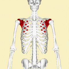
Pectoral (Shoulder) Girdle |
|
|
|
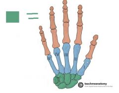
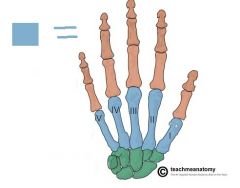
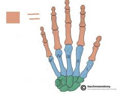
Carpals |
|
|
|
Metacarpals |
|
|
|
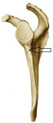
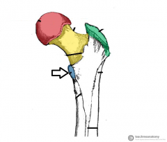
Infraglenoid Tubercle |
|
|
|
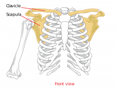
Clavicle |
|
|
|
Scapula |
|
|
|
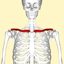
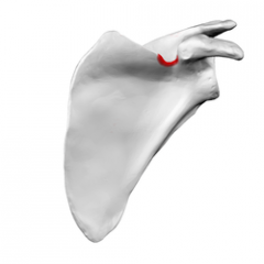
Glenoid Cavity |
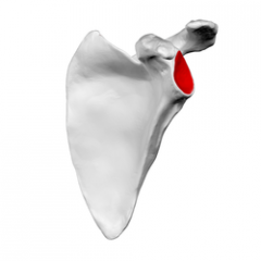
A shallow socket that articulates with the head of the humerus
|
|
|
Spine (Of scapula) |
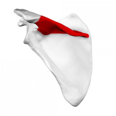
A ridge of bone on the posterior surface that is easily felt through the skin |
|
|
Acromion |
The Lateral end of the spine of the scapula that articulates with the clavicle to form the AC joint
|
|
|
Coracoid Process |
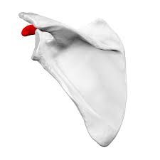
Projects above the glenoid cavity as a hooklike process; helps attach the biceps brachii muscle
|
|
|
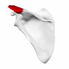
Suprascapular notch |
Small notch located medial to the coracoid process that allows for the passage of blood vessels and a nerve
|
|
|
Phalanges |
|
|
|
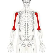
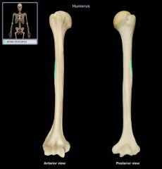
Humerus |
|
|
|
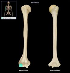
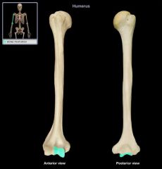
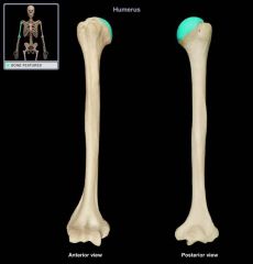
Greater Tubercle |
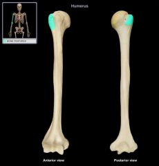
Large lateral prominence; site of the attachment of rotator cuff muscles
|
|
|
Lesser Tubercle |
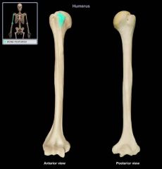
Small medial prominence; site of attachment of rotator cuff muscles
|
|
|
Intertubercular sulcus |
A groove separating the greater and lesser tubercles; the tendon of the biceps brachii lies in this groove
|
|
|
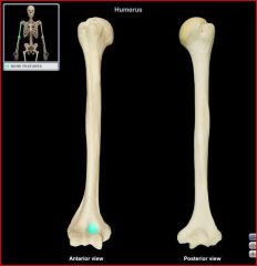
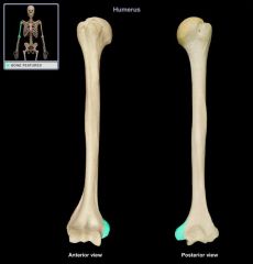
Deltoid Tuberosity |
A roughened area about midway down the shaft of the lateral humerus; site of attachment of the deltoid muscle
|
|
|
Radial Fossa |
Small lateral depression; receives the head of the radius when the forearm is flexed
|
|
|
Coronoid Fossa |
Small medial anterior depression; receives the coronoid process of the ulna when the forearm is flexed
|
|
|
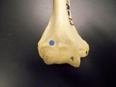
Capitulum |
A rounded lateral condyle that articulates with the radius
|
|
|
Trochlea |
A flared medial condyle that articulates with the ulna
|
|
|
Lateral Epicondyle |
Small condyle proximal to the capitulum
|
|
|
Medial Epicondyle |
rough condyle proximal to the trochlea
|
|
|
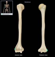
Olcranon Fossa |
Large distal posterior depression that accommodates the olecranon of the ulna
|
|
|
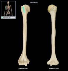
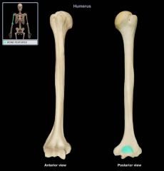
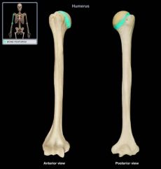
head of Humerus |
|
|
|
Anatomical Neck of Humerus |
|
|
|
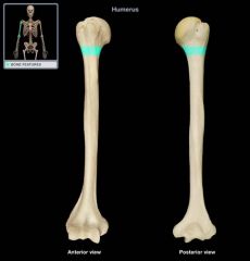
Surgical Neck |
|
|
|
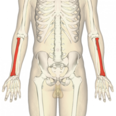
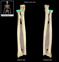
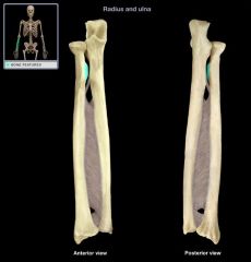
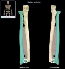
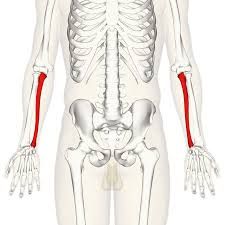
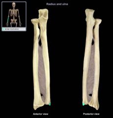
Radius |
|
|
|
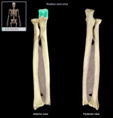
Head of Radius |
Proximal end of the radius that forms part of the proximal radioulnar joint and articulates with the capitulum of the humerus
|
|
|
Radial Tuberosity |
Medial prominence just below the head of the radius; site of attachment of the biceps brachii
|
|
|
Radial Styloid Process |
Distal prominence; site of attachment for ligaments that travel to the wrist
|
|
|
Ulnar Notch |
small distal depression that accomodates the head of the ulna, forming the distal radioulnar joint
|
|
|
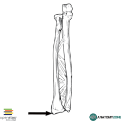
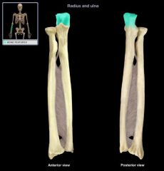
Ulna |
|
|
|
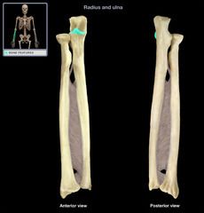
Olecranon |
Prominent process on the posterior proximal ulna; articulates with the olecranon fossa of the humerus when the forearm is extended
|
|
|
Trochlear Notch |
Deep notch that separates the olecranon and the coronoid process; articulates with the trochlea of the humerus
|
|
|
Coronoid Process |
shaped like a point on a crown; articulates with the trochlea of the humerus
|
|
|
Radial Notch |
Small proximal lateral notch that articulates with the head of the radius; forms part of the proximal radioulnar joint
|
|
|
Head of Ulna |
Slim distal end of the ulna; forms part of the distal radioulnar joint
|
|
|
Ulnar Styloid Process |
Distal pointed projection; located medial to the head of the ulna
|
|
|
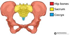
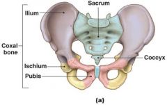
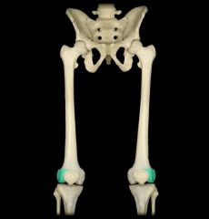
Pelvic (Hip) Girdle |
|
|
|
Ossa Coxae (Coxal bones) |
|
|
|
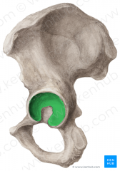
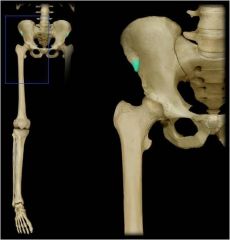
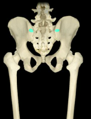
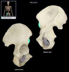
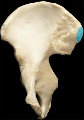
Acetabulum |
|
|
|
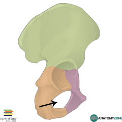
Obturator Foramen |
|
|
|
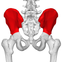
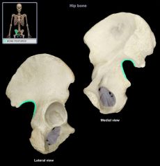
Ilium |
|
|
|
Anterior Superior iliac spine |
The blunt anterior end of the iliac crest
|
|
|
Posterior superior iliac spine |
The sharp posterior end of the iliac crest
|
|
|
Anterior inferior iliac spine |
Small projection located just below the anterior superior iliac spine
|
|
|
Posterior Inferior iliac spine |
Small projection located just below the posterior superior iliac spine
|
|
|
Greater sciatic notch |
Deep notch located inferior to the posterior inferior iliac spine; allows the sciatic nerve to enter the thigh
|
|
|
Iliac fossa |
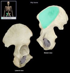
Shallow depression below the iliac crest; forms the internal surface of the ilium
|
|
|
Auricular Surface |
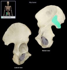
Rough medial surface that articulates with the auricular surface of the sacrum, forming the sacroiliac joint
|
|
|
Sacroiliac Joint |
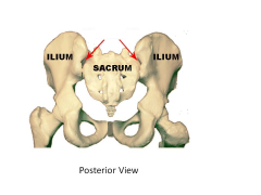
|
|
|
Ischium |
|
|
|
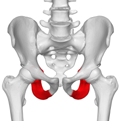
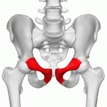
Ischial Tuberosity |
Rough projection that receives the weight of our body when we are sitting
|
|
|
Pubis |
|
|
|
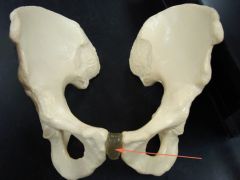
Pubic symphysis |
|
|
|
Patella |
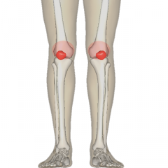
|
|
|
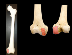
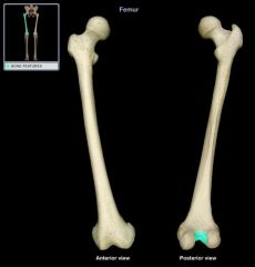
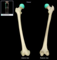
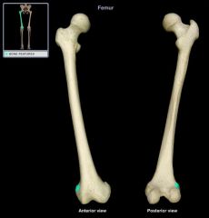
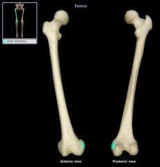
Femur |
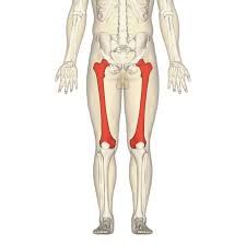
|
|
|
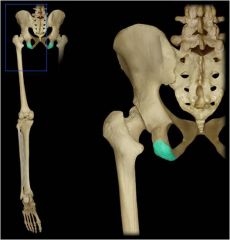
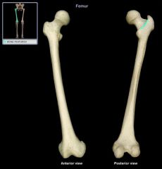
Neck of femur |
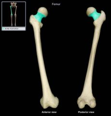
Weakest part of the femur, the usual fracture site of a broken hip
|
|
|
Greater Trochanter |
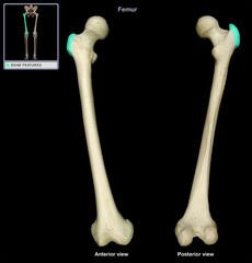
Large lateral projection; serves a site for muscle attachment on the proximal femur
|
|
|
Lesser Trochanter |
Large posteromedial projection; serves a site for muscle attachment on the proximal femur
|
|
|
Intertrochanteric Crest |
Prominent ridge of bone that connects the two trochanters posteriorly |
|
|
Gluteal Tuberosity |
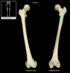
Thin ridge of bone located posteriorly; serves as a site for muscle attachment on the proximal femur
|
|
|
Linea Aspera |
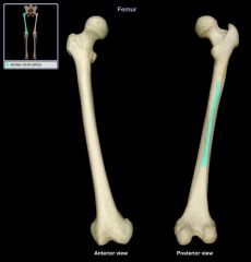
Long vertical ridge of bone on the posterior shaft of the femur
|
|
|
Medial condyle |
Distal "wheel shaped" projections that articulate with the tibia, each condyle has a corresponding epicondyle
|
|
|
Lateral Condyle |
Distal "wheel shaped" projections that articulate with the tibia, each condyle has a corresponding epicondyle
|
|
|
Intercondylar Fossa |
Deep depression located between the condyles and beneath the popliteal surface
|
|
|
Head of Femur |
|
|
|
Lateral Epicondyle |
|
|
|
Medial Epicondyle |
|
|
|
Patellar surface |
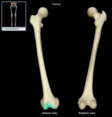
Smooth distal anterior surface between the condyles; articulates with the patella
|
|
|
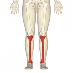
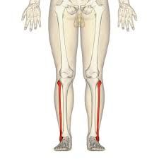
Tibia |
|
|
|
Lateral condyle |
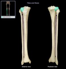
slightly concave surface that articulates with the lateral condyle of the femur; the inferior region of this condyle articulates with the fibula to form the superior tibiofibular joint
|
|
|
Medial Condyle |
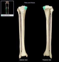
Slightly concave surface that articulates with he medial condyle of the femur
|
|
|
Intercondylar eminence |

Irregular projection located betweent he two condyles
|
|
|
Tibial tuberosity |
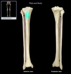
Roughened anterior surface; site of patellar ligament attachment
|
|
|
Anterior Border |
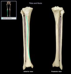
Sharp ridge of bone easily palpated because it is close to the surface
|
|
|
Medial Malleolus |
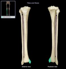
forms the medial bulge of the ankle
|
|
|
Fibula |
|
|
|
Head of Fibula |

Proximal end of the fibula that articulates with the tibia to form the inferior tibiofibular joint |
|
|
Lateral Malleolus |
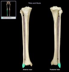
Forms the lateral bulge of the ankle and articulates with the talus
|
|
|
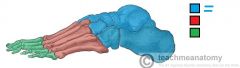
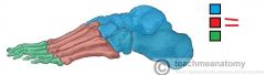
Tarsal |
|
|
|
Metatarsals |
|
|
|
Phalanges |

|
|
|
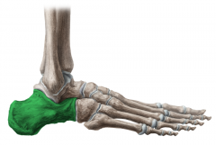
calcaneus |
|