Reading...
![]()
Play button
![]()
Play button
![]()
Use LEFT and RIGHT arrow keys to navigate between flashcards;
Use UP and DOWN arrow keys to flip the card;
H to show hint;
A reads text to speech;
181 Cards in this Set
- Front
- Back
|
Distinguish between sensory input, integration and motor output.
|
The nervous system is the fundamental control system of the body. It takes in information, processes the information and elicites commands of action that are carried out by muscles or glands.
- sensory input - to monitor what goes on in the external environment as well as within the body, the nervous system gathers sensory information called sensory input. This input begins in sensory recepts which are specialized cells located outside the central nervous system in the periphery that transduce some form of energy (e.g. mechanical, electromagnetic or chemical) that originates inside or outside the body. Sensory neurons carry this information into the central nervous system - integration - to intepret the sensory information and select an appropriate response to the stimulus, the information must be processed. The processing of sensory information and the selection of a response is called integration and occurs entirely in the central nervous system - motor output - once the information is processed, the nervous system elicits a response by activating an effector organ, which may be either muscles or glands. This response is called motor output. Motor output originates in the central nervous system, but the response is elicited by actions of the periphery All three basic functions (sensory input, integration and motor output) result from electrictal signals in cells called neurons which are sometimes referred to as nerve cells. Integration results when electrical signals are transferred among neurons at sites called synapses |
|
|
Relate the terms "sensory input", "integration" and "motor output" to sensory neurons, association neurons, interneurons and motor neurons.
|
- sensory neurons - bring information to the central nervous system about the skin, skeletal muscles and joints. Because this information is coming from the body, the nerve cells carrying the information are called somatic afferents
- interneurons - important for processing sensory (afferent) information coming from the periphery and going into the central nervous system - motor neurons - the motor division is also called the efferent (from the latin meaning to bring out) division |
|
|
Distinquish between the central nervous system and the peripheral nervous system.
|
- central nervous system - consisting of the brain and spinal cord
- peripheral nervous system - consisting of the portion of the nervous system located outside of the central nervous system Much of the peripheral nervous system consists of nerves that carry both sensory and motor information |
|
|
Describe where the neurons in the central and peripheral nervous systems are located and relate their positions to the concept of the central and peripheral nervous systems.
|
a
|
|
|
Define afferent and efferent and relate these terms to the concept of the central and peripheral nervous systems.
|
Both may be found in the CNS and the PNS:
- afferent - (latin: to carry forward) sensory - sending to - efferent - motor - to carry away |
|
|
Define visceral and somatic.
|
- visceral - pertaining to the soft internal organs of the body, especially those contained within the abdominal and thoracic cavities
- somatic - of or relating to the portion of the vertebrate nervous system that regulates voluntary movement |
|
|
Define caudal with respect to the central nervous system.
|
relatively distant from the forehead, especially in reference to the brain and spinal cord
|
|
|
What branches of the nervous system effect function in the visceral and somatic regions?
|
a
|
|
|
Define dorsal with respect to the central nervous system.
|
toward the back (spinal side) of the body
|
|
|
Within the visceral branch, distinguish between sympathetic and parasympathetic nervous systems.
|
the autonomic nervous system is subdivided into two systems:
- sympathetic - tends to increase the activity of an organ - parasympathetic - tends to decrease activity |
|
|
Define ventral with respect to the central nervous system.
|
pertaining to the front of the body, the regions of the chest and abdomen
|
|
|
Define rostral with respect to the central nervous system.
|
relatively close to the forehead, especially in reference to structures of the brain and spinal cord
|
|
|
Name the four major divisions of the brain and briefly describe their function.
|
a
|
|
|
Draw an outline of the spinal cord and label the gray and white matter. Draw the dorsal and ventral horns. Show which regions are sensory and which are motor.
|
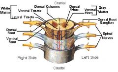
a
|
|
|
What is gray matter? What is white matter? What is the function of each?
|
- gray matter - is a collection of cell bodies and unmyelinated processes
- white matter - comprised of myelinated axons - is white because myelin contains large amounts of lipid that acts as an electrical insuling layer of the axon |
|
|
Trace the pathway of sensory information from the periphery to association neurons in the central nervous system to motor output.
|
1. sensory recepts initiate a response in the sensory neuron that conducts toward the central nervous system in the spinal cord
2. the sensory information enters the central nervous system through the dorsal root and the central process of the dorsal root ganglion neuron extends into the dorsal horn of the spinal cord 3. for one form of sensory information (light touch or joint position) the snesory neuron travels rostrally in the spinal cord and forms a synpase in the brain stem where it communicates with an interneuron that sends a process to the thalamus. in the thalamus, this neuron synapses onto nerve cells that extend process to the cerebral cortex. specifically, these thalamic neurons send processes to the primary samatosensory cortex which is located in the rostral portion of the parietal lob, just caudal to the central sulcus 4. association neurons (also known as interneurons) send processes from the sensory cortex to the association areas in the cerebral cortex of the frontal lobe. the association areas interpret the sensory information and send information to the motor and premotor areas 5. upper motor neurons are located in the primary motor cortex and receive information from the association regions. upper motor neurons extend descending procersses carrying efferent information down the corticospinal tracts to synapse on the lower motor neurons 6. activation of the lower motor neurons leads to the contraction of muscles and the initiation of movement remember that afferent information entering the right hand side of the cord will make its way to the left cerebral hemisphere, and that efferent information from the motor cortex in the left cerebral hemisphere will make its way back to the right side of the spinal cord |
|
|
What are the parts of the neuron in the Bodian classification of the neuron?
|
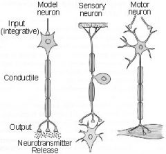
According to the Bodian classification of neuron structure, neurons have three functionally similar regions in common:
1. an input region 2. a conducting region 3. a secretory region |
|
|
Wha is the function of each of the regions of the Bodiean classification of the neuron?
|
According to the Bodian classification of neuron structure, neurons have three functionally similar regions in common:
1. an input region sometimes referred to as a receptive or dendritric region 2. a conducting region that carries action potentials. this is the long distance communication part of the cell 3. a secretory region sometimes called the output region where neurotransmitter is released |
|
|
Where does protein synthesis occur in neurons?
|
The cell body is the protein factory of the neuron. The cell body contains the nucleus and most of the protein synthesis in the neuron occurs in the cell body. There is some protein synthesis in the dendrites, but very little in the axons. The Nissl substance is characteristic of neurons and is an elaborated form of the rough ER
|
|
|
How do proteins synthesized in the cell body find their way to the axon terminal?
|
a
|
|
|
Describe the structure of the axon. Compare the organization of a myelinated and unmyelinated axon.
|
a
|
|
|
Describe the structure of the axon terminal. What are synaptic vesicles? What are contained in synaptic vesicles?
|
a
|
|
|
How do the synaptic vesicles release their contents?
|
a
|
|
|
Describe the function and location of motor neurons, sensory neurons and interneurons. Where are the cell bodies for these neurons located?
|
a
|
|
|
Define afferent and efferent in terms of motor neurons and sensory neurons.
|
a
|
|
|
What are oligodendrocytes, where are they found, and what is their function?
|
- in the central nervous system are responsible for forming the myelin sheaths around axons
- myelin forms the insulating layers around neurons so that the action potentials can conduct more rapidly - are responsible for forming the white matter in the central nervous system - they can envelope several axons and wrap them with myelin - myelination proceeds when processes of oligodendrocytes wrap around the axon foring layer upon concentric layer of myelin |
|
|
What are astrocytes, where are they found, and what is their function?
|
Astrocytes are important in regulating the concentration of ions, particularly K+.
Because the volume of extracellular space is small in the central nervous system, electrical activity in cells can change the local concentration of extracellular ions; one of the roles of astrocytes is to regulate the ion concentrations by pumping ions. Astrocytes are also thought to be important in regulating the concentration of neurotransmitters in the extracellular space since neuronal activity can also lead to increases in the extracellular concentration of neurotransmitters. The position of astrocytic processes wrapped around capillaries indicates that these cells are important in regulating the movement of substances between the central nervous system and the blood |
|
|
What are microglia, what is their origin, and what is their function?
|
Cells that are thought to be modified macrophages that are derived from a white blood cell called a monocyte.
They do not originate from neuroectodermal cells as do all neuronal and glial cells. |
|
|
What are Schwann cells, what is their function, where are they found, and how are they different from oligodendrocytes?
|
Schwann cells are the myelin producing cells in the peripheral nervous system.
When Schwann cells myelinate an axon, they envelope a single axon. They extend multiple wraps around the axon to form numerous layers of myelin. They clean up debris after peripheral nerve damage and support regeneratio of peripheral nerves. This is in contrast to eoligodendrocytes which inhibit regeneration of the central nervous system |
|
|
Distinguish between cations and anions. What is the charge of each type, and give examples of each.
|
a
|
|
|
Distinguish between permeant ions and impermeant ions in a neuron.
|
a
|
|
|
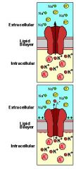
In a neuron at rest, how are the ions in a neuron distributed? Where is the concentration of Na+ highest? Where is the concentration of K+ highest? Where is the concentration of impermeant anions highest?
|
a
|
|
|
What is the relationship between the impermeant anions and K+?
|
See membrane potential generation
|
|
|
What is meant by a Na+ selective ion channel? What is meant by a K+ selective ion channel?
|
a
|
|
|
Define a leak channel. Define a voltage-gated channel.
|
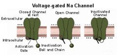
- leak channel - a K+ channel that is always open
- voltage-gated channel - charge-sensitive channels - local increase in + charge causes channel to open; this is usually a cascading effect |
|
|
How do the K+ leak channels and the impermeant anions serve to separate charge?
|
a
|
|
|
What is the relationship between the separation of charge and the formation of a potential?
|
a
|
|
|
What is the unit of measure for the separation of charge?
|
a
|
|
|
Define a voltage-gated channel and contrast it with a leak channel.
|
See action potential gradient diagram
|
|
|
How does the voltage-gated Na+ channel open?
|
a
|
|
|
What moves through the voltage-gated Na+ channel?
|
a
|
|
|
Define electrical current in terms of the open channel and the voltage.
|
a
|
|
|
What is the effect on the membrane potential when the channel opens and Na+ ions flow through it?
|
a
|
|
|
Why does the Na+ current stop flowing?
|
a
|
|
|
How is the potential across the membrane restored to rest? Describe the role of the voltage-gated K+ channel and the K+ current.
|
a
|
|
|
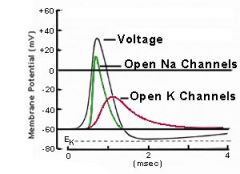
Graph the change in voltage versus time during an action potential. Relate the opening of voltage-gated Na+ and K+ channels to the following terms:
- threshold - rising phase - Na+ current - Na+ channel closing - K+ channel opening - falling phase - K+ current - undershoot |

- threshold - balance between Na+ influx and K+ efflux
|
|
|
What makes up the CNS?
|
1. spinal cord
- contained within the vertebrae 2. brain stem - extension of the brain - controls movement in the head and neck - receives sensory input from the head and neck - plays a role in governing efferents - plays a role in monitoring visceral afferents 3. diencephelon a. thalamus - serves as sensory relay site b. hypothalamus - plays a role in regulation of body function (hungry, thirsty, hot, cold, etc.) - regulation of hormonal release from adenohypophesis 4. cerebrum - cerebral cortex (outer shell) - composed of six layers of neurons |
|
|
Diagram the nervous system as a machine.
|
input -> analysis -> output
(sensory) (association (motor) functions) |
|
|
Define sensory transduction
|
conversion of one form of energy to another. for example, conversion of magnetic energy to cellular energy (movement of ions across biological membranes)
|
|
|
What are the three forms of energy?
|
1. electromagnetic (light)
2. mechanical (touch, hearing, etc.) 3. chemical (taste, smell) |
|
|
Define afferents
|
to carry forward - sending information into the CNS
sensory: - somatic afferents (from body - concious) - visceral afferents (interal organs - unconcious) |
|
|
Define efferents
|
to carry away from (effect - to carry a response)
motor: - somatic efferents - body muscles - concious - visceral efferents - unconcious muscle movement |
|
|
Describe the basic diagram of CNS vs. PNS
|
sensory -> CNS -> motor
(afferents) (efferents) CNS - brain - spinal cord PNS - sensory (afferents) - motor (efferents) |
|
|
Define neuron doctrine
|
the nervous system is a collection of separate cells connected and their chemical synapses
|
|
|
Define the cells of the nervous system
|
1. neurons
- electrically active cells 2. glia - once thought to be just glue - support cells a. astrocytes - responsible for regulating ion concentrations in the space around neurons - responsible for regulating concentration of neurotransmitters around neurons b. oligodendrocytes - produce myelin - the insulating wrap around neurons c. "microglia" - actually macrophages and not glia - cells of the immune system d. Schwann cells - glial cells in the pheripheral nervous system |
|
|
Describe axoplasmic transport
|
the long-distance movement of proteins and memory
a. package protein in vesicles b. transport vesicles on microtubules |
|
|
What are microtubles made of?
|
polymers of a protein called tubulin
|
|
|
Describe the anatomical organization of a generic neuron.
|
Label the:
- dendrite - cell body - axon - conductile region - input region - output region - axon terminal - secretory region - synaptic vesicles |
|
|
Define passive membrane
|
a membrane that lacks voltage-gated channels
|
|
|
Define myelin
|
the insulating layer around axons
|
|
|
List some characteristics of astrocytes
|
(glial cells) maintain a favorable environment for neurons
1. maintain a constant ion concentration in the extracellular space (act as K+ sponges) 2. take up excess neurotransmitter (act as neurotransmitter sponges) |
|
|
Define microglial
|
(macrophages)
- dendrite cell (cell of the immune system) - extension of the immune system in the central nervous system |
|
|
Where do glial cells occur?
|
- glial cells are found in the central nervous system
- in the peripheral nervous system, glial cells are called Schwann cells |
|
|
Define voltage gate
|
a voltage gate is a charge-sensitive channel
a local increase in positive charge causes the channel to open |
|
|
What are the neurotransmitters that open ligand-gated ionotropic channels
|
1. acetylcholine
- neuromuscular junction - autonomic nervous system - approximately 8 receptors 2. glutamate - most abundant neurotransmitter in the CNS - 12 receptors 3. serotonin |
|
|
What is an EPSP?
|
excitatory postsynaptic potential
|
|
|
What is an IPSP?
|
inhibitory postsynaptic potential
|
|
|
What is a trigger zone?
|
a cluster of Na+ and K+ voltage-gated channels
|
|
|
Define synaptic integration
|
EPSPs and IPSPs work together to permit a decision by the neuron to fire an action potential
|
|
|
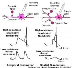
Define spatial summation
|
the summation of synaptic potentials from separated sites on the input region
|
|
|
Define ganglion
|
collection of neuronal cell bodies in the peripheral nervous system
|
|
|
Describe what becomes of current (ions) injected into a cylinder of axoplasm surrounded by axon membrane at the site of current injection, and at 1, 3 and 5 millimeters away from the injection site. Assume that there are no voltage-gated channels in the membrane.
|
a
|
|
|
Describe what becomes of current (ions) injected into a cylinder of axoplasm surrounded by axon membrane at the site of current injection, and at 1, 3 and 5 millimeters away from the injection site. Assume that there are no voltage-gated channels in the membrane. Frame these answers in terms of voltage, current and resistance.
|
a
|
|
|
Compare the electrical events in a cylinder of axoplasm and axon membrane which lacks voltage-gated ion channels to a leaky garden hose.
|
a
|
|
|
Describe the distribution of voltage-gated Na+ and K+ channels along the length of an unmyelinated axon.
|
a
|
|
|
Describe propagation of the action potential along the length of an unmyelinated axon.
|
See propagation of unmyelinated neurons
|
|
|
Describe the structure of a myelinated axon. What are nodes of Ranvier and internodal regions? What cell types are involved in myelination in the central and peripheral nervous systems?
|
a
|
|
|
Compare the distribution of voltage-gated Na+ and K+ channels along the length of a myelinated and unmyelinated axon.
|
a
|
|
|
Compare the propagation of action potentials in myelinated and unmyelinated axons.
|
See propogation of myelinated neurons
|
|
|
Why does myelinated increase the rate of action potential propagation along the axon?
|
a
|
|
|
What is the term that describes the rate of propogation?
|
a
|
|
|
Using a sensory neuron as an example, describe how a generator potential is initiated.
|
a
|
|
|
Describe how the generator potential leads to the production of an action potential. In your describe, include the concepts of stretch-activated channel, graded potential, trigger zone, voltage-gated Na+ channels and threshold.
|
a
|
|
|
What are the differences between passively propagated potentials (graded potentials) and action potentials?
|
a
|
|
|
Describe how graded potentials add together (summate) at the trigger zone to initiate an action potential.
|
a
|
|
|
What is the literal definition of synapse?
|
a
|
|
|
Define presynaptic cell
|
a
|
|
|
Define postsynaptic cell
|
a
|
|
|
Define synaptic vesicle
|
a
|
|
|
Define exocytosis
|
a
|
|
|
Define neurotransmitter
|
a
|
|
|
Define voltage-gated Ca2+ channel
|
a
|
|
|
Define neurotransmitter release
|
a
|
|
|
Define ligand-gated channel
|
a
|
|
|
Define postsynaptic potential
|
a
|
|
|
Make a drawing of a chemical synapse. Show the position of each of the following structures:
- presynaptic cell - postsynaptic cell - synaptic vesicle - exocytosis - neurotransmitter - voltage-gated Ca2+ channel - neurotransmitter release - ligand-gated channel - postsynaptic potential |
a
|
|
|
List in chronological order the sequence of events from the arrival of an action potential in the teldendrion to the release of the neurotransmitter.
|
a
|
|
|
What is the role of intracellular Ca2+ in neurotransmitter release?
|
a
|
|
|
What is the source of this Ca2+ and how does it get into the terminal?
|
a
|
|
|
How is the synaptic vessicle membrane retrieved following exocytosis?
|
a
|
|
|
Define ligand
|
a
|
|
|
Define receptor
|
a
|
|
|
Define ligand-gated channel
|
a
|
|
|
Define ionotropic receptor
|
a
|
|
|
Define excitatory postsynaptic potential (EPSP)
|
a
|
|
|
What does the neurotransmitter bind to on the postsynaptic cell?
|
a
|
|
|
Describe the effect on the membrane potential in the postsynaptic cell when neurotransmitter binds to a receptor that opens a cation channel.
|
a
|
|
|
Describe the relationship between the amplitude of the EPSP and the number of ionotropic channels that are opened by neurotransmitter.
|
a
|
|
|
Why are these postsynaptic potentials said to be graded?
|
a
|
|
|
Related the initiation of EPSPs in the dendritic region of the neuron with the initiation of action potentials at the trigger zone in the axon.
|
a
|
|
|
What is an inhibitory postsynaptic potential?
|
a
|
|
|
How is an inhibitory postsynaptic potential generated?
|
a
|
|
|
What are the inhibitory neurotransmitters?
|
a
|
|
|
When an inhibitory transmitter binds to the ligand-gated receptor, what is the charge on the ion that usually flows through these channels?
|
a
|
|
|
How do EPSPs and IPSPs interact at the trigger zone to affect action potential generation?
|
a
|
|
|
Give two examples of inhibitory neurotransmitters that open ionotropic anion channels.
|
a
|
|
|
Describe how EPSPs summate to elicit an action potential.
|
a
|
|
|
Describe what hapens to the amplitude of an EPSP between the time when it is initiated in the dendritic region of a neuron and when it arrives at the trigger zone.
|
See membrane properties influnce action potential generation
|
|
|
Describe spatial summation.
|
a
|
|
|
Describe the effects of inhibition on spatial summation.
|
a
|
|
|
Draw a spinal cord in cross section showing dorsal, ventral, medial and lateral orientations.
|
See spinal cord organization
|
|
|
Draw a spinal cord in cross section labeling the gray matter.
|
a
|
|
|
Draw a spinal cord in cross section labeling the white matter.
|
a
|
|
|
Describe why the gray matter is gray and the white matter is white.
|
a
|
|
|
Draw a spinal cord in cross section placing the dorsal root ganglion in the proper place.
|
a
|
|
|
What types of neurons have their cell bodies located in the dorsal root ganglion? Where is the input region of these neurons? Where is the output region of these cells? Describe action potential initiation and conduction in these neurons.
|
a
|
|
|
Draw a spinal cord in cross section labeling the dorsal and ventral horns.
|
a
|
|
|
What types of neurons have their cell bodies in the dorsal horn?
|
a
|
|
|
The cell bodies of what types of neurons are located in the ventral horn?
|
a
|
|
|
Draw a spinal cord in cross section labeling the dorsal and ventral roots. What type of information is carried in each of these structures?
|
a
|
|
|
What is the spinal nerve and what type of information is carried in spinal nerves?
|
a
|
|
|
Define proprioception.
|
a
|
|
|
Where is the input zone for the sensory neurons that are responsible for initiating the reflex arc?
|
see reflex arc
|
|
|
How is the action potential in the stretch receptor neuron initiated?
|
see neuron structure
|
|
|
What type of neuron does the sensory neuron synapse onto, and where in the spinal cord is this synapse located? What is the effect of bringing this neuron to threshold? Where does this neuron form synapses?
|
a
|
|
|
What type of interneuron does the sensory neuron innervate in the spinal cord? What neurons does this interneuron innervate?
|
a
|
|
|
Describe the reflex arc of the patellar tendon reflex from its initiation in the muscle spindle to the response in the extensor (quadriceps) and flexor (hamstring) muscles.
|
a
|
|
|
Why is the autonomic system called the involuntary nervous system?
|
a
|
|
|
What are the two divisions of the autonomic nervous system?
|
see overview of autonomic nervous system
|
|
|
What is meant by the statement that the sympathetic brance of the autonomic nervous system is responsible for the flight or fright response? What responses are typical of activing this system?
|
a
|
|
|
How do the sympathetic responses compare with the responses of the parasympathetic nervous system?
|
a
|
|
|
What is the function of the preganglionic neuron in the automnomic nervous system?
|
a
|
|
|
What is the function of he postganglionic neuron in the autonomic nervous system?
|
a
|
|
|
How are the preganglionic neurons in the sympathetic and parasympathetic brances of the autonomic nervous system similar? How are they different?
|
see overview of autonomic nervous system
|
|
|
Where are the cell bodies for the preganglionic sympathetic neurons located
|
see gross anatomy of the sympathetic system
|
|
|
Where are the cell bodies for the postganglionic sympathetic neurons located?
|
a
|
|
|
Where are the cell bodies for the preganglionic parasympathetic neurons located?
|
a
|
|
|
Where are the cell bodies for the post ganglionic parasympathetic neurons located?
|
a
|
|
|
Where are the synapses between pre and post ganglionic neurons in the sympathetic system?
|
a
|
|
|
Where are the synapses between pre and post ganglionic neurons in the parasympathetic system?
|
a
|
|
|
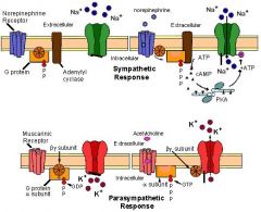
What is the neurotransmitter at the synapse between the preganglionic and post ganglionic neurons in both brances of the autonomic nervous system?
|
see autonomic transmitter and receptors
|
|
|
What is the neurotransmitter in the postganglionic sympathetic neurons?
|
a
|
|
|
What is the receptor for the neurontransmitter in the post ganglionic sympathetic neurons?
|
a
|
|
|
What is the neurotransmitter in the postganglionic parasympathetic neurons?
|
a
|
|
|
What is the receptor for the neurotransmitter in the postganglionic parasympathetic neurons?
|
a
|
|
|
Describe the effects of the sympathetic and parasympathetic postganglionic neurotransmitters on their respective receptors.
|
a
|
|
|
How are the sympathetic and parasympathetic postganglionic neurotransmitters receptors different from the ionotropic recepts?
|
a
|
|
|
Define GTP.
|

see autonomic metabotropic recepts
|
|
|
Define GTP binding protein.
|
see autonomic metabotropic recepts
|
|
|
Define adenelyl cyclase.
|
see autonomic metabotropic recepts
|
|
|
Define cyclic AMP.
|
see autonomic metabotropic recepts
|
|
|
Define protein kinase A.
|
see autonomic metabotropic recepts
|
|
|
Define protein phosphorylation.
|
see autonomic metabotropic recepts
|
|
|
Define phosphatase.
|
see autonomic metabotropic recepts
|
|
|
Define protein dephosphorylation.
|
see autonomic metabotropic recepts
|
|
|
Using the terms below, describe the sequence of events between the binding of neurotransmitter to a metabotropic receptor and the opening of a channel:
- GTP - GTP binding protein - adenelyl cyclase - cyclic AMP - protein kinase a - protein phosphorylation - phosphatase - protein dephosphorylation |
see autonomic metabotropic receptors
|
|
|
How do ionotropic receptors respond when they bind to neurotransmitter?
|
a
|
|
|
Describe how sensory information from the baroreceptors in the carotid arteries affects the heart beat.
|
a
|
|
|
If there is an increase in stretch, what information is fed through the Nucleus of the Solitary Tract to reduce the heart rate and strength of contraction?
|
a
|
|
|
What branch of the autonomic nervous system is used to reduce the heart rate?
|
a
|
|
|
If the stretch on the baroreceptors is reduced, what brance of the autonomic nervous system is used to increase the strength of contraction?
|
a
|
|
|
Information from the sensory cortex along with centers deep in the brain including the basal nuclei initiate activity in the premotor regions of the brain.
|
a
|
|
|
The premotor regions in the frontal cortex appear to be important for planning the execution of movement.
|
a
|
|
|
Action potentials from neuron in the prefrontal cortex convey information to the motor cortex.
|
a
|
|
|
How is the homunculus arranged in the motor cortex?
|
see somatotopy in the motor cortex
|
|
|
What are the neurons in the motor cortex called that initiate the movement?
|
a
|
|
|
What is the name of the descending tract that contains the axons of neurons that synapse onto lower motor neurons?
|
see motor cortex and the corticospinal tract
|
|
|
Where do the axons of neurons that synapse onto the lower motor neurons decussate?
|
a
|
|
|
What are the pyramids and where are they located?
|
a
|
|
|
Related the homunculus of the motor cortex to the general position in the spinal cord of the targets for the upper motor neurons.
|
a
|
|
|
Discribe how activation of an upper motor neuron leads to the activation of a lower motor neuron.
|
a
|