![]()
![]()
![]()
Use LEFT and RIGHT arrow keys to navigate between flashcards;
Use UP and DOWN arrow keys to flip the card;
H to show hint;
A reads text to speech;
67 Cards in this Set
- Front
- Back
|
What is an Articulation? |
An articulation, or joint, is the place where bone meets another bone, cartilage, or teeth. They vary in stability and mobility and are classified into categories based on their joint structure and their mobility |
|
|
Fibrous Structure |
Bones are held together by dense regular connective tissue |
|
|
Cartilaginous Structure |
Bones joined by cartilage |
|
|
Synovial Structure |
Bones separated by fluid filled cavity. Most joints. |
|
|
Function of Synarthrosis |
Immobile joint |
|
|
Function of Amphiarthrosis |
Slightly mobile joint |
|
|
Function of Diarthrosis |
Freely mobile joint |
|
|
How are the fibrous joint categorized? |
Gomphosis Suture And syndesmosis |
|
|
Gomphosis |
Is a fibrous structure. Peridoral membranes hold tooth to bony jaw. Example of this joint is tooth to jaw. It is Synarthrosis (immobile) |
|
|
Suture |
It is a fibrous joint. Dense regular connective tissue connects skull bones. An example is the lamboid suture, which connects the occipital and parietal bones. Is Synarthrosis, immobile. |
|
|
Syndesmosis |
A fibrous joint. Dense regular connective tissue fibers between the bones. Examples is the articulation between the radius and ulna, and tibia and fibula. Is Amphiarthrosis, slightly mobile. |
|
|
How are the cartilaginous joints categorized? |
Synchondrosis and Symphysis |
|
|
Synchondrosis |
Part of the cartilaginous structure. When there's hyaline cartilage between bones. Found in the epiphyseal plates in growing bones, and the costochondral joints. Is Synarthrosis, immobile. |
|
|
Symphysis |
One of the cartilaginous structures. It is a fibrocartilage pad between bones. Examples are the pubic symphysis and intervertebral disc articulations. Are Amphiarthrosis, slightly mobile. |
|
|
How are synovial joints categorized? |
Most joints are synovial so they can be categorized several ways and are all Diarthrosis or freely mobile. If they are uniaxial, they are either a plane joint, a hinge joint, or a pivot joint. If they are biaxial, they are either condylar or saddle. If they are multiaxial, they are ball and socket joints. |
|
|
Uniaxial Joints |
Plane joint, hinge joint, and pivot joint |
|
|
Plane joint. |
Is a uniaxial synovial joint. Is a flattened or slightly curved face that slides across one another. Examples are the intercarpal joints and intertarsal joints. Are Diarthrosis or freely mobile. |
|
|
Hinge Joint |
Is a uniaxial synovial joint. Where a convex feature of one bone fits into concave depression of another bone. Examples are elbow joint, knee joint, and IP (interphalangeal) joints. Are Diarthrosis or freely mobile |
|
|
Pivot Joint |
Is a uniaxial synovial joint. Bones with rounded surfaces fits into a ring formed by a ligaments and another bone. Example is at the atlantoaxial joint. Is Diarthrosis or freely mobile. |
|
|
Biaxial joints |
Condylar and saddle |
|
|
Condylar joint |
Is a biaxial synovial joint. Oval articular surface on one bone closely interfaces with a depressed oval surface on another bone. Examples are MP(metacarpophalangeal or metatarsophalangeal) joints. Is Diarthrosis or freely mobile. |
|
|
Saddle joint |
Is a biaxial synovial joint. Is a Saddle shaped articular surface on one bone closely interfaces with a saddle-shaped surface on another bone. Is found at the articulation between carpal and first metacarpal bone. Is Diarthrosis or freely mobile. |
|
|
Ball and socket joint |
Is the only multiaxial synovial joint. When a rounded head of one bone rests within a cup-shaped depression in another bone. Examples are the glenohumeral joint and hip joint. Is Diarthrosis or freely mobile |
|
|
General anatomy of Synovial joints |
Articular capsule, articular cartilage, joint cavity, ligaments, sensory nerves, and blood vessels. There are also accessory structures that surround the joint thay function in reducing friction and filling spaces, called the bursae, tendon sheaths and fat pads. |
|
|
Articular Capsule (general anatomy of Synovial joints) |
Has two layers. The other fibrous layer which is made of dense regular connective tissue, it strengthens the joint. The inner synovial membrane secretes the synovial fluid. |
|
|
Articular Cartilage (general anatomy of Synovial joint) |
Articular surfaces in synovial joints are covered by hyaline cartilage, it reduces friction and acts as a shock absorber. |
|
|
Joint Cavity (general anatomy of Synovial joints) |
The space between articulating bones. Contains a small amount of Synovial fluid that lubricates and nourishes (and the chondrocytes) of the articular cartilage. Absorbs shock during compression of the joint. |
|
|
Ligaments (general anatomy of Synovial joints) |
Connect bone to bone, is made of dense regular connective tissue. Strengthen and reinforces the capsule. |
|
|
Sensory nerves (general anatomy of Synovial joints) |
Detects pain and amount of stretch in a joint |
|
|
Blood vessels (general anatomy of a synovial joint) |
Nourish tissues in the joint |
|
|
Bursae |
Sacs outside most synovial joints where ligaments, muscles, tendons and/or bones rub. Contains synovial fluid. |
|
|
Tendon sheaths. |
Elongated bursae around tendons, particularly in confined areas (wrist and ankle) where tendons rub each other |
|
|
Fat pads |
Packing material, also provides some protection |
|
|
Synovial joints movements |
Gliding motion, angular motion, rotational motion, and special movements |
|
|
Gliding motion |
When two opposing articular surfaces slide past each other in almost any direction, the amount of movement is slight. Slides back and forth and side to side. Angle between bones does not change. Occurs mainly in plane joints. |
|
|
Angular motion |
Increases or decreases the angle between bones. Seven different types: flexion, extension, hyperextension, lateral flexion, abduction, adduction, circumduction |
|
|
Flexion |
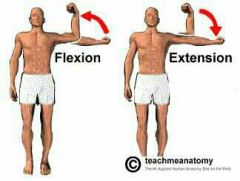
The angle between articulating bones decreases |
|
|
Extension |

An angular motion. The angle between articulating bones increase. |
|
|
Hyperextension |

Extension movement continues past the anatomic position. Is a angular movement. |
|
|
Lateral Flexion |

The vertebral column moves (bends) in a lateral direction along a coronal plane. |
|
|
Abduction |

Movement away from the midline. An angular movement. |
|
|
Adduction. |

Movement of a bone toward the midline. An angular movement. |
|
|
Circumduction |

A continuous movement that combines flexion, abduction, extension, and adduction in succession. The distal end of the limb or digital movies in a circle. An angular movement. |
|
|
Pronation |

Rotation of the forearms where the palm is turned posteriorly. A rotational movement. |
|
|
Supination |

Rotation of the forearms in which the palm is turned anteriorly. A rotational movement. |
|
|
Depression (movement) |

Is a special movement. Movement of a body part inferiorly. |
|
|
Elevation |

A special movement. Movement of a body part superiorly. |
|
|
Dorsiflexion |

Ankle joint movement where the dorsum(superior surface) of the foot is brought closer to the anterior surface of the leg. A special movement. |
|
|
Plantar flexion |

A special movement. Ankle joint movement whereby the sole of the foot is brought towards the posterior surface of the leg |
|
|
Inversion |

A special movement. Twisting motion of the foot that turns the sole medially or inward. |
|
|
Eversion |

A special movement. Twisting motion of the foot that turns the sole laterally or outward. |
|
|
Protraction |
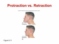
A special movement. Anterior movement of a body part from anatomic position. |
|
|
Retraction |

A special movement. Posterior movement of a body part from anatomic position. |
|
|
Opposition |

A special movement. Special movement of the thumb across the palm and toward the fingers to permit grasping and holding of an object. |
|
|
Some joints in depth: the axial skeleton |
Included the tempotomandibular joint and the invertebral articulations |
|
|
Tempotomandibular Joint |
Diathrotic hinge joint between mandibular and temporal bone with: Articular disc and ligaments: sphrnomandibular, stylomandibular, and tempotomandibular (lateral) |
|
|
Intervertebral articulations |
-Amphiarthrosis between vertebral bodies; diarthroses between articular processes -vertebral bodies separated by intervertebral discs with outer anulus fibrosus and inner nucleus pulposus -ligaments: anterior and posterior longitudinal |
|
|
Joints of the pectoral girdle and upper limbs |
Sternoclavicular Acromuoclavicular Glenohumeral (shoulder) joints Elbow Radiocarpal (wrist) joints |
|
|
Stericlavicular Joint |
Diarthrotic Saddle Joint between manubrium of sternum and sternal end of the clavicle -ligaments: Anterior and posterior sternoclavicular Costoclavicular Interclavicular |
|
|
Acromioclavicular Joint |
Diarthrosis between acromial end of clavicle and acromion of scapula Ligaments: acromioclavicular and coracoclavicular |
|
|
Glenohumeral Joint |
Also known as the shoulder joint Diarthrotic ball and socket joint between the head of humerus and glenoid cavity of scapula Features: -fibrocartilaginous glenoid labrum -ligaments: coracoacromial, coracohumeral, glenohumeral, transverse humeral -rotator cuff muscles -bursae subacromual, subcoracoid, subdeltoid, and subscapular |
|
|
Elbow Joint |
Diarthrotic hinge joint composed of humeroulnar and humeroradial joint -ligaments: radial (lateral) collateral and ulnar (medial) collateral. Anular. |
|
|
Radiocarpal (wrist) joints |
Diarthrotic condylar joint between: -distal articular surface of radius and -three proximal carpal bones: Scaphoid, lunate, and triquetrum. |
|
|
Joints of the pelvis girdle and lower limbs |
Hip (coxal) joint Knee joint Talocrural (ankle) joints Joints of the foot |
|
|
Knee joint |
Diarthrotic hinge joint between the femur, tibia, and patella. Largest most complex joint in the body with: Medial and lateral menisci, Ligaments: Patellar, fibular (lateral) collateral, tibial (medial) collateral, anterior and posterior cruciate (ACL and PCL) |
|
|
Talocrural (ankle) joint |
Diathrotic hinge joint composed of two articulations: Between distal end of tibia and the talus Between distal end of fibula and the lateral aspect of the talus Ligaments: deltoid, lateral and anterior and posterior tibiofibular |
|
|
Joints of the foot |
Four different types of diarthroses: -intertarsal: plane joint between tarsals -tarsometatarsal: plane joint between distal tarsal bones and metatarsals -metatarsophalangeal (MP): condylar joint between metatarsal and proximal phalanges Interphalangeal (IP): hinge joint between phalanges |