![]()
![]()
![]()
Use LEFT and RIGHT arrow keys to navigate between flashcards;
Use UP and DOWN arrow keys to flip the card;
H to show hint;
A reads text to speech;
12 Cards in this Set
- Front
- Back
|
Posterior triangle boundaries (include roof & floor)
What divides it into occipital and subclavian triangles? |
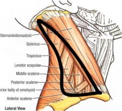
SCM, trapezius muscle, clavicle
Roof: Skin, fascia, platysma Floor: splenius capitis, levator scapulae, scalenus medius, scalenus anterior
omohyoid muscle |
|
|
Contents of posterior triangle |
branches of cervical plexus (C2-C4); accessory nerve (11th cranial nerve), external jugular vein, subclavian artery and vein, brachial plexus.
(Image) |
|
|
Sternocleidomastoid |
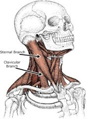
Origin: Sternum, medial third clavicle insertion: MAstoid process Action: unilaterally - tilts head to side, rotation. Nerve: accessory 11th cranial nerve --> sensory info to C2 |
|
|
Trapezius |
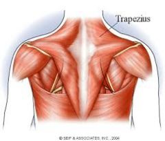
Origin: c7, all thoracic vertbrae. medial part of superior nuchal line, external occipital protuberance, ligamentum nuchae insertion: Action: Middle - braces shoulders. upper - raise shoulder tip, lower - pull medial scapula downwards. movement of shoulder and arms Nerve: motor - accessory nerve sensory c3,c4 |
|
|
Digastric |
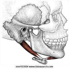
anterior - posterior belly joined by tendon. Origin: Anterior belly - lower border of Md (digastric fossa). Posterior - notch medial aspect mastoid process (digastric notch). Insertion: intermediate tendon superior border hyoid bone. Action: depression Md - open mouth. retrude Md. lift hyoid bone during swallow. Nerve: anterior - trigeminal (inferior alveolar). posterior - facial |
|
|
Infrahyoid muscles |
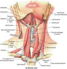
omohyoid: superior belly - hyoid -->intermediate tendon deep to SCM. inferior - tendon --> lateral across posterior triangle --> scapula. sternohyoid: (superficial) sternum, hyoid bone Thyrohyoid: (deep) thyroid cartilage --> hyoid bone Sternothyroid: (deep) thyroid cartilage --> sternum |
|
|
Larynx - describe. function. components |
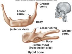
upper airway. Speech. hyoid bone, thyroid cartilage, cricoid cartilage |
|
|
Hyoid bone |
|
|
|
thyroid cartilage |
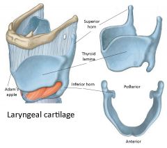
Two Laminae that join anteriorly to form laryngeal prominence (adam's apple) |
|
|
Cervical Fascia - structure - function - Layers |
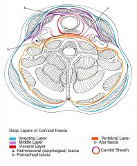
s: connective tissue f: surrounds muscles, BVs, nerves. binding and allows sliding over each other. l: superficial - subcutis blending with reticular layer of dermis. areolar CT, adipose deep- dense fibrous. Investing: wraps SCM, trapezius. vertebral: wraps vertebra and vertbral muscles. Visceral: wraps the oesophagus and trachea. cranial heath: wraps common carotid, internal jugular vein, vagus nerve (10th). |
|
|
splenius capitus |
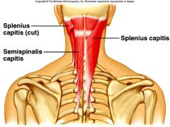
o: Nuchal ligament and spinous process of C7-T3 I: mastoid process bs: aorta nn: Posterior ramus of spinal nerves C3and C4 a: Extend, rotate, and laterally flex the head |
|
|
Scalenes |
O: Cervical vertebrae (CII-CVII) I: First and second ribs A: Ascending cervical artery N: Cervical nerves (C3-C6) Action: Elevation of first and second ribs |