Reading...
![]()
Play button
![]()
Play button
![]()
Use LEFT and RIGHT arrow keys to navigate between flashcards;
Use UP and DOWN arrow keys to flip the card;
H to show hint;
A reads text to speech;
209 Cards in this Set
- Front
- Back
|
What kind of heart does a primitive fish without lungs have?
What are its chambers called? |
Tubular Embryonic Heart with 4 Chambers
1. Sinus Venosus 2. Atrium 3. Ventricle 4. Conus Arteriosus |
|
|
In adult primitive fish the heart is ____________ and receives and pumps _________.
|
a. Folded up on itself
b. Blood low in oxygen content |
|
|
What body part evolved early in Vertebrate evolution as an accessory to gills?
|
Lungs
|
|
|
What can some amphibians and fishes use? (Two things)
|
Lungs and Gills
|
|
|
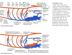
Ductus Arteriosus
|
Part of 6th aortic arch, dorsal to gills
|
|
|
When is the ductus arteriosus valve open?
|
When using gill respiration and not lungs
|
|
|
When is the ductus arteriosus valve closed?
|
When using lungs and not gills
|
|
|
Amphibian Atrium
|
Divided into right (oxygen poor) and left (oxygen rich)
|
|
|
Amphibians have ___ ventricle
|
One
|
|
|
In the Amphibian ventricle, what happens to do the blood?
|
The timing of entry into the ventricle alternates so that there is little mixing of the oxygenated and deoxygenated blood.
|
|
|
Lizards and Turtles have ______ atria and _____ ventricle(s).
|
a. Two
b. One ventricle that is partly divided |
|
|
How do the circulation systems of lizards and turtles work?
|
Due to the partly divided ventricle, they can adjust blood flow through the lungs depending on the resistance of the lungs (holding breathe/slow breathing) to match the extent that the lungs are being used.
|
|
|
Birds and mammals are _______ while lizards, and amphibians are ________.
|
a. Endothermic
b. Ectothermic |
|
|
Birds and mammals have _______ atria and _________ ventricles.
|
a. Two
b. Two They possess a four-chambered heart. |
|
|
Endotherms have _______ resting and ______ metabolic rates which produces a high demand for ______.
|
a. High resting
b. Active metabolic rates c. O2 They must have high resting if they want high activity metabolism |
|
|
Birds and mammals have similar adaptations due to _________ and not ___________.
|
a. Convergent evolution
b. Homology (common ancestry) |
|
|
Mammals and birds have _________ circulation which creates a need for __________ BP.
How is this provided? |
a. Systemic Circulation
b. Higher blood pressure Produced by thicker ventricle wall due to greater ventricular muscularization |
|
|
In mammals and birds, the ________ is reduced in size and incorporated into the _________ and functions as a ___________.
|
a. Sinus venosus
b. Right atrium c. Pacemaker |
|
|
Sinoatrial Node
|
- Creates the action potential propagation of the pacemaker (right atrium)
- Contracts to atrioventricular node which moves through atrioventricular bundles (Purkinje fibers) |
|
|
Pacemaker
|
- Incorporated into wall of right atrium
- Modified cardiac muscle |
|
|
Purkinje Fibers
|
- Modified cardiac muscle
- Located in the walls of ventricle - Transmits action potential from pacemaker to all parts of ventricle |
|
|
Where do cardiomyocytes originate?
|
Splanchnic layer - Lateral Plate Mesoderm
and induced by BMPs (bone morphogenic protein) from endoderm |
|
|
What do cardiomyocytes form?
|
The embryonic heart
|
|
|
Genetic control of development is __________.
|
a. Conserved
- Genes that influence the development of the mammalian heart are the same throughout vertebrates (and even some other animals). |
|
|
Are the components of the heart and aortic arches controlled by the same genes?
What does this mean? |
No, each is under different specific genetic control, and thus can be defective separately, accounting for different congenital defects.
|
|
|
What is the pathway of cardiomyocytes?
|
Splanchnic mesoderm (then converge along ventral midline and form a crescent then tube) --> blood vessels --> 1st vitelline veins --> tubular heart --> cardiac looping (repositioning of ventricles and atria) --> divide into chambers called cardiac cushions (swellings of cells)
|
|
|
Cardiac Cushions
|
Endocardial (endothelial) cells migrate into and form cardiac valves between the atria/ventricles and the ventricles/pulmonary arteries/aorta
|
|
|
Day 21 of development
|
Morphic tube developed (no identifiable chambers yet) but cell fates are determined
|
|
|
_________ cells help separate the _________ and the __________ from the area of the ___________ and the __________.
|
Neural crest cells help separate the pulmonary trunk and the aorta (CT) from the area of the conus arteriosus and ventral aorta (AS).
|
|
|
Neural Crest Cells (Genetic blueprint paper)
**3 Things** |
- Invade ventral aorta and conus arteriosus (AS)
- Separate pulmonary trunk from aorta (CT) -Remodel remaining aortic arches into: aorta, proximal subclavian, internal carotid and pulmonary arteries. |
|
|
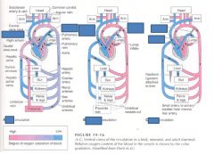
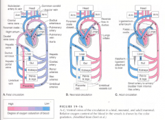
Foramen ovale
(Fetal v. Neonate v. Adult Circulation) |
Fetal - foramen ovale flows oxygenated blood from placenta (to right atrium) to left atrium
Neonate - Foramen ovale closed Adult - Becomes fossa ovalis (no hole) |
|
|
Ductus Arteriosus
(Fetal v. Neonate v. Adult Circulation) |
Fetal - deoxygenated blood flows from right ventricle to (pulmonary artery) and ductus arteriosus through the aorta, umbilical arteries, and finally the placenta
Neonate - Ductus arteriosus is reversed in flow direction, oxygenated blood from right atrium flows through ductus arteriosus to the liver/gut/kidneys/pelvis and legs, and mixes with deoxygenated blood to the lungs Adult - Ductus arteriosus is nonexistant, becomes ligamentum arteriosum |
|
|
|
|
|
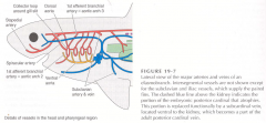
Aerial and aquatic breathing in capable fish and amphibians.
|
|
|
|
Zygote
1. What does it become and through what mechanism? |
Fertilized egg
Undergoes cleavage to become a blastula |
|
|
Blastula
1. What does it become and through what mechanism? |
Hollow ball of cells
Undergoes gastrulation to form a gastrula |
|
|
Gastrula
- What is it composed of and through what mechanism is it formed? |
Composed of three germ layers
- Ectoderm, mesoderm, and endoderm |
|
|
Blastocyst
|
Mammalian version of a blastula
Forms the embryo and some extraembryonic membranes |
|
|
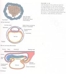
Trophoblast
- What is it, what does it do, where does it go? |
Cells forming the outer layer of a blastocyst (extraembryonic membrane)
Provide nutrients to the embryo Develops into the fetal part of the placenta |
|
|
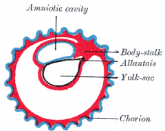
Amniotes (reptiles) have _____ extraembryonic membranes.
|
Four
1. Amnion 2. Yolk Sac 3. Allantois 4. Chorion |
|
|
Amnion
(amniotic extraembryonic membrane) |
Forms a protective environment around the blastula
|
|
|
Yolk Sac (amniotic extraembryonic membrane)
1. Which reptiles are they ONLY ones who have this? 2. Fxn now and for mammals? |
Amphibians and fish
Provides nutrients for the embryo Mammalian Fxn - early source of red blood cells and pathway for early gamete migration |
|
|
Allantois
(amniotic extraembryonic membrane) |
Waste resevoir of blastula
|
|
|
Chorion
(amniotic extraembryonic membrane) |
Wall of allantois; fetal part of placenta
|
|
|
Maternal Part of Placenta
|
Wall of the Uterus
|
|
|
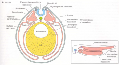
Neurulation
|
Ectoderm
1. Neural crest ectoderm moves to other parts of the body (makes up skull) 2. Neural placode - neuroectoderm 3. Epidermal ectoderm Notochord - from chordamesoderm Mesoderm 1. Somites (Paraxial mesoderm) 2. Intermediate mesoderm 3. Lateral plate mesoderm |
|
|
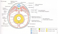
Mesoderm differentiation
|
Paraxial mesoderm (Somites)
1. Dermatome 2. Myotome 3. Schlerotome Intermediate mesoderm 1. Gonadal and Nephric ridge Lateral Plate mesoderm 1. Somatic layer 2. Ceolom cavity 3. Splanchnic layer - Peritineum (outside of body cavity) |
|
|
Dermatome (forms)
|
Dermis
|
|
|
Myotome (forms)
|
Axial and appendicular musculature
|
|
|
Schlerotome (forms)
|
Vertebral column and occipital (1/4) of skull
|
|
|
Splanchnic Lateral Plate Mesoderm (forms)
|
Heart, blood vessels and blood cells
Visceral muscles (gut tube) Visceral peritoneum (organs inside coating) - Thin clear tissue that lines the cavity |
|
|
Peritineum (forms)
|
Appendicular Skeleton
|
|
|
Embryonic Development Stages
|
1. Gastrulation (3 germ layers simultaneously)
2. Extraembryonic membrane formation (trophoblast) 3. Neurulation 4. Mesoderm differentiation (early organogenesis) |
|
|
In mammalian development, cells move from the ______ through the Primitive Streak.
|
Ectoderm
|
|
|
In mammalian development, the ______ and _________ layers of a gastrula cavitate to form the _________ and the ____________.
|
Ectoderm and endoderm cavitate to form the amniotic sac and yolk sac
|
|
|
In mammalian development, the _______ of the trophoblast connects the embryo to the trophoblast which the ________ embryonic layer travels through in order to surround the ____________ and coat the inside of the ___________.
|
The stalk of the trophoblast connects the embryo to the trophoblast
Mesoderm goes through the stalk to coat the inside of the trophoblast |
|
|
In mammalian development, the ________ undergoes ________ evagination of __________ by growing through the stalk of the trophoblast.
|
The allantois undergoes endodermal evagination of the archenteron by growing through the trophoblastic stalk.
|
|
|
Fetal Part of Placenta - Chorioallantoic Placenta
|

Trophoblast - Chorion, Allantois and Mesoderm
|
|
|
In mammalian development, the yolk sac eventually becomes divided into the ___________ and the __________.
|

Archenteron and still the Yolk Sac
|
|
|
|
|
|
Urbilaterians
|
Only possess one hox gene
|
|
|
Chordate Characteristics
|
1. Notochord
2. Dorsal Hollow Nerve Cord 3. Post-anal tail 4. Pharyngeal slits 5. One complete set of Hox genes |
|
|
3 Components of skull
|
Chondrocranium - Surrounds brain/sense organs
Splanchnocranum - Branchial/Pharyngeal/ Gill Arches Dermatocranium - Most Superficial layer |
|
|
Chondrocranium (Craniate)
- Components |
1. Nasal Capsule
2. Orbit 3. Otic Capsule 4. Base of Cranium |
|
|
Splanchnocranium (Bony Fish)
|
Palatoquadrate and Mandibular ends ossify into quadrate and articular
2nd Arch (Hyomandibular) ossifies 3rd-7th Arch are branchial arches |
|
|
Gnathostomes Attributes
|
Jaws evolved
1st Branchial arch (Mandibular) becomes Jaws - Palatoquadrate becomes top, Mandibular bottom 2nd Branchial arch - Hyoid Arch and Hyomandibula |
|
|
Sharks (Cartilaginous fishes) Attributes
|
Posses chondrocranium and Jaws
No Dermatocranium |
|
|
Dermatocranium
|
Bony Fish posses over chondrocranium and splanchnocranium
|
|
|
Nasal Capsule Analogous in Humans to ____________.
|
Ethmoid
|
|
|
Orbit Analogous in Humans to ____________.
|
Orbit
|
|
|
Otic Capsule Analogous in Humans to ____________.
|
Petrous Part of Temporal (Prootic and Opisthotic)
|
|
|
Base of Cranium Analogous in Humans to ____________.
|
Sphenoid (Presphenoid, base occipitals)
|
|
|
Neurulation occurs.....
|
Simultaneously w/ extraembryonic membrane formation, and mesoderm differentiation
|
|
|
Neurogenetic cells
|
Neural tube, neural crest, neural placode
|
|
|
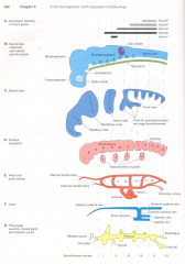
Neural Tube Morphogenesis
|
- Prosencephalon (forebrain)
- Mesencephalon (midbrain) - Rhombencephalon (hindbrain) |
|
|
Prosencephalon
|
Forebrain
2 Bilateral evaginations make optic vessels - Optic (Optic Nerve) and Optic Cup (Retina) |
|
|
Neural Placode Cells
|
1. Nasal Placode - olfactory epithelium
2. Otic Placode - inner ear 3. Ganglia associated w/ Cranial nerves |
|
|
Neural Crest Cells
|
1. Starts out as epithelium and changes into mesenchyme cells
2. Forms most of the cranium (splanch, chondro (2/3), dermato) 3. Peripheral nervous system and autonomic nervous system ganglia 4. Dentine/Dental pulp (Stem cells) 5. Alveolar bones of jaw 6. Periodontal ligaments |
|
|
Mesenchyme
|
Star-shaped mobile cells
|
|
|
Ganglia
|
Group of nerve cells outside of the central nervous system
|
|
|
Vertebral Column and Posterior 1/3 of Chondrocranium
|
Sclerotome PAM
|
|
|

|
|
|
Branchiomeres (Pharyngeal/gill arches and pouches) have __________.
|
1. Each has their own skeletal elements (splanchnocranium)
2. Each has their own muscles 3. Each has their own blood supply (aortic arches) 4. Each has their own nerve supply |
|
|
Cranial Development
|
Mostly neural crest cells w/ some sclerotome LPM (occipital region)
|
|
|
Cranial development is _____________ conserved in morphology and __________ variable.
|
Less conserved in morphology, and more variable
|
|
|
New Head Hypothesis
|
1. Neural crest cells are responsible for the evolution of the head
2. Derived from craniates |
|
|
Neural Crest Genesis
|
Neural crest induced to form cells at neural plate and epidermal ectoderm border from the chordamesoderm signals
|
|
|
Neural Crest Migration/Exodus
|
The cells follow a stereotyped pathway dependent on the site of origin and signals (guidance cues) from neighboring cells along the way
|
|
|
Neural crest cell migration (is/is not) Intrinsic
|
Neural crest cell migration is NOT intrinsic
- It is not genetically encoded for and depends on location and neighboring signals |
|
|
Neural Crest Differentiation
|
It is induced, with possible intrinsic values dependent on species (Duck & Quail beaks)
|
|
|
How do appendicular and axial skeletons with different embryonic origins differ?
|
They are all of mesodermal origin, and are fairly conserved in morphology (bones stay roughly the same)
|
|
|
Induction of Cells
|
Through morphogens or growth factors produced in cells, these then turn on (induce) different homeobox genes
|
|
|
Noden's Experiment
|
Inadvertently took a piece of neural tube with the neural crest cells, so the transferred mandibular cells formed a second mandible where the hyoid was supposed to form. Experimental mistake - neural crest cells are NOT stereotyped/intrinsic
|
|
|
If you remove mandibular arch neural crest cells and place them where the hyoid arch is, will they form the hyoid arch?
|
Yes, neural crest cells are naive and depend on induction for their differentiation
|
|
|
What tissues induce neural crest differentiation?
|
Neural tube cells
Overlying and underlying ectoderm Isthmus (Midbrain/Hindbrain boundary) |
|
|
Neurocristopathy
|
Class of pathologies arising from neural crest cell defects
|
|
|
Craniofacial defect
|
A neurocristopathy
1. Clefting between cranial bones (no fusion) 2. Abnormal fusions of cranial bones |
|
|
Bone Clefts are caused by...
|
Teratogens which disrupt morphogens (like Shh) which inhibits the cell growth at sutures
|
|
|
Teratogen
|
A chemical that disrupts normal development
1. Cyclopamine - causes cleft palate 2. Alcohol |
|
|
Abnormal bone fusion
|
Caused by defects in dura mater which secretes morphogens (BMP) that regulate osteogenesis (suture fusion)
|
|
|
Development of Appendicular Skeleton
|
1. Establishment
2. Condensation 3. Joint Formation |
|
|
Establishment of Appendicular Skeleton
|
Outward growth of limb bud in stereotyped positions as an aggregation of lateral plate mesoderm (only two arms/legs etc.)
1. Induced by itself (LPM) 2. Ectoderm |
|
|
Condensation of Appendicular Skeleton
|
Recruits cells from limb buds in order to form a bone
|
|
|
Joint Formation of Appendicular Skeleton
|
Condenses and flattens cells at joint locations which leads to cell death
|
|
|
Connective Tissues
|
1. Extracellular materials
2. Loose connective tissue 3. Dense connective tissue 4. Cartilage 5. Bone Cells |
|
|
Extracellular Materials
|
1. Collagen and elastic fibers matrix
2. Other molecules that affect the consistency of the matrix (more fluid or more solid) |
|
|
Loose connective tissue
|
A few fibroblasts which produce lots of loosely arranged fibers and matrix
Ex. Epidermis |
|
|
Dense Connective Tissue
|
Densely packed with fibers
1. Ligaments 2. Tendons |
|
|
Ligaments
|
Dense connective tissue with tightly packed fibers arranged in layers
|
|
|
Tendons
|
Dense connective tissue with tightly packed fibers arranged in cables
|
|
|
Cartilage
|
1. More rigid than dense connective tissue
2. Lots of fibers, but not arranged into layers/cables; cells are microscopically tightly packed |
|
|
The appendicular skeleton and much of the cranium form as _____________.
|
Cartilage replacement bone
|
|
|
Cartilage replacement bone
|
1. Neural crest (migrate to location or nearby mesoderm)
2. Mesenchymal cells (might to location and transform into chondroblasts which produces cartilage) 3. Adult cartilage (few chondrocytes located in lacumen) |
|
|
Chondroblast
|
Cell which originates from a mesenchymal stem cell and forms chondrocytes (cartilage cells) which are embedded in the matrix
|
|
|
Bone Cells
|
Osteocytes and Osteoblasts
1. Dense fibers, matrix of polysaccharides and collagen which bind hydroxipatite (calcium) and make a very dense, strong connective tissue; Calcification |
|
|
Calcification
|
The binding of calcium (hydroxiapatite) which makes a very dense, strong connective tissue of bone
|
|
|
Cartilage replacement bone is turned into bone by...
|
Cartilage precursors are invaded by osteoblasts (matrix and fibers) and undergo calcification
|
|
|
Membrane bone
|
Dermatocranium
1. Neural crest cells transform into osteoblasts within the dermis (connective tissue) 2. Membrane bone forms directly as bone, no cartilage precursor |
|
|
Membrane bone forms __________ as __________.
|
Membrane bone forms directly as bone
|
|
|
In bone, blood is _________ vascularized, while in cartilage it is _____________ vascularized.
|
Blood is highly vascularized in bone, but not in cartilage
|
|
|
Highly vascularized means that there is ____________ blood supply.
|
A great blood supply
|
|
|
Limb Bud Development
|
Signals from the LPM and Ectoderm
1. Start the position of limb bud 2. Initiate different skeletal elements - Proximal, Middle, Distal |
|
|
Proximal Limb Elements
|
Humerus, Femur
|
|
|
Middle Limb Elements
|
Radius and Ulna
Tibia and Fibula |
|
|
Distal Limb Elements
|
Hands and Feet
|
|
|
Apical Ectodermal Ridge
|
Forms from the ectodermal cells at the distal end of each limb bud
Acts as a signaling center to ensure proper limb development |
|
|
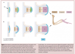
Apical Ectodermal Ridge PZ Model
|
Progressive Zone of AER
1. Directly beneath AER 2. Cells measure length of time in PZ - Short: proximal - Medium: middle - Long: Distal 3. PZ is not intrinsic (depends on time spent) 4. Signals travel from AER to FGF morphogen to specific Hox genes in proximal/middle/distal elements |
|
|
FGF Morphogen
|
Fibroblast Growth Factor
Induces cell outgrowth w/ a positive feedback loop between Intermediate Mesoderm, LPM, and surface ectoderm |
|
|
ZPA (Zone of Polarizing Activity)
|
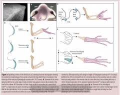
1. Active in distal element
2. Determines digit identity 3. Removal and grafting results in duplication of digits |
|
|
IDM (Interdistal Mesenchyme)
|

1. Determines the number of phalanges in each digit
2. Removal results in decreased number of phalanges |
|
|
Circulatory System Origin
|
Splanchnic Lateral Plate Mesoderm
|
|
|
Circulatory System Development
|
Angioblasts (embryonic blood vessel cells) migrate and differentiate
|
|
|
Blood Vessels (Histology)
|
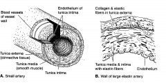
1. Tunica intima
2. Tunica media 3. Tunica externa |
|
|
Tunica Intima
|

1. Endothelial cells (epidermal cells which line blood vessels and the heart)
2. Can include elastic fibers |
|
|
Tunica Media
|

Connective tissue and smooth muscle of a blood vessel
|
|
|
Tunica Externa
|

Connective tissue and collagen fibers of a blood vessel
|
|
|
Shark Blood Vessels
|
Blood travels first from the ventral aorta, then to the ventral branches of the aortic arch.
From the ventral branches, it creates gill circulation which picks up oxygen and gets rid of CO2. It then moves from the gills to the dorsal branches of the aortic arches, and then to the dorsal aorta. |
|
|
Every living vertebrae has lost the aortic arch number ________, which can be still found in sharks.
|
Every living vertebrae has lost Aortic Arch 1
|
|
|
Tetrapod Blood Vessel Development (Frog)
|
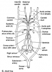
Lost aortic arch #1,2,5
3rd Aortic Arch - Internal carotid arteries 4th Aortic Arch (Left) - Dorsal Aorta 4th Aortic Arch (Right) - Right Subclavian Artery 6th Aortic Arch - Pulmonary Arteries |
|
|
The umbilical vein (mammals) carries __________ blood.
|
The umbilical vein carries oxygenated blood.
|
|
|
Fetal Circulation Pathway
|

1. Umbilical Vein carries oxygenated blood
2. Liver is bypassed through ductus venosus 3. Oxygenated and deoxygenated blood is mixed in the cadual vena cava 4. The blood passes into the right atrium and through the formen ovale into the left atrium **Blood from the pulmonary artery mostly bypasses lungs through ductus arteriosus (fetuses don't breathe) |
|
|
Neonatal Circulation Pathway (differences from fetal)
|
1. Blood pressure closes the foramen ovale
2. Blood now flows to the lungs even though ductus arteriosus is still open 3. Aortic blood travels through the ductus arteriosus to the lungs where it picks up more oxygen (from inflated lungs) which saturates the hemoglobin to release more oxygen 4. Days after birth the ductus arteriosus closes and hemoglobin transforms into adult stage |
|
|
Neonate
|
Newborn child or mammal
|
|
|
Fetal Hemoglobin holds _______ O2 than adults, and releases it _________ easily. Therefore _________ oxygen is released from fetal hemoglobin than adults.
|
Fetal Hemoglobin holds more O2 than adults, and doesn't release it as easily. Therefore more oxygen is released from fetal hemoglobin than adults.
|
|
|
Adult Blood Circulation (difference from neonate)
|
The ligamentum arteriosum is formed from the ductus arteriosus that becomes filled up with cells
|
|
|
Why do chordates/urbilaterians have symmetry on the outside?
|
1. Hydrodynamics (due to first fishy ancestor)
2. Locomotion (in general) works better with symmetry |
|
|
Urbilaterians
|
Bilaterally symmetrical ancestor
- Symmetrical on inside AND outside |
|
|
Chordates (symmetry)
|
1. Symmetrical on outside
2. Asymmetrical on inside |
|
|
Why are chordates asymmetrical on the inside?
|
Because as we develop to be greater in total size, we need a rapidly increasing digestive system in order to keep the same surface area ratio for digestion and absorption
|
|
|
How do the gut sizes of herbivores, omnivores, and carnivores compare?
|
Herbivores > Omnivores > Carnivores
|
|
|
As our digestive system expands, how do we keep our internal balance?
|
The organs distribute themselves to have symmetrical weight
Left side: spleen, stomach, heart and some lung/liver Right: Most of the lung/liver |
|
|
Chordate Asymmetrical Internal Development
|
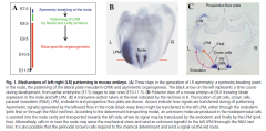
1. Node located in the anterior end of primitive streak
2. In the node are cilia which beat circularly to the left and morphogens in the node are directed leftward 3. Morphogens turn on developmental genes and create a cascade (nodal circuit) which eventually turns on PitX2, which causes the organs on the left to develop differently than the right |
|
|
Nodal Circuit
|

Cascade of genes "turning on" caused by developmental genes turned on by morphogens from the node of the primitive streak
Last gene in cascade is PitX2 |
|
|
PitX2 gene
|

1. Last gene to be turned on in the nodal circuit
2. Causes the organs on the left to develop differently than the ones on the right 3. Causes cell death OR bigger growth on left side (dependent on interacting genes) |
|
|
Is the left or right side of a mammal the default side? I.E. if PitX2 was not expressed, what would develop?
|
The right side is default. If PitX2 is missing, then the body has stereoisomeric right sides.
|
|
|
Which direction do the cilia in the node of the primitive streak beat?
|

The cilia beat to the left and push the morphogens in the node leftward
|
|
|
Why do the node cilia beat to the left?
|
1. Accident of initial evolution
2. Protein helices twist to the right because they are using left amino acids which makes the cilia beat to the left |
|
|
What does PitX2 result in on the left side?
|
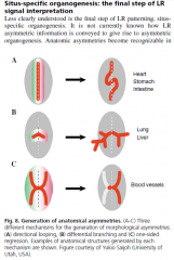
1. Directional looping of the heart
2. Directional looping of the digestive system 3. Differences in the size of lung/liver branching 4. Regression (Cell death) of some organs leaving them on only one side of the body (Spleen) |
|
|
Directional Looping of the Digestive System (from PitX2)
|
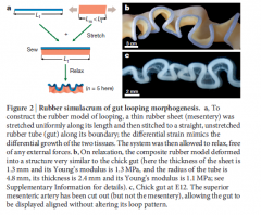
1. Endoderm forms inner lining (epithelium)
2. Mesoderm forms involuntary muscles and outer mesentery 3. Embryo gut tube supported by dorsal mesentery which becomes asymmetrically thick and folds the gut tube 4. As development continues, dorsal mesentery thins out but is very elastic and elongation of gut tube creates small folds Due to MECHANICAL differences in growth/elasticity of dorsal mesentery/gut tube |
|
|
Asymmetric Dorsal Mesentery Among Species
|
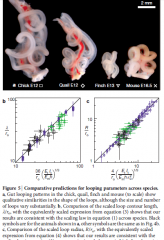
Tension between dorsal mesentery (shorter, more elastic) and intenstine (longer, more pliable) is stereotyped among species
1. Chick and quail are closely related 2. Mouse is more rightly packed b/c it's softer It is an EVOLVABLE anatomy |
|
|
Evolvable Anatomy
|
Very few developmental genes affecting anatomy, so evolution of that element is easier
|
|
|
Production of Villi in Small Intestine
|
1. As internal epithelium of gut (endoderm) expands (cell division), the surrounding circular and longitudinal smooth muscles impose mechanical stress causing it to buckle into ridges and villi
2. This buckling keeps the surface area of the epithelium high |
|
|
Purpose of Artery and Vein Branching
|
1. There is no exact branching of more extended vessels
2. The only thing that matters is that the target tissue gets enough circulation |
|
|
Blood Vessel Development Pathway
|
1. Initially, more vessels than necessary sprout from aorta and follow cues from the environment to the target organ
2. Later, some extra vessels are pruned due to oxygen tension of the target organs (too many vessels) **Evolvable Anatomy - Vessels develop as a need-only basis for the organs |
|
|
Finger Prints
|
Example of Asymmetrical Development
1. Pattern does not matter, only fxn of gripping does 2. Frozen Accident of Development |
|
|
Nerves as Evolvable Anatomy and Asymmetry
|
1. Nerves evolved first so blood vessels use their chemical pathway to help develop
2. Nerves also sprout and prune because the target organs secrete limiting amounts of neurotropic factors required for neuronal survival |
|
|
Lethal Asymmetry
|
Aorta meets illiac veins in abdomen which allows for crushing of the veins (vericose veins?)
|
|
|
Lung Development (mammalian)
|
1. Ventral out-pockets of endoderm (airway epithelium)
2. Right side has larger diameter so more secondary bronchioles, and more lobes (more letters more lobes) 3. Blood vessels follow behind the development of bronchioles 4. At the alveoli, gas exchange occurs |
|
|
Mouse Lung Development Branching Patterns
|
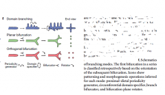
Three Branching Patterns
1. Domain 2. Planar Bifurcation 3. Orthogonal Bifurcation |
|
|
Mouse Lung Branching Locations
|
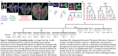
1. Domain Branching - Forms initial scaffold
2. Planar Bifurcation - Forms edges of lobes 3. Orthogonal Bifurcation - Fills in interior spaces |
|
|
The differentiation of alveoli occurs...
|
The differentiation of alveoli occurs after the bronchial branching is complete
|
|
|
Differentiation of Lateral Plate Mesoderm
|
1. Coelomic cavity and mesenteries form by splitting LPM
2. Somatic layer forms outer wall (Parietal serosa, peritoneum, pleura, and pericardium) 3. Splanchnic layer forms epithelium covering organs (Visceral sera, peritoneum, pleura, and pericardium/epicardium) 4. Vental (bottom)/Dorsal(top) mesenteries formed by joining of LPM |
|
|
Serosa (Mesenteries)
|
Simple Squamos Epithelium with a layer of connective tissue beneath
|
|
|
Early Development of Mesenteries
|
LPM expands dorsally and ventrally to create dorsal and ventral mesenteries (two layers of epithelium and connective tissue)
|
|
|
Median Development of Mesenteries
|
1. Liver grows within ventral mesentery
2. Ventral mesentery divides into lesser omentum and falciform ligament 3. Pancreas grows within dorsal mesentery |
|
|
Latest Development of Mesenteries
|
1. Most of ventral mesentery is lost
2. Most organs suspended by dorsal mesentery only 3. Heart loses dorsal and ventral mesenteries 4. Kidney remains retroperitoneal (no mesentery) |
|
|
Dorsal Mesentery which Suspends Organs
|
1. Mesovarian - suspends ovaries
2. Mesorchium - suspends testes 3. Mesogaster - supports the stomach - Free edge draps from greater curvature to greater omentum |
|
|
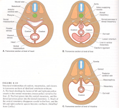
Other Mesenteries of Body Cavity (Fish Development)
|
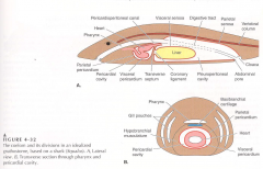
1. Transverse septum forms as the liver (visceral peritoneum) grows and touches the body wall (parietal peritoneum) to separate the liver from the heart
2. Coronary ligament connects liver to transverse septum 3. There are two cavities: pleuroperitoneum and pericardial |
|
|
Other Mesenteries of Body Cavity (Tetrapod Development)
|
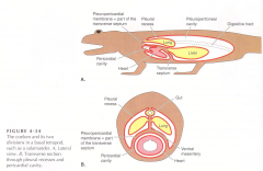
1. Pleuropericardial membrane is now completely seperate from pleuroperitoneal cavity
2. Transverse septum separates the heart from the liver, digestive system and lungs 3. Heart migrates caudually towards lungs while the enlarging lungs and pleural cavities grow laterally and ventrally to surround pericardial cavity and heart |
|
|
Other Mesenteries of Body Cavity (Reptile and Mammalian Development)
|
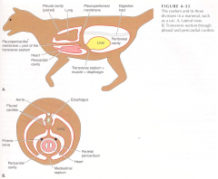
1. Dorsal pleuroperitoneal membranes expand ventrally to separate pleural and peritoneal cavities
2. Lungs separate into two cavities, separate from heart and guts 3. Four cavities overall: 2 pleural, 1 pericardial, 1 peritoneal |
|
|
Other Mesenteries of Body Cavity (Only Mammalian Development)
|

1. Somatic muscles grow into new pleuroperitoneal septum and become the diaphragm
2. The lungs expand ventrally carrying the pleural peritoneum membrane with it, creating a septum between the lungs, resulting in two pleural cavities 3. Space between two pleural cavities called the Mediastinum 4. Pericardial cavity becomes the parietal pericardium and pleura which is called the pericardial sac |
|
|
Diaphragm is innervated and receives blood from...
|

The diaphragm is innervated by the phrenic nerve (spinal nerve) and phrenic vein/artery
|
|
|
Mediastinum
|

The area between the two pleural cavities of mammals; Contains the pericardial cavity, the heart, the esophagus, major arteries and veins, and the phrenic plus other nerves
|
|
|
Pericardial Sac
|
Composed of the parietal pleura and pericardium which used to be the pericardial cavity (In mammals)
|
|
|
Digestion (Gut) Development
|
1. Archenteron (endoderm) forms lining of digestive tract and derivatives
- Respiratory passages, secretory cells of liver, pancreas, pharyngeal pouches 2. Urinary bladder and allantois evagination of floor of hindgut 3. Lumen of gut is first broadly connected to yolk sac - Differentiates into foregut, midgut, hindgut and lengthens |
|
|
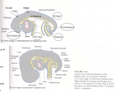
Early Embryo Digestion
|

1. Stomodeum and proctodeum, pharynx, liver primordium, foregut, midgut, hindgut, yolk sac, allantois
|
|
|
Later Embryo Digestion
|

1. Oral cavity, pharyngeal pouches, lung, esophagus, stomach, liver, pancreas, yolk stalk, small and large instestine, cloaca, allantoic stalk
2. Stomodeum invaginates at front of embryo and extends toward archenteron to form oral cavity - Oral plate between stomodeum and oral cavity breaks down to make the oral cavity and pharynx continuous 3. Protodeum invaginates and is separated from the archenteron by the cloacal membrane which breaks down to make the cloaca 4. Cloaca becomes divided into rectum (dorsal) and urogenital passages (ventral) |
|
|
Cloaca
|

1. Embryonic chamber which receives digestive, urinary, and reproductive tracts.
2. Dorsal part contributes to make the rectum and the ventral part contributes to the urogenital passages |
|
|
Development of Embryonic Circulation Summary
|
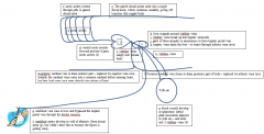
1. Blood vessels from Splanch LPM develop adjacent to yolk sac and form two vitelline veins
2. Vitelline veins empty into tubular heart 3. Ventral aorta of tubular heart extends forward and develops into 6 pairs of aortic arches 4. Aortic arches extend through the gills to the paired dorsal aorta 5. Paired dorsal aorta unite into a single dorsal aorta which continues caudally and gives off branches to supply body 6. Liver expands around vitelline veins 7. Vitelline veins break up into hepatic sinusoids in which parts atrophy, or anastomose to form hepatic portal vein 8. Hepatic veins drain the liver to the heart through the inferior vena cava 9. Posterior cardinal veins form to drain posteriorly but are replaced by inferior vena cava 9b. Initially cardinal veins form a common cardinal before entering heart but vena cava is direct 10. Umbilical vein runs to liver and bypasses hepatic portal by ductus venosus 11. Umbilical artery develops in the wall of allantois from dorsal aorta |
|
|
Lungs initially developed from the gut because....
|
The organism needed to obtain a bigger bubble of air for faster gas exchange
1. Early fish vertebrates just "gulped" in air and held it in the gut to diffuse oxygen through the gut wall |
|
|
Anastomose
|
Joining of blood vessels that previously split apart
1. How the hepatic portal vein is created from hepatic sinusoids (created from vitelline veins breaking up) |
|
|
Oral Cavity Embryonic Origin
|
Ectodermal Stomodeum (depression between pericardium and brain)
Composed of 1. Hard Palate (roof) 2. Soft Palate (mammalian adaptation to breathe while eating) 3. Tongue (endoderm epithelium) |
|
|
Tongue
|
1. Endoderm epithelium
2. Muscular (somites invaded by taste buds) 3. Shape is circular in humans (thin and long in dogs, etc.) |
|
|
Pharynx
|
1. Nasopharynx
2. Oropharynx 3. Laryngeal pharynx 4. Paired pharyngeal pouches |
|
|
Paired Pharyngeal Pouches (Pharynx) Create...
|
1. Auditory Tube (Eustachian Tube
2. Tonsils 3. Parathyroid Glands 4. Thymus |
|
|
Epiglottis
|
1. Mammalian innovation to decrease choking
2. Food has to bypass glottis to get to the esophagus so epiglottis prevents breathing in food |
|
|
Larynx Descent
|

1. In humans, descends 4-6 mo. after birth in order to allow the horizontal and vertical vocal tractways to be similar lengths
2. Descent's main purpose is to increase language production 3. Makes vowel sounds and gutteral H's 4. Increases the probabilty of choking |
|
|
Larynx in Other Mammals and Infants
|
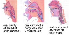
1. Epiglottis abuts the soft palate, lining up internal nares with trachea
2. Liquid flows around either side of epiglottis into esophagus (in order to drink and breathe at the same time) |
|
|
Development of GI Tract
|

1. Endoderm Formation
2. Endoderm Gastrulation 3. Looping of Tract due to Dorsal Mesentery 4. Radial Patterning of Gut Tube 5. Splanch LPM encircles gut tube to form connective tissue and circular/longitudinal muscle layers 6. Cranial neural crest migrates to the gut the colonize AP axis as autonomic ganglia 7. DV axis and right left patterning occur with gut organs (pancreas, tonsils..?) budding from stereotyped positions |
|
|
Endoderm Formation of GI Tract Development
|
In multicellular animals the endoderm and ectoderm evolved first while the mesoderm evolved millions of years later
|
|
|
Endoderm Gastrulation of GI Tract Development
|
1. Endoderm converges at midline to cover entire A-P axis
2. Endodermal sheet then folds 3. Specialized regions of gut arise and accessory organs: thryoid, lungs, liver, pancreas due to interaction of endoderm with surrounding mesoderm 4. Mesoderm induces differentiation of endodermal cells and HOX genes are expressed |
|
|
Radial Patterning of Gut of GI Tract Development
|
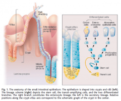
1. Stem cells of the epithelium form mature cells by migrating
2. Enterocytes secrete enzymes and absorb nutrients (VILLI TIPS) 3. Goblet cells secrete mucus (VILLI TIPS) 4. Paneth cells secrete antimicrobial peptides (CRYPTS of small intestine) 4. Enteroendocrine cells secrete hormones (VILLI TIPS) 5. At the villi tips, old cells are shred into lumen after undergoing apoptosis |
|
|
The large intestine __________ have paneth cells and villi.
What does it have? Hint: This is where cancer starts. - Why does cancer start? Which intestine has a greater chance of cancer? |
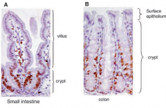
The large intestine does not have Paneth cells and villi. It does have crypts, which are larger than the small intestine's. Cancer starts in the crypts due to the division of stem cells (after 3-4 divisions they differentiate). The colon has a 70x greater chance of cancer than the small intestine.
|
|
|
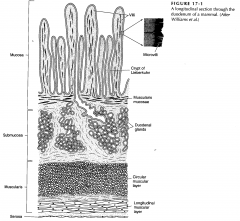
Layers of the Small Intestine of a Mammal
|
|
|
|
How does the GI Tract handle bacteria?
|

1. Immunity and inflammation
2. Beneath epithelium immune cells monitor and deal with invading bacteria 3. At the same time, commensal microbes are benefical (gut flora) |
|
|
Later Embryonic Digestion (Esophagus)
|
1. Aminotes have a long esophagus that is collapsed at rest
2. Lumen opens when food is present 3. Striated muscle (voluntary) |
|
|
Three Types of Muscles
|
1. Striated Muscle
2. Smooth Muscle 3. Cardiac Muscle |
|
|
Later Embryonic Digestion (Stomach)
|
1. Evolved as a storage area
2. Had HCl as a bacteriocidal mechanism and to preserve food 3. In humans it functions as initial digestion and churning with pepsinogens (degrades food into peptides) |
|
|
Later Embryonic Digestion (Intestines)
|
1. Site of digestion (small) and absorption (large)
2. Small intestine (duodenem, jejunum, ileum) - Receives bile from the liver (duct) to neutralize HCl - Involved in fat digestion (lipases) - Pancreas secretes pepsinogens 3. Large intestine (colon) - Water absorption and consolidation of feces - Colonic Bacteria to produce vitamin K 4. Cecum (Between large and small intestine) - Appendix (3-18 cm varies among humans) - Vestigial (may be present or not due to mutations) - Also fxns to replace normal commensal bacteria after diarrhea |