Reading...
![]()
Play button
![]()
Play button
![]()
Use LEFT and RIGHT arrow keys to navigate between flashcards;
Use UP and DOWN arrow keys to flip the card;
H to show hint;
A reads text to speech;
203 Cards in this Set
- Front
- Back
|
|
|
|
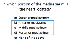
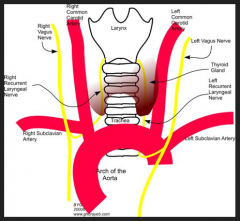
What are the contents of the mediastinum?
|

|
|
|
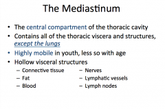
What is the mediastinum? What does it contain? When is it more mobile? What are the hollow visceral structures?
|
|
|
|
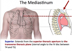
What is the extent of the superior mediastinum?
|
|
|
|
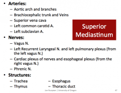
What arteries, nerves, and structures are in the superior mediastinum?
|
|
|
|
|
|
|
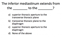
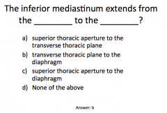
What is the extent of the inferior mediastinum?
|
|
|
|
What is contained in the anterior and posterior portions of the inferior mediastinum?
|
Inferior Mediastinum:
Middle - Heart - Pericardium - Phrenic N - Vagus N - Roots of the Great Vessels: Ascending Aorta, Pulmonary Trunk, and Superior Vena Cava |
|
|
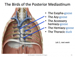
What are the "birds" of the posterior mediastinum?
|
|
|
|
|
|
|
|
|
|
|
|
|
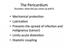
What is the function of the pericardium? (5)
|
|
|
|
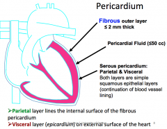
What makes up the pericardium? (5)
|
|
|
|
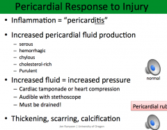
How does the pericardium respond to injury?
|
|
|
|
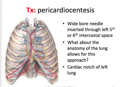
What is pericardiocentesis?
|
|
|
|
Where are the pericardial sinuses?
|
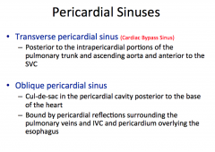
|
|
|
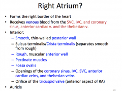
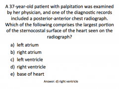
What are the borders of the right atrium? What structures can be found here?
|
|
|
|
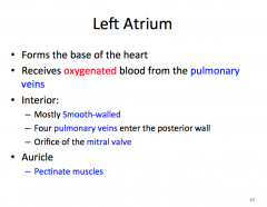
Where is the left atrium found? Where does it receive blood from? What structures are found here?
|
|
|
|
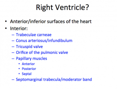
Where is the right ventricle found? What structures can be found here?
|
|
|
|
Where is the left ventricle found? What is the difference between it and the right ventricle? What structures are found here?
|
|
|
|
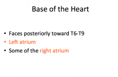
What does the base of the heart face? What does it contain?
|
|
|
|
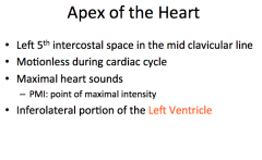
What are the borders of the apex of the heart? What is special about its motion? What is special about the noise it makes?
|
|
|
|
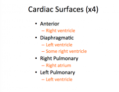
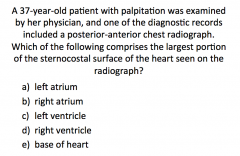
Name the cardiac surfaces
|
|
|
|
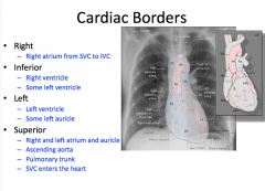
What are the cardiac borders? right, inferior, left, superior
|
|
|
|
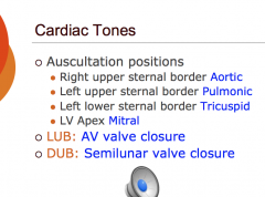
What are the auscultation positions?
|
|
|
|
|
|
|
Where is the mitral valve found?
|
posterior to sternum at 4th costal cartilage
|
|
|
Where is the pulmonic valve found?
|
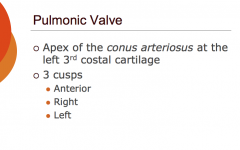
|
|
|
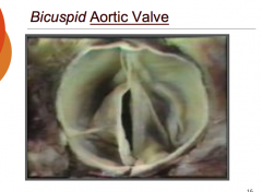
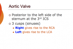
Where is the aortic valve found?
|
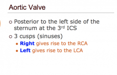
|
|
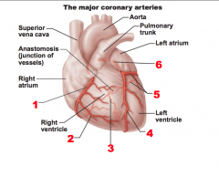
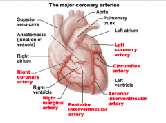
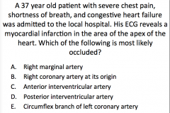
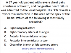
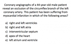
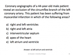
Name these major coronary arteries
|
|
|
|
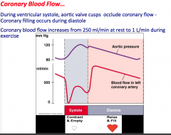
How does coronary blood flow change from systole to diastole? How about during exercise?
|
|
|
|
Where can you find the aortic valve?
|
|
|
|
|
|
|
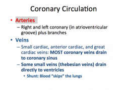
Coronary circulation: arteries? veins? Where do most veins drain? Where do smaller veins drain?
|
|
|
|
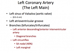
Left Coronary artery: borders? where is it? branches?
|
|
|
|
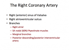
Right Coronary Artery: borders? where is it? branches?
|
|
|
|
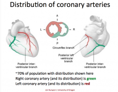
What is the distribution of the coronary arteries?
|
|
|
|
|
|
|
How do materials cross the capillary wall?
|
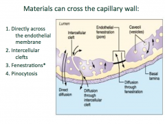
|
|
|
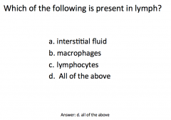
What is the function of the lymphatic system?
|
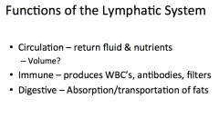
|
|
|
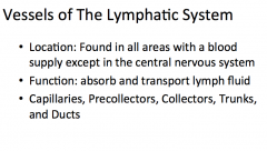
What are the vessels of the lymphatic system and what is their function?
|
|
|
|
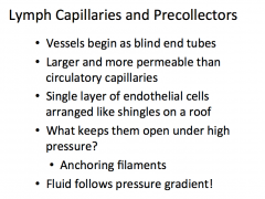
What are lymph capillaries and precollectors?
|
|
|
|
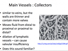
What are the main vessels of the lymphatic system and what are they similar to? What is their job?
|
|
|
|
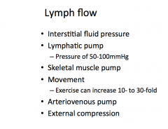
How does lymph flow?
|
|
|
|
|
|
|
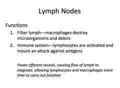
What is the function of the lymph nodes?
|
|
|
|
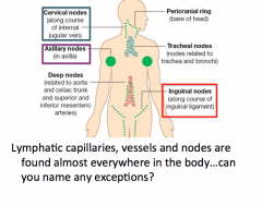
Where are lymph nodes located?
|
|
|
|
Where is the spleen and what structures are around it?
|
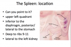
|
|
|
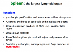
What is the function of the spleen?
|
|
|
|
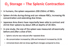
What is the splenic contraction?
|
|
|
|
|
|
|
Never Let Monkeys Eat Bananas
|

|
|
|
|
|
|
|
|
|
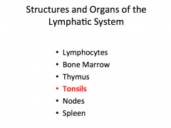
What are the structures and organs of the lymphatic system?
|
|
|
|
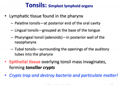
How many different tonsils are there (4)? What are tonsillar crypts? What is their job?
|
|
|
|
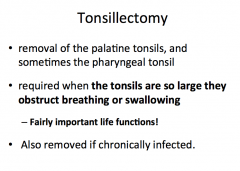
What is a tonsillectomy?
|
|
|
|
what type of muscle is the rectus femoris?
|
bipennate
|
|
what type of muscle is the extensor digitorum longus?
|
unipennate
|
|
|
what type of muscle is the orbicularis occuli?
|
circular
|
|
|
what type of muscle is the rectus abdominis?
|
quadrate
|
|
|
what type of muscle is the omohyoid?
|
digastric
|
|
|
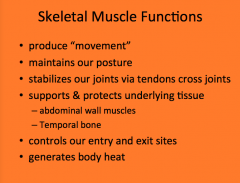
Skeletal Muscle Functions (6)
|
|
|
|
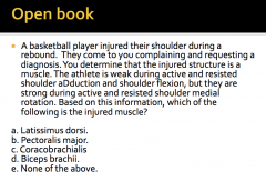
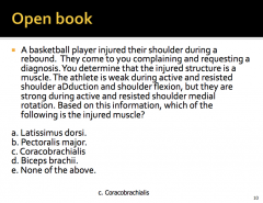
What "special tests" would you used to identify injured tissue? (3) Give an example of how you would perform each test.
|
"Range of motion" testing
|
|
|
what are the two types of isotonic contraction?
|
There are two types of isotonic contractions: (1) concentric and (2) eccentric. In a concentric contraction, the muscle tension rises to meet the resistance, then remains the same as the muscle shortens. In eccentric, the muscle lengthens due to the resistance being greater than the force the muscle is producing.
|
|
|
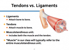
what is a musculotendinous unit?
|
|
|
|
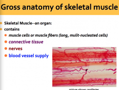
What is "contained" in a skeletal muscle? (4)
|
|
|
|
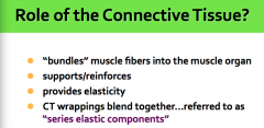
What is the role of connective tissue? (4)
|
|
|
|
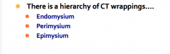
What is the hierarchy of CT wrappings? (3)
|
|
|
|
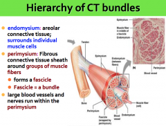
Describe endomysium and perimysium
|
|
|
|
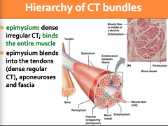
Describe epimysium
|
|
|
|
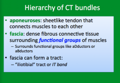
What is aponeurosis? What is fascia?
|
|
|
|
|
|
|
|
|
|
What is a condyle?
|
A rounded protuberance at the end of some bones, forming an articulation with another bone.
|
|
|
What is a perforation?
|
an aperture passing through or into something
|
|
|
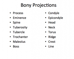
What are some examples of bony projections?
|
|
|
|
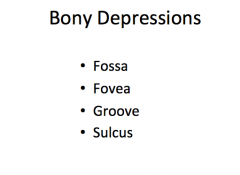
What are some examples of bony depressions? (4)
|
|
|
|
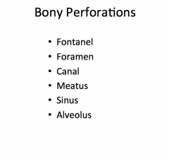
What are some examples of bony perforations? (6)
|
|
|
|
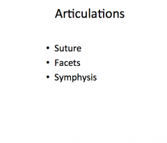
What are some examples of bony articulations? (3)
|
|
|
|
Where does ossification begin regarding growth of long bones?
|
ossification begins in the middle of the bones
|
|
|
True or False, bone cells muliply to produce growth in length of long bones
|
False
|
|
|
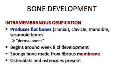
Describe intramembranous ossification (4)
|
|
|
|
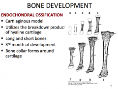
Describe endochondral ossification (5)
|
|
|
|
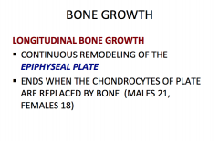
Describe longitudinal bone growth (2)
|
|
|
|
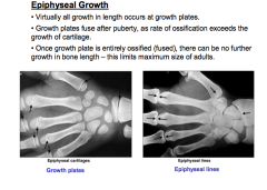
Describe epiphysial growth (3) When does growth cease and why?
|
|
|
|
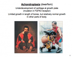
What is achondroplasia? Why does it occur? Where is the mutation?
|
|
|
|
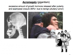
What is acromegaly? Why does it occur?
|
|
|
|
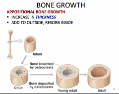
What to you call an increase in the thickness of a bone?
|
|
|
|
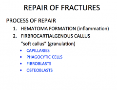
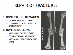
First two steps in the process of repair of fractures
|
|
|
|
Last two steps in the repair of fractures
|
|
|
|
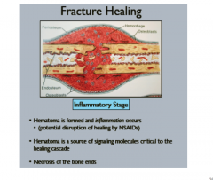
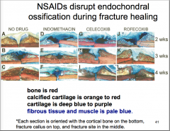
Fracture Healing: Inflammatory Stage (3) include explanation of NSAIDs
|
|
|
|
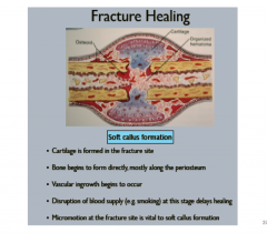
Fracture Healing: Soft Callus Formation (5)
|
|
|
|
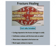
Fracture Healing: Hard Callus Formation (4)
|
|
|
|
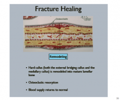
Fracture Healing: Remodeling (3)
|
|
|
|
What is the side effects of using NSAIDs? How does it change the healing process?
|
|
|
|
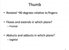
Thumb: flexes/extends in which plan? abducts/adducts in which plane?
|
|
|
Name the movement
|
Inversion
|
|
|
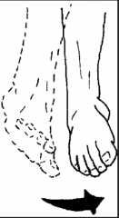
Name the movement
|
Eversion
|
|
|
Name the movement
|
plantarflexion
|
|
|
Name the movement
|
dorsiflexion
|
|
|
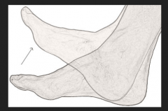
what is happening during pronation/supination?
|
Distal radius rolls over distal ulna; proximal radius spins in it's place
|
|
|
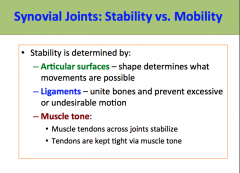
What is stability of synovial joints determined by? (3)
|
|
|
|
What are the four joints of the pectoral girdle?
|
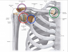
|
|
|
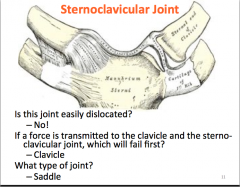
Sternoclavicular joint: is it easily dislocated? If a force is transmitted to the clavicle and the sternoclavicular joint, which will fail first? What type of joint?
|
|
|
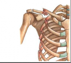
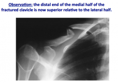
how would you describe this clavicle fracture?
|
|
|
|
|
|
|
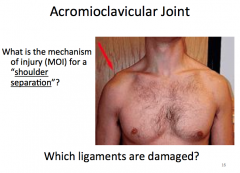
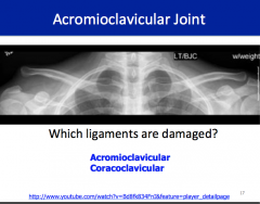
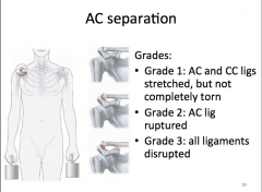
How is AC separation graded?
|
|
|
|
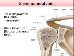
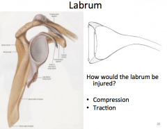
What is the glenoid labrum? How congruent is the glenohumeral joint?
|
|
|
|
How would the labrum be injured?
|
|
|
|
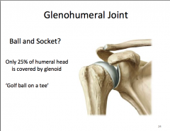
What type of joint is the glenohumeral joint? How much of the humeral head is covered by glenoid?
|
|
|
|
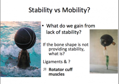
What provides stability to joints of the pectoral girdle?
|
|
|
|
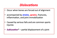
What is a disclocation? What is it accompanied by? Cause? What is partial displacement called?
|
|
|
|
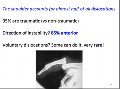
How many dislocations are accounted for by the shoulder? Direction of instability?
|
|
|
|
|
|
|
|
|
|
|
|
|
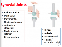
Ball and socket joint movements vs hinge joint movements?
|
|
|
|
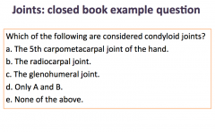
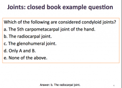
Condyloid joint movements vs saddle joint movements
|
|
|
|
pivot joint movements vs plane/gliding joint movements
|
|
|

|
C
|
|
|
|
|
|
|
|
|
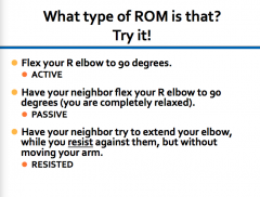
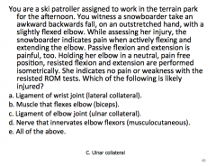
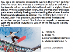
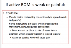
Weak and painful active ROM: muscles, nerves and ligaments
|
|
|
|
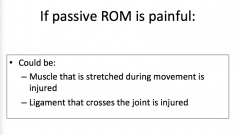
Passive ROM is painful?
|
|
|
|
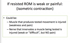
If resisted ROM is weak or painful (isometric contraction)
|
|
|
|
|
|
|
|
|
|
|
|
|
|
|
|
|
|
|
|
|
|
|
|
|
Describe the boundaries of the superior mediastinum
|
extends from superior thoracic aperture to transverse thoracic plane (sternal angle to IV disc between T4/T5)
|
|
|
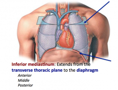
Describe the boundaries of the inferior mediastinum
|
transverse thoracic plane to diaphragm; subdivided into anterior, posterior, and middle portions
|
|
|
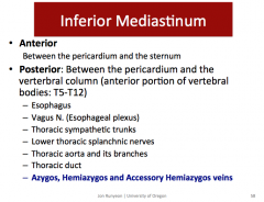
Describe the boundaries of the anterior mediastinum
|
between the pericardium and sternum
|
|
|
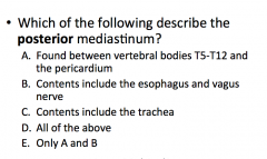
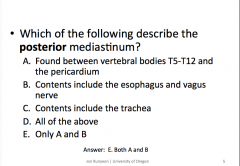
Describe the boundaries of the posterior mediastinum
|
between the pericardium and anterior portion of vertebral bodies (T5-T12)
|
|
|
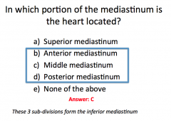
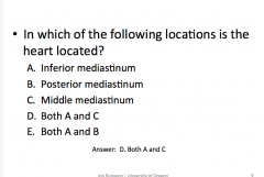
Describe the boundaries of the middle mediastinum
|
location of the heart
|
|
|
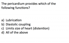
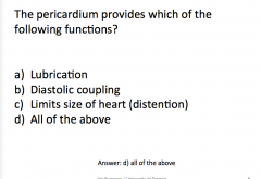
What are the functions of the pericardium? (5)
|
- Protection
- Lubrication - Prevents infection spreading - Limits size of the heart (limits gross acute distention); protects against overfilling of heart chambers and overstretching of cardiac muscle - Diastolic coupling |
|
|
What are the unique features of the right atrium? (8)
|
- smooth/thin posterior wall
- sulcus terminalis - rough/muscular anterior wall - pectinate muscles - fossa ovalis - openings for superior and inferior vena cava and coronary sinus - tricuspid valve - auricle |
|
|
What are the unique features of the left atrium? (6)
|
- base of heart
- mostly smooth-walled - auricle - pectinate muscles - openings for 4 pulmonary veins - mitral valve |
|
|
What are the unique features of the right ventricle? (7)
|
- anterior/inferior surface of heart
- trebeculae carneae - conus arteriosus - tricuspid valve - opening for pulmonic valve - 3 papillary muscles (anterior, posterior, and septal) - moderator band/septomarginal trebecula |
|
|
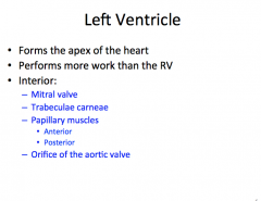
What are the unique features of the left ventricle?
|
- forms apex of heart
- performs majority of work - mitral valve - trabeculae carneae - 2 papillary muscle groups (anterior and posterior) - opening for aortic valve - thick walls |
|
|
What anatomical feature of the heart prevents the SA node from causing direct ventricular contraction?
|
Fibrous skeleton of the heart
|
|
|
What doesn't the entire heart contract with the SA node?
|
The fibrous skeleton acts as an electrical insulator between the atria and ventricles, facilitating their independent contraction
|
|
|
Which coronary artery supplies the AV and SA nodes in the majority of people?
|
Right coronary: AV (node) most people) & SA node in ~60%
Left coronary: SA node in ~40% of people & AV bundle is supplied largely by LCA |
|
|
Where do the postsynaptic fibers begin and end?
|
originate in upper thoracic and cervical ganglia of sympathetic trunks, travel to the heart after passing through the cardiac plexus, and finally innervate the SA & AV nodes, heart muscle, and coronary arteries. In addition, indirect sympathetic stimulation stems from the adrenal gland (via blood stream)
|
|
|
What is the general effect of sympathetic stimulation of the heart?
|
Increases heart rate, increase force of contraction, and dilates coronary arteries (via beta-2 receptors on coronary vessels)
|
|
|
Where do the presynaptic fibers originate and end?
|
Presynaptic parasympathetic fibers are bundles of axons from the vagus nerve, which originates in the medulla oblongata. These fibers travel through the cardiac plexus to innervate the intrinsic ganglia (postsynaptic cell bodies) of the heart wall, which project the majority of their fibers to the SA and AV nodes
|
|
|
Where are the postsynaptic fibers located?
|
heart wall...mostly wall of right atrium and interatrial septum
|
|
|
What is the general effect of parasympathetic stimulation of the heart?
|
Slows heart rate
|
|
|
Describe how lymphatic vessels progress. Where are the smallest? And the largest? Where do they start? What do they transport?
|
- capillaries are the smallest - they are blind ended and carry lymph; can be found in most cascular beds, with the notable exception of the CNS, bone and teeth
- superficial and deep lymphatic vessels collect lymph from capllaries - lymphatic vessels pass lymph through many lymph nodes, progressively draining lymph into larger vessels - the large lymphatic vessels drain into the larger lymphatic trunks - lymphatic trunks converge to form the thoracic ducts and right lymphatic duct (which drain directly to the venous system) |
|
|
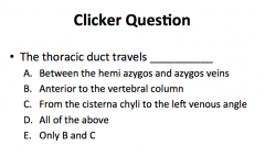
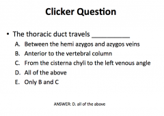
Where are the two ducts locate? Where do they terminate (drain their contents) and from where do they receive lymph?
|
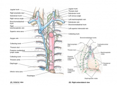
- right lymphatic duct empties into the right subclavian vein, receives lymph from R upper extremity, thorax, head an neck
- thoracic ducts empties into L subclavian vein, receives lymph from the rest of the body (L upper extremity, thorax, and head; all of abdomen, pelvis and both lower limbs) |
|
|
where are the three tonsils located?
|
- palatine tonsils: posterolateral oropharynx ("tonsils")
- pharyngeal tonsils: roof and posterior wall of nasopharynx ("adenoids") - lingual tonsil: root of the tongue - tubal tonsil: lymphoid tissue around opening of auditory tube |
|
|
What is the primary structural difference between a lymphatic nodule and an organ?
|
organs are surround by connective tissue capsule
|
|
|
What is the function of a lymph node?
|
maturation of new lymphocytes, filtration of lymph, initiation of immune response
|
|
|
Where is the thymus? Differentiate how it looks in a child/fetus vs an adult--what is its function? How does it change as you age?
|
Thymus is located anterior and superior to the heart, just posterior to the manubrium of the sternum, within the superior mediastinum; most active in childhood, diminished function as we age; progressively replaced by fat. Site of formation and maturation of t-lymphocytes, an important component of the immune system
|
|
|
What is the function of the spleen?
|
- produce lymphocytes
- initiates immune response - regulates blood volume - filters blood Located in the abdominal cavity below the diaphragm: Left Upper Quadrant (LUQ) |
|
|
name the structures of the upper respiratory tract and name one function
|
external nose, nasal cavity and pharynx
clean, warm and humidify air |
|
|
name the structures of the lower respiratory tract
|
larynx, trachea, bronchi and lungs
|
|
|
name two functions of the vocal folds
|
protects airway and produce sound
|
|
|
Describe the relationship between the respiratory and terminal bronchioles
|
One terminal bronchiole divides into multiple respiratory bronchioles. Each respiratory bronchiole leads directly to an alveolar sac
|
|
|
What are the structures (5) and functions of the nasal cavity?
|
- choana (internal nares)
- nares (external) - nasal septum - inferior, middle and superior nasal concha (turbinates) - inferior, middle and superior nasal meatus Improve the quality of the air before it reaches the lower respiratory system: humidify, warm and filter |
|
|
what are the boundaries of each portion of the pharynx?
|
- nasopharynx: internal nares to plane of soft palate
- oropharynx: soft palate to upraised epiglottis - laryngopharynx: upraised epiglottis to larynx (cricoid cartilage) |
|
|
What causes change in length of the thoracic cavity?
|
contraction or flattening of the diaphragm increases length
relaxation and elevation causes a decrease in length |
|
|
what causes change in width and depth of the thoracic cavity?
|
contraction of the intercostal muscles
|
|
|
describe the movement of the sternum when the ribs are elevated, and in which dimension the movement occurs
|
sternum moves anteriorly and superiorly, increasing the depth of the thoracic cavity
|
|
|
When the volume of the thoracic cavity increases, what happens to the pressure within?
|
the pressure decreases
|
|
|
Describe flexion/extension: plane? angles?
|
flexion is movement in the sagittal plane (anterior-posterior plane) that reduces the angle between articulating elements
extension increases the angle between articulating elements, also in the sagittal plane flexion of the elbow brings your hand to your bicep extension of the elbow reverses this process |
|
|
Describe adduction/abduction: movement? plane?
|
abduction is movement from the longitudinal axis of the body in the frontal plane
adduction is movement toward the longitudinal axis of the body, in the frontal plane swinging your leg to the side of your body is abduction bringing your leg back to the anatomical position is adduction |
|
|
Describe internal/external rotation (or medial/lateral rotation)
|
internal (or medial) rotation is the movement of the anterior aspect of a limb rotating inward, toward the ventral surface of the body
rotating my head from looking to my right, back to the anatomical position, is an example of internal rotation external (or lateral) rotation is movement of the anterior aspect away from the ventral surface of the body moving my head from the anatomical position to a position looking over my shoulder is external rotation rotation occurs, in this case, in the transverse plane |
|
|
Describe circumduction
|
the circular movement of a joint that the distal end of the limb traces an arc or complete circle
an example of this movement is tracing a circle, in the air, with your arm held straight could occur in any anatomical plane, depending on the joint involved moving the shoulder through circumduction results in movement through the sagittal, frontal, and transverse planes |
|
|
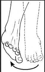
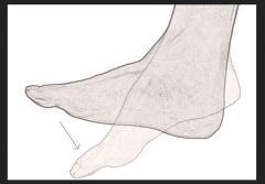
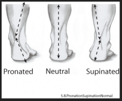
Foor special movements: Describe plantar flexion/dorsiflexion, inversion/eversion, supination/pronation
|
dorsiflexion is flexion of the ankle and elevation of the toes towards the shin "digging in your heels"
plantar flexion is the opposite...."pointing your toes" eversion is twisting the foot that turns the sole outward inversion turns the sole inward-->rolling an ankle pronation of the foot: combination of eversion and abduction that results in lowering of medial margin of foot..."flat footed" supination of the foot: results in raising the medial margin of the foot |
|
|
Hand special movements: opposition
|
special movement of the thumb, producing pad-to-pad contact of the thumb with palm or any other finger
the reverse is called reposition |
|
|
Special forearm movements: supination/pronation
|
twisting the wrist and hand from palm facing front to palm facing back, rotating the radius across the anterior surface of the ulna, is called pronation
the opposite movement, turning the palm forward, is called supination |
|
|
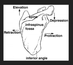
Special scapular movements: protraction/retraction, elevation/depression, rotation
|
protraction is the movement of a body part anteriorly (forward) in the horizontal plane
retraction is the movement of a body part posteriorly elevation of the scapula rasised the shoulders superiorly (shrugging) depression moves the shoulders inferiorly (down) rotation is the turning or revolving og the bone around its longitudinal axis |
|
|
Special trunk movements
|
lateral flexion or side bending
occurs when vertebral column bends to the side |
|
|
Special shoulder movements: horizontal adduction/abduction
|
horizontal adduction occurs when the shoulder is flexed and the same arm is brought across the body, towards midline
horizontal abduction occurs when the shoulder is flexed and the same arm is moved away from the body, out to the side |
|
|
describe a ball and socket joint and provide an example
|
allows movement in multiple axes and planes: flexion/extension, abduction/adduction, medial/lateral rotation, circumduction; tri-axial or multi-axial joint; example is shoulder and hip joints
|
|
|
describe a hinge joint and give an example
|
flexion and extension only, in single plane; monoaxial joint; examples are elbow and interphalangeal joint
|
|
|
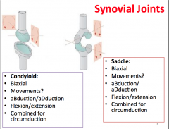
describe a saddle joint and give an example
|
abdction/adduction & flexion/extension are possible; biaxial joints/movement in two plants (frontal and sagittal); combined movement can produce circumduction; carpometacarpal joint at base of 1st digit (thumb) is a saddle joint
|
|
|
describe a condyloid joint and give an example
|
abduction/adduction & flexion/extension are possible; biaxial joints, movement in two planes (frontal and sagittal), movement in sagittal plane is usually greater, circumduction also possible (more restricted that that of saddle joints); metacarpophalangeal joints (knuckels) are an example of this joint
|
|
|
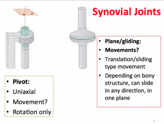
describe a pivot joint and give an example
|
rotation only, around central axis; uniaxial; example is central atlantoaxial joint between atlas (C1) and (C2)
|
|
|
describe a plane/gliding joint and give an example
|
slight movement in plane of relatively flat bone surfaces; depending on location of planar joint, movement can be in any direction
examples are between clavicle and scapula (acromioclavicular joint), between carpal and tarpal bones |
|
|
Describe the cartilaginous model of bone growth
|
model of bone (in fetal period) made of cartilage (chondroblasts, growth cells); bone eventually replaces cartilage via ossification/calcification
|
|
|
Describe the primary ossification center
|
where bone tissue replaces cartilage in main body of bone; diaphysis is bone formed from primary ossification center
|
|
|
Describe the secondary ossification center:
|
occurs at various parts of bone, often at ends of long bones
begins at birth or later, depending on bone ossification centers contain capillaries and osteoblasts |
|
|
Describe calcification of bone
|
deposition of calcium salts within tissue
|
|
|
What is ossification?
|
formation of bonr
|
|
|
Describe the epiphysial plate
|
"growth plate" separates epiphysis from diaphysis; hyaline cartilage
|
|
|
Describe the epiphysial line:
|
dense osseous seam formed when diaphysis fuses with epiphyses, replacing growth plates (and all cartilage) with bone, terminating longitudinal bone growth
|
|
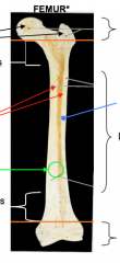
Identify the proximal and distal epiphysis, diaphysis, metaphysis, spongy (trabecular) bone, compact (cortical) bone, medullary cavity, epiphyseal line and periosteum
|
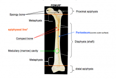
Identify the proximal and distal epiphysis, diaphysis, metaphysis, spongy (trabecular) bone, compact (cortical) bone, medullary cavity, epiphyseal line and periosteum
|
|
|
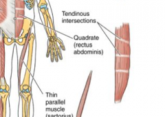
flat skeletal muscle
|
also called convergent
muscle fibers based over broad area, come together at common attachment site; pectoralis muscles are an example |
|
|
unipennate skeletal muscle
|
muscle cells all on same side of tendon; feather like arrangement of fasicles
ex: extensor digitorum muscle (extends fingers) |
|
|
bipennate skeletal muscle
|
muscle fibers on both sides of tendon
ex: rectus femoris muscle |
|
|
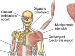
multipennate skeletal muscle
|
muscle fibers along branched tendon
ex: deltoid muscle (triangle muscle covering superior surface of shoulder joint) |
|
|
circular skeletal muscle
|
fibers are arranged concentrically around an opening
contraction decreases diameter of opening ex: orbicularis oris of mouth |
|
|
quadrate skeletal muscle
|
four sided muscle
ex: pronator quadratus |
|
|
fusiform skeletal muscle
|
fascicles (bundles of muscle fibers) run parallel to long axis of muscle
most skeletal muscles are fusiform spindle shaped with round thick belly ex: biceps brachii |
|
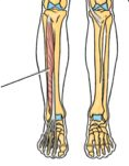
label the nerves/arteries
|
|
|
|
Where is the sulcus terminalis?
|
The separation of the right atrial pectinate muscle from the sinus venarum is indicated externally by a groove, the terminal sulcus (sulcus terminalis or crista terminalis), which extends from the front of the superior vena cava to the front of the inferior vena cava, and represents the line of union of the sinus venosus of the embryo with the primitive atrium. On the internal aspect of the right atrium, corresponding to the sulcus terminalis is the crista terminalis.
The superior border the sulcus terminalis designates the transverse plane in which the SA node resides. The inferior border designates the transverse plane in which the AV node resides. |
|
|
What is the function of the septomarginal trabecula?
|
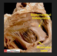
The septomarginal trabecula (also known as moderator band) is a muscular band of heart tissue found in the right ventricle. It is well-marked in sheep and some other animals, and frequently extends from the base of the anterior papillary muscle to the ventricular septum.
This septomarginal trabecula is important because it carries part of the right bundle branch of the AV bundle of the conduction system of the heart to the anterior papillary muscle. This shortcut across the chamber of the ventricle seems to facilitate conduction time, allowing coordinated contraction of the anterior papillary muscle; (see electrical conduction system of the heart). From its attachments it was thought to prevent overdistension of the ventricle, and was named the "moderator band". The moderator band is often used by radiologists and obstetricians to more easily identify the right ventricle in prenatal ultrasound. |