Reading...
![]()
Play button
![]()
Play button
![]()
Use LEFT and RIGHT arrow keys to navigate between flashcards;
Use UP and DOWN arrow keys to flip the card;
H to show hint;
A reads text to speech;
271 Cards in this Set
- Front
- Back
|
Anatomy
|
the study of the structure of body parts and their relationships to one another
|
|
|
Physiology
|
the study of the function of the body’s structural machinery
|
|
|
Physiology
|
- Considers the operation of specific organ systems
Ex. Neurophysiology – workings of the nervous system - Cardiovascular – operation of the heart and blood vessels - Focuses on the functions of the body, often at the cellular or molecular level - Understanding physiology also requires a knowledge of physics, which explains: electrical currents , blood pressure, muscle/bone movement |
|
|
Principle of Complementarity
|
- Function always reflects structure
- What a structure can do depends on its specific form |
|
|
Levels of Structural Organization
|
- Chemical – atoms combined to form molecules
- Cellular – cells are made of molecules - Tissue – consists of similar types of cells - Organ – made up of different types of tissues - Organ system – consists of different organs that work closely together - Organism – made up of the organ systems |
|
|
Integumentary System
|
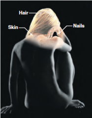
- Forms the external body covering
- Composed of the skin, sweat glands, oil glands, hair, and nails - Protects deep tissues from injury and synthesizes vitamin D |
|
|
Skeletal System
|
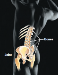
Composed of bone, cartilage, and ligaments
- Protects and supports body organs - Provides the framework for muscles - Site of blood cell formation - Stores minerals |
|
|
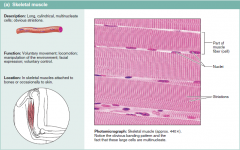
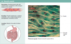
Muscular System
|
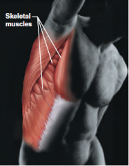
- Composed of muscles and tendons
- Allows manipulation of the environment, locomotion, and facial expression - Maintains posture - Produces heat |
|
|
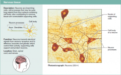
Nervous System
|
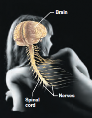
- Composed of the brain, spinal column, and nerves
- Is the fast-acting control system of the body - Responds to stimuli by activating muscles and glands |
|
|
Cardiovascular System
|
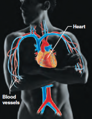
- Composed of the heart and blood vessels
- The heart pumps blood - The blood vessels transport blood throughout the body |
|
|
Lymphatic System
|
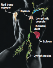
- Composed of red bone marrow, thymus, spleen,
lymph nodes, and lymphatic vessels - Picks up fluid leaked from blood vessels and returns it to blood - Disposes of debris in the lymphatic stream - Houses white blood cells involved with immunity |
|
|
Respiratory System
|
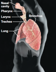
- Composed of the nasal cavity, pharynx, trachea, bronchi,
and lungs - Keeps blood supplied with oxygen and removes carbon dioxide |
|
|
Digestive System
|
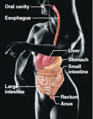
- Composed of the oral cavity, esophagus, stomach,
small intestine, large intestine, rectum, anus, and liver - Breaks down food into absorbable units that enter the blood - Eliminates indigestible foodstuffs as feces |
|
|
Urinary System
|
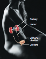
- Composed of kidneys, ureters, urinary bladder, and urethra
- Eliminates nitrogenous wastes from the body - Regulates water, electrolyte, and pH balance of the blood |
|
|
Male Reproductive System
|
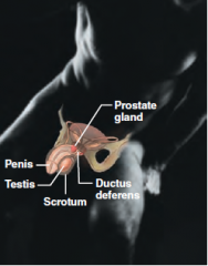
- Composed of prostate gland, penis, testes, scrotum, and ductus deferens
- Main function is the production of offspring - Testes produce sperm and male sex hormones - Ducts and glands deliver sperm to the female reproductive tract |
|
|
Female Reproductive System
|
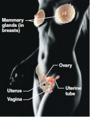
- Composed of mammary glands, ovaries, uterine tubes, uterus, and vagina
- Main function is the production of offspring - Ovaries produce eggs and female sex hormones - Remaining structures serve as sites for fertilization and development of the fetus - Mammary glands produce milk to nourish the newborn |
|
|
Organ Systems Interrelationships
|
No system stands alone. Examples:
- The integumentary system protects many systems of the body from the external environment - Digestive and respiratory systems, in contact with the external environment, take in nutrients and oxygen - Nutrients and oxygen are distributed by the blood for metabolic processes - Metabolic wastes are eliminated by the urinary and respiratory systems |
|
|
Homeostasis
|
- ability to maintain a relatively stable internal environment in an ever-changing outside world
- The internal environment of the body is in a dynamic state of equilibrium - Chemical, thermal, and neural factors interact to maintain homeostasis |
|
|
Homeostatic Control Mechanisms
|
Variables produce a change in the body
The three interdependent components of control mechanisms: - Receptor – monitors the environments and responds to changes (stimuli) - Control center – determines the set point at which the variable is maintained - Effector – provides the means to respond to stimuli |
|
|
Homeostatic Imbalance
|
- Disturbance of homeostasis or the body’s normal equilibrium
- Overwhelming the usual negative feedback mechanisms allows destructive positive feedback mechanisms to take over |
|
|
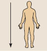
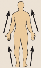
Anatomical Position
|
Body erect, feet slightly apart, palms facing forward, thumbs point away from body
|
|
|
Superior
|
Toward the head end
|
|
|
Inferior
|
Away from the head end or toward the lower part of a structure or the body
|
|
|
Anterior
(ventral) |
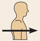
Toward or at the front of the body; in front of
Anterior refers to the leading portion of the body (abdominal surface in humans, head in a cat), but ventral specifically refers to the “belly” of a vertebrate animal, so it is the inferior surface of four legged animals |
|
|
Posterior
(dorsal) |
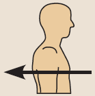
Toward or at the back of the body; behind
Although the dorsal and posterior surfaces are the same in humans, the term dorsal specifically refers to an animal’s back. Thus, the dorsal surface of four-legged animals is their superior surface. |
|
|
Medial
|
Toward or at the midline of the body; on the inner side of
|
|
|
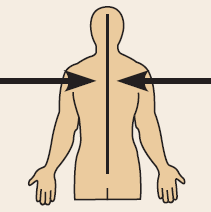
Lateral
|
Away from the midline of the body; on the outer side of
|
|
|
Superficial
|
Toward or at the body surface
|
|
|
Deep
|
Away from the body surface; more internal
|
|
|
Distal
|
Farther from the origin of a body part or the point of attachment of a limb to the body trunk
|
|
|
Distal
|
Farther from the origin of a body part or the point of attachment of a limb to the body trunk
|
|
|
Proximal
|
Closer to the origin of the body part or the point of attachment of a limb to the body trunk
|
|
|
Dorsal cavity
|
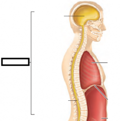
- protects the nervous system, and is divided into two subdivisions
|
|
|
Cranial cavity
|
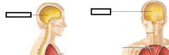
within the skull; encases the brain
|
|
|
Vertebral cavity
|
- runs within the vertebral column; encases the spinal cord
|
|
|
Ventral cavity
|
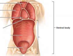
- houses the internal organs (viscera), and is divided into two subdivisions
|
|
|
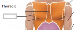
Thoracic
Abdominopelvic |
Thoracic cavity is subdivided into two pleural cavities, the mediastinum, and the pericardial cavity
|
|
|
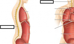
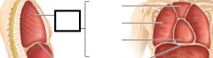
Pleural cavities
|
– each houses a lung so that lungs are independant
|
|
|
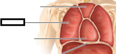
Mediastinum
|
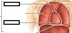
– contains the pericardial cavity; surrounds the remaining thoracic organs
|
|
|
Pericardial cavity
|
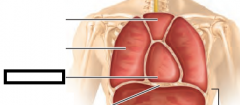
– encloses the heart
|
|
|
Abdominopelvic cavity
|
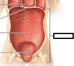
- is separated from the superior thoracic cavity by the dome-shaped diaphragm
- It is composed of two subdivisions - Abdominal cavity – contains the stomach, intestines, spleen, liver, and other organs - Pelvic cavity – lies within the pelvis and contains the bladder, reproductive organs, and rectum |
|
|
Ventral Body Cavity Membranes
|
- Parietal serosa lines internal body walls
- Visceral serosa covers the internal organs - Serous fluid separates the serosae - Name of membrane ties to organ Pleura – lungs Pericardial – heart Peritoneum – abdominal cavity, contains subdivision (omentum, mesenteric, mesocolon) |
|
|
Oral and digestive
|
– mouth and cavities of the digestive organs
|
|
|
Nasal
|
–located within and posterior to the nose
|
|
|
Orbital
|
– house the eyes
|
|
|
Middle ear
|
– contains bones (ossicles) that transmit sound vibrations
|
|
|
Synovial
|
– joint cavities
|
|
|
Abdominopelvic Quadrants
|
Right upper,
Left upper, Right lower, Left lower |
|
|
AbdominoPelvic Regions
|
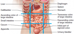
- Rt hypochondriac - (below rib cartilage) Right lobe of liver
- Epigastric – left liver lobe, most of stomach - Lt hypochondriac – small portion of stomach and transverse colon - Rt lumbar – gallbladder, ascending colon, - Umbilical (belly button) –small intestines, most of transverse colon - Lt lumbar – portion of descending colon, some small intestines - Rt iliac (Inguinal) - cecum, appendix - Hypogastric (Pubic) - small intestines, urinary bladder, rectum - Lt iliac - portion of descending colon, some small intestines |
|
|
Ribosomes
|
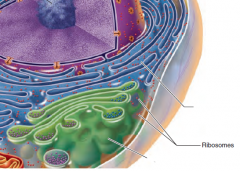
Actual site of protein synthesis
Non-membranous(rRNA, mRNA, tRNA) |
|
|
ee
|
ee
|
|
|
Golgi
|
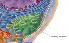
Packages proteins and other substances for export.
Membranous "UPS" of cell |
|
|
Lysosomes
|
Digest worn out cell organelles and foreign substances.
Membranous |
|
|
Peroxisomes
|
Contain oxidase enzymes to detoxify
Membranous clean up ... |
|
|
Mitochondria
|
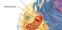
Oxidize foodstuffs to Produce ATP
Membranous |
|
|
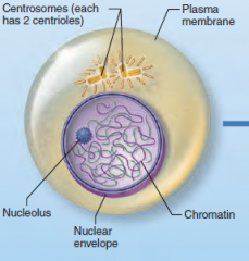
Centrioles
|
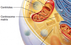
Direct formation of mitotic spindle during cell division
Non-Membranous |
|
|
Nucleus
|
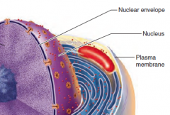
Contains nuclear envelope, nucleoli, chromatin, and distinct compartments rich in specific protein sets
Gene-containing control center of the cell Contains the genetic library with blueprints for nearly all cellular proteins Dictates the kinds and amounts of proteins to be synthesized |
|
|
Plasma Membrane
|

Double layer of lipids (phospholipids, cholesterol...) with embedded proteins. Proteins may extend entirely through the lipid bilayer or protrude on only one face. Most externally facing proteins and some lipids have attached sugar groups.
External cell barrier, acts in transport of substances into or out of the cell. Maintains a resting potential. Externally facing proteins act as receptors (for hormones, neurotransmitters...), transport proteins, and in cell-to-cell recognition. |
|
|
Nuclear Envelop
|

Selectively permeable double membrane barrier containing pores
Encloses jellylike nucleoplasm, which contains essential solutes Outer membrane is continuous with the rough ER and is studded with ribosomes Pore complex regulates transport of large molecules into and out of the nucleus |
|
|
Cyoskeletal
|
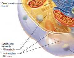
The “skeleton” of the cell
Dynamic, elaborate series of rods running through the cytosol Consists of microtubules, microfilaments, and intermediate filaments |
|
|
Cell cycle phases
|
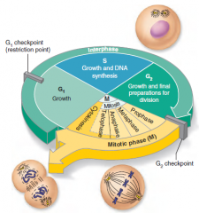
1. Interphase 22hrs
2. Mitosis/cell division- starts when cell volume increases 1.5hrs 3. Cytokinesis- pinching in 2 of the cell .5hrs |
|
Name the phase and describe it.
|
Interphase
G1Phase - Growth phase S Phase -DNA is replicated (92 chromasomes) G2 Phase - Prep for mitosis. Starts bulging Nucleus still present |
|
|
Mitosis
|

Asexual
1. Prophase 2. Metaphase 3. Anaphase 4. Telaphase |
|
|
Chromasomes vs Chromatid
|
Each chromasome has 2 chromatids
|
|

Name the phase and describe it.
|
-Nuclear membrane begins to break apart(egg yolk breaks)
-Chromosomes become visible -Centrioles separate -Spindle fibers form 'chromatin (loose spaghetti) spirals to form chromosomes(spagetti spun on fork) |
|
Name the phase and describe it.
|
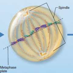
-Chromosomes attach to spindle
-Chromasomes line up across the center. -Centrioles separate (* astors) |
|
Name the phase and describe it.
|
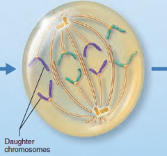
-Spindle fibers shorton pulling centromeres apart
-Sister Chromatids separate to opposite poles becoming chromosomes -Shortest phase |
|

Name the phase and describe it.
|
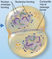
-Chromosomes gather at opposite ends
-Nuclear membrane forms enclosing chromosomes -Cytoplasm pinches inward to prepare for cytokinesis |
|
|
Major Elements of the Human Body
|
Oxygen (O)
Carbon (C) Hydrogen (H) Nitrogen (N) |
|
|
Deoxyribonucleic Acid (DNA
|
- Double-stranded helical molecule found in the nucleus of the cell
- Replicates itself before the cell divides, ensuring genetic continuity - Provides instructions for protein synthesis - Complementary Bases: --- adenine (A) and thymine (T) --- guanine (G) and cytosine (C) |
|
|
Nucleic Acids
|
- Two major classes – DNA and RNA
- Five nitrogen bases contribute to nucleotide structure – adenine (A), guanine (G), cytosine (C), thymine (T), and uracil (U) - Composed of carbon, oxygen, hydrogen, nitrogen, and phosphorus - Their structural unit, the nucleotide, is composed of N-containing base, a pentose sugar, and a phosphate group |
|
|
Ribonucleic Acid (RNA)
|
- Single-stranded molecule found in both the nucleus and the cytoplasm of a cell
- Three varieties of RNA: --- messenger RNA, --- transfer RNA, --- ribosomal RNA - Complementary Bases: --- adenine (A) and uracil (U) --- guanine (G) and cytosine (C) |
|
|
Proteins
|
Macromolecules composed of combinations of 20 types of amino acids bound together with peptide bonds
Amino Acids Building blocks of protein, containing an amino group and a carboxyl group Amino group NH2 Carboxyl groups COOH |
|
|
Amino Acids
|
- Building blocks of protein, containing an amino group and a carboxyl group
- Amino group NH2 - Carboxyl groups COOH |
|
|
Structural Levels of Proteins
|
- Primary – amino acid sequence
- Secondary – alpha helices or beta pleated sheets - Tertiary – superimposed folding of secondary structures - Quaternary – polypeptide chains linked together in a specific manners |
|
|
Fibrous and Globular Proteins
|
Fibrous proteins
--- Extended and strand-like proteins --- Very stable & insoluble in water Examples: keratin, elastin, collagen, and certain contractile fibers Globular proteins --- Compact, spherical proteins with tertiary and quaternary structures --- Water soluble, regulatory, stability of some can be effected by adverse conditions Examples: antibodies, hormones, and enzymes |
|
|
Characteristics of Enzymes
|
- Most are globular proteins that act as biological catalysts
- Holoenzymes consist of an apoenzyme (protein) and a cofactor (usually an ion or vitamin derivative) - Enzymes are chemically specific - Frequently named for the type of reaction they catalyze - Enzyme names usually end in -ase - Lower activation energy |
|
|
Mechanism of Enzyme Action
|
- Enzyme binds with substrate
- Product is formed at a lower activation energy - Product is released |
|
|
Protein Denaturation
|
- Reversible unfolding of proteins due to drops in pH and/or increased temperature
- Irreversibly denatured proteins cannot refold and are formed by extreme pH or temperature changes |
|
|
Factors Influencing Rate of Chemical Reactions
|
- Temperature – chemical reactions proceed quicker at higher temperatures
- Particle size – the smaller the particle the faster the chemical reaction - Concentration – higher reacting particle concentrations produce faster reactions - Catalysts – increase the rate of a reaction without being chemically changed - Enzymes – biological catalysts |
|
|
Acids and Bases
|
Acids:
- Acids release H+ and are therefore proton donors HCl --> H+ + Cl – - Acidic solutions have higher H+ concentration and less OH-, therefore a lower pH - Acidic: pH 0–6.99 Bases: - Bases release OH– and are proton acceptors NaOH --> Na+ + OH– - Alkaline solutions have lower H+ concentration and more OH-, therefore a higher pH - Basic: pH 7.01–14 |
|
|
The body's main electrolytes
|
NaCl,
CaCO3 (calcium carbonate), KCl (potassium chloride). CaPO4 (bones/teeth) - most plentiful Calcium- Found as a salt in bones and teeth. Its ionic (Ca21) form is required for muscle contraction, conduction of nerve impulses, and blood clotting. Phosphorus- Part of calcium phosphate salts in bones and teeth. Also present in nucleic acids, and part of ATP. Potassium- Its ion (K1) is the major positive ion (cation) in cells. Necessary for conduction of nerve impulses and muscle contraction. Sodium- As an ion (Na1), sodium is the major positive ion found in extracellular fluids (fluids outside of cells). Important for water balance, conduction of nerve impulses, and muscle contraction. Chlorine- Its ion (chloride, Cl2) is the most abundant negative ion (anion) in extracellular fluids. |
|
|
Carbohydrates
|
- Contain carbon, hydrogen, and oxygen
- Their major function is to supply a source of cellular food Examples: Monosaccharides or simple sugars Disaccharides or double sugars Polysaccharides or polymers of simple sugars |
|
|
Simple diffusion
|
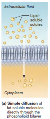
Kinetic energy
- Net movement of molecules from an area of their higher concentration to an area of their lower concentration, that is, along their concentration gradient(no carrier protein) - nonpolar and lipid-soluble substances - Diffuse directly through the lipid bilayer - Diffuse through channel proteins EX. Fats, oxygen, carbon dioxide move through the lipid bilayer of the membrane |
|
|
Facilitated diffusion
|
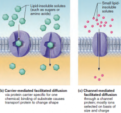
Kinetic energy
Same as simple diffusion, but the diffusing substance is attached to a lipid-soluble membrane CARRIER PROTEIN(carriermediated facilitated diffusion) or moves through a membrane channel (channelmediated facilitated diffusion) Glucose and some ions move into cells |
|
|
Osmosis
|
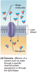
- Occurs when the concentration of a solvent is different on opposite sides of a membrane
- Diffusion of water(SOLVENT) across a semipermeable membrane - Osmolarity – total concentration of solute particles in a solution - Tonicity – how a solution affects cell volume EX. Movement of water into and out of cells directly through the lipid bilayer of the membrane or via membrane channels (aquaporins) |
|
|
Filtration
|
- The passage of water and solutes through a membrane by hydrostatic pressure
- Pressure gradient pushes solute-containing fluid from a higher-pressure area to a lower-pressure area |
|
|
Solute Pump
(Primary active transport) |
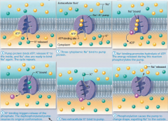
ATP
Transport of substances against a concentration (or electrochemical) gradient. Performed across the plasma membrane by a solute pump, directly using energy of ATP hydrolysis. |
|
|
Secondary active transport Ion concentration
|
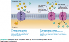
Ion conc gradient maintained with ATP
Cotransport (coupled transport) of two solutes across the membrane. Energy is supplied indirectly by the ion gradient created by primary active transport. Symporters move the transported substances in the same direction; antiporters move transported substances in opposite directions across the membrane. |
|
|
Phagocytosis
|
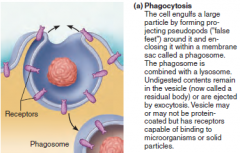
ATP
- “Cell eating”: A large external particle (proteins, bacteria, dead cell debris) is surrounded by a “seizing foot” and becomes enclosed in a vesicle (phagosome). EX. In the human body, occurs primarily in protective phagocytes (some white blood cells and macrophages) |
|
|
Pinocytosis
|
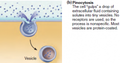
(fluid-phase endocytosis)
ATP - Plasma membrane sinks beneath an external fluid droplet containing small solutes. Membrane edges fuse, forming a fluid-filled vesicle. Occurs in most cells; important for taking in dissolved solutes by absorptive cells of the kidney and intestine |
|
|
Vesicular Transport
|
Fluids containing large particles and macromolecules are transported across cellular membranes inside membranous sacs called vesicles.
|
|
|
Exocytosis
|
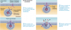
ATP
Secretion or ejection of substances from a cell. The substance is enclosed in a membranous vesicle, which fuses with the plasma membrane and ruptures, releasing the substance to the exterior. EX. Secretion of neurotransmitters, hormones, mucus, etc.; ejection of cell wastes |
|
|
Selectively Permeable
|
allows only select substances to enter & leave the cell
|
|
|
The cell membrane
|
- Gatekeeper of the Cell
- Composed of Phospholipid Bilayer: --- Hydrophillic Head Polar; faces outside of membrane --- Hydrophobic Tails Non-polar, fatty acid chains facing inside of membrane - Integral Proteins assist in the transport of lg. substances in/out of cell; marks the cell as “self” - Cholesterol/ Carbohydrates provides support & flexibility of cell |
|
|
Effects of Solutions of Varying Tonicity
|
- Isotonic – solutions with the same solute concentration as that of the cytosol
- Hypertonic – solutions having greater solute concentration than that of the cytosol(cause crenation) - Hypotonic – solutions having lesser solute concentration than that of the cytosol (burst/lyse) |
|
|
Cilia
Flagellum |
- Whip-like, motile cellular extensions on exposed surfaces of certain cells
- Move substances in one direction across cell surfaces Flagellum Like a cilium, but longer; only example in humans is the sperm tail. Propels the cell. |
|
|
What is the trigger for cell division
|
Your bodies way of initiating/ starting CELL DIVISION is to increase the volume inside of the cell increasing cytoplasm, organelles, & DNA.
|
|
|
The four types of tissues
|
- Epithelial - Coverings and linings/Glandular epithelium
- Connective - support - Muscle - motion - Nerve - control |
|
|
Epithelial Tissue
|
- Cellularity – composed almost entirely of cells
- Special contacts – form continuous sheets held together by tight junctions and desmosomes - Polarity – apical and basal surfaces - Supported by connective tissue – reticular and basal laminae - Avascular but innervated – contains no blood vessels but supplied by nerve fibers - Regenerative – rapidly replaces lost cells by cell division |
|
|
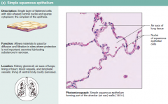
Simple Squamous
|
- Single layer of flattened cells with disc-shaped nuclei and sparse cytoplasm
- Functions Diffusion and filtration Provide a slick, friction-reducing lining in lymphatic and cardiovascular systems Present in the kidney glomeruli, lining of heart, blood vessels, lymphatic vessels, and serosae |
|
|
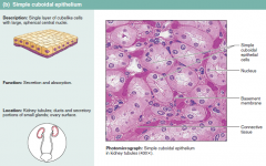
Simple Cuboidal
|
- Single layer of cube-like cells with large, spherical central nuclei
- Function in secretion and absorption - Present in kidney tubules, ducts and secretory portions of small glands, and ovary surface |
|
|
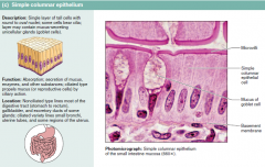
Simple Columnar
|
- Single layer of tall cells with oval nuclei; many contain cilia
- Goblet cells are often found in this layer - Function in absorption and secretion - Nonciliated type line digestive tract and gallbladder - Ciliated type line small bronchi, uterine tubes, and some regions of the uterus - Cilia help move substances through internal passageways |
|
|
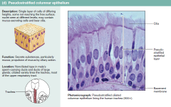
Pseudostratified Columnar
|
- Single layer of cells with different heights; some do not reach the free surface
- Nuclei are seen at different layers - Function in secretion and propulsion of mucus - Present in the male sperm-carrying ducts (nonciliated) and trachea (ciliated) |
|
|
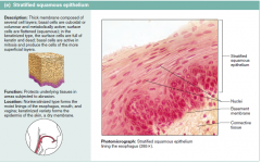
Stratified Squamous
|
- Thick membrane composed of several layers of cells
- Function in protection of underlying areas subjected to abrasion - Forms the external part of the skin’s epidermis (keratinized cells), and linings of the esophagus, mouth, and vagina (nonkeratinized cells) |
|
|
Stratified Cuboidal and Columnar
|
Stratified cuboidal
- Quite rare in the body - Found in some sweat and mammary glands - Typically two cell layers thick Stratified columnar - Limited distribution in the body - Found in the pharynx, male urethra, and lining some glandular ducts - Also occurs at transition areas between two other types of epithelia |
|
|
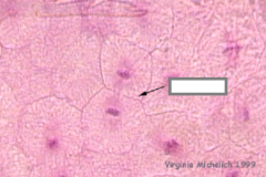
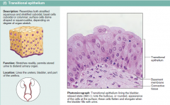
Transitional
|
- Several cell layers, basal cells are cuboidal, surface cells are dome shaped
- Stretches to permit the distension of the urinary bladder - Lines the urinary bladder, ureters, and part of the urethra |
|
|
Connective Tissue
|
- Found throughout the body; most abundant and widely distributed in primary tissues
--- Connective tissue proper --- Cartilage --- Bone --- Blood - Connective tissues have: --- Mesenchyme as their common tissue of origin --- Varying degrees of vascularity --- Nonliving extracellular matrix, consisting of ground substance and fibers - Structural Elements of Connective Tissue --- Ground substance – unstructured material that fills the space between cells --- Fibers – collagen, elastic, or reticular --- Cells – fibroblasts, chondroblasts, osteoblasts, and hematopoietic stem cells |
|
|
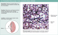
Fibers
|
- Collagen – tough; provides high tensile strength
- Elastic – long, thin fibers that allow for stretch - Reticular – branched collagenous fibers that form delicate networks |
|
|
Special connections between cells
|
- Tight Junctions - Protein molecules in adjacent cell membranes fuse, forming an impermeable connection
--- Keeps enzymes, etc. within the proper tissue (digestive tract) - Desmosomes - Strands of protein extend and connect adjacent cell membranes; helps resist tearing --- stresses --- neck of the uterus --- Heart muscle --- skin - Gap Junctions - Cell membranes connected by hollow cylinders that allow transmission of small molecules (electrically excitable tissue) |
|
|
What is Adipose and what does it do?
|
- Matrix similar to areolar connective tissue with closely packed adipocytes
- Reserves food stores, insulates against heat loss, and supports and protects - Found under skin, around kidneys, within abdomen, and in breasts - Local fat deposits serve nutrient needs of highly active organs |
|
|
Connective Tissue (Function)
|
- Binding and support
- Protection - Insulation - Transportation |
|
|
What is the most abundant tissue in the body?
|
Connective
Collagen is most abundant among 3 fibers Hyaline cartilage is the most abundant cartillage |
|
|
Connective tissue Cells
|
- Fibroblasts – connective tissue proper
- Chondroblasts – cartilage - Osteoblasts – bone - Hematopoietic stem cells – blood - White blood cells, plasma cells, macrophages, and mast cells |
|
|
-blast versus -cyte
|
The immature (undifferentiated) cells, indicated by the suffix blast (literally, “bud” or “sprout,” but the suffix means “forming”), are actively mitotic cells that secrete the ground substance and the fibers characteristic of their particular matrix
Mature, less active mode, indicated by the suffix cyte The mature cells maintain the health of the matrix. However, if the matrix is injured, they can easily revert to their more active state to repair and regenerate the matrix. (The blood-forming stem cells are always actively mitotic.) |
|
|
Endocrine gland
|
- Ductless glands that produce hormones
- Secretions include amino acids, proteins, glycoproteins, and steroids |
|
|
Exocrine Gland
|
- More numerous than endocrine glands
- Secrete their products onto body surfaces (skin) or into body cavities - Examples include mucous, sweat, oil, and salivary glands - The only important unicellular gland is the goblet cell - Multicellular exocrine glands are composed of a duct and secretory unit |
|
|
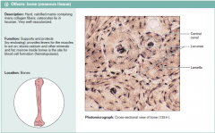
Cartilage types
|
Hyaline Cartilage
- Amorphous, firm matrix with imperceptible network of collagen fibers - Chondrocytes lie in lacunae - Supports, reinforces, cushions, and resists compression - Forms the costal cartilage - Found in embryonic skeleton, the end of long bones, nose, trachea, and larynx Elastic Cartilage - Similar to hyaline cartilage but with more elastic fibers - Maintains shape and structure while allowing flexibility - Supports external ear (pinna) and the epiglottis Fibrocartilage Cartilage - Matrix similar to hyaline cartilage but less firm with thick collagen fibers - Provides tensile strength and absorbs compression shock - Found in intervertebral discs, the pubic symphysis, and in discs of the knee joint |
|
|
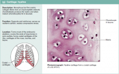
Hyaline Cartilage
|
Hyaline Cartilage
- Amorphous, firm matrix with imperceptible network of collagen fibers - Chondrocytes lie in lacunae - Supports, reinforces, cushions, and resists compression - Forms the costal cartilage - Found in embryonic skeleton, the end of long bones, nose, trachea, and larynx |
|
|
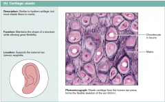
Elastic Cartilage
|
- Similar to hyaline cartilage but with more elastic fibers
- Maintains shape and structure while allowing flexibility - Supports external ear (pinna) and the epiglottis |
|
|
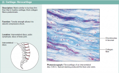
Fibrocartilage Cartilage
|
- Matrix similar to hyaline cartilage but less firm with thick collagen fibers
- Provides tensile strength and absorbs compression shock - Found in intervertebral discs, the pubic symphysis, and in discs of the knee joint |
|
|
Extracellular matrix
|
Connective tissues are largely nonliving extracellular matrix (ma9triks; “womb”), which separates, often widely, the living cells of the tissue.
Together ground substance and fibers make up the extracellular matrix. Ground Substance --- Interstitial (tissue) fluid --- Adhesion proteins – fibronectin and laminin --- Proteoglycans – glycosaminoglycans (GAGs) Functions as a molecular sieve through which nutrients diffuse between blood capillaries and cells Fibers --- Collagen – tough; provides high tensile strength --- Elastic – long, thin fibers that allow for stretch --- Reticular – branched collagenous fibers that form delicate networks |
|
|
Bone matrix vs Cartilage
|
Cartilage
--- Gel-like ground substance --- Fibers: collagen, elastic fibers in some Bone --- Gel-like ground substance --- calcified with inorganic salts --- Fibers: collagen |
|
|
inflammation, characterized by:
|
- Dilation of blood vessels
- Increase in vessel permeability - Redness, heat, swelling, and pain |
|
|
Tissue Repair
|
1. Inflammation is the body’s response to injury. Tissue repair begins during the inflammatory process. It may lead to regeneration, fibrosis, or both
2. Organization and restored blood supply --- The blood clot is replaced with granulation tissue 2. Regeneration and fibrosis --- Surface epithelium regenerates and the scab detaches --- Fibrous tissue matures and begins to resemble the adjacent tissue Results in a fully regenerated epithelium with underlying scar tissue |
|
|
How well does each tissue regenerate
|
Strong Regeneration:
--- Epithelial --- Bone --- Areolar Connective --- Dense-Irregular Connective --- Blood-forming Moderate Regeneration: --- Skeletal Muscle --- Cartilage Weak Regeneration: --- Skeletal Muscle --- Cartilage NO Regeneration: --- Cardiac Muscle --- Nervous Tissue |
|
|
Epithelial Membranes:
- Cutaneous Membrane - Mucous Membrane |
Epithelial Membranes:Cutaneous Membrane
- Cutaneous – skin Mucous Membrane - Mucous – lines body cavities open to the exterior (e.g., digestive and respiratory tracts) - Serous – moist membranes found in closed ventral body cavity |
|
|
Scar Tissue
|
- Strong, but lacks flexibility; It is mostly composed of collagen fibers.
- Not able to perform the normal functions of the original tissue! |
|
|
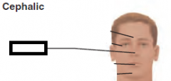
Nasal
|
|
|
|
Orbital
|
Eyes
|
|
|
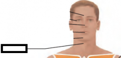
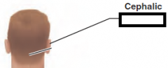
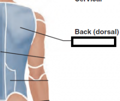
Cervical
|
|
|
|
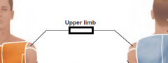
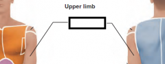
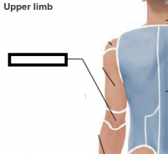
Acromial
|
|
|
|
Axillary
|
|
|
|
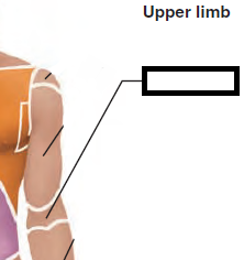
Brachial
|
|
|
|
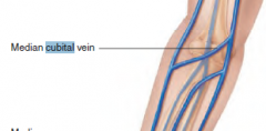
Antecubital
|
|
|
|
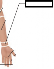
Carpal
|
|
|
|
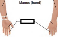
Digital
|
|
|
|
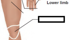
Patellar
|
|
|
|
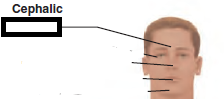
Frontal
|
|
|
|
Buccal
|
|
|
|
Femoral
|
|
|
|
Otic
|
|
|
|
Olecranal
|
|
|
|
Scapular
|
|
|
|
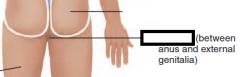
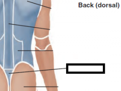
Perineal
|
|
|
|
Sacral
|
|
|
|
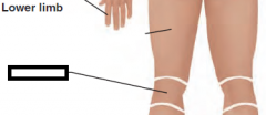
Popliteal
|
|
|
|
Plantar
|

|
|
|
Median/Midsaggittal Plane
|

A sagittal plane that lies exactly in the midline
|
|
|
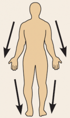
Saggittal Plane
|
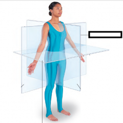
A vertical plane that divides the body into right and left parts.
|
|
|
Parasaggittal Plane
|

All other sagittal planes, offset from the midline, are parasagittal planes (para = near)
|
|
|
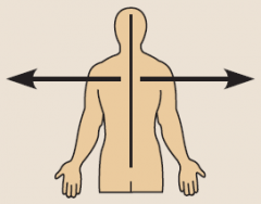
Frontal/Coronal Plane
|
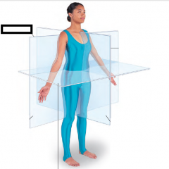
Frontal planes, like sagittal planes, lie vertically. Frontal
planes, however, divide the body into anterior and posterior parts. A frontal plane is also called a coronal plane |
|
|
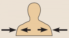
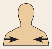
Transverse Plane
|
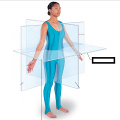
... or horizontal, plane runs horizontally from
right to left, dividing the body into superior and inferior parts. Many different transverse planes exist, at every possible level from head to foot. A transverse section is also called a cross section. |
|
|
Olecranon Process
|
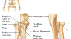
Upper forearm
|
|
|
Acromion Process
|
Shoulder
|
|
|
Popliteal Fossa
|
Lower femoral
|
|
|
Cubital Fossa
|
Front of elbow
|
|
|
Microvilli
|
Tubular extensions of the plasma membrane;
contain a bundle of actin filaments. Increase surface area for absorption |
|
|
Nucleolus
|
Dense spherical (non-membrane-bounded) bodies, composed of ribosomal RNA and proteins.
Site of ribosome subunit manufacture |
|
|
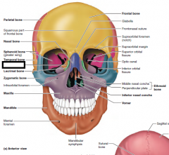
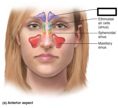
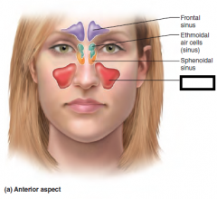
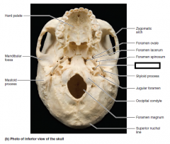
sinus
|
|
|
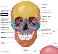
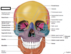
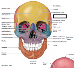
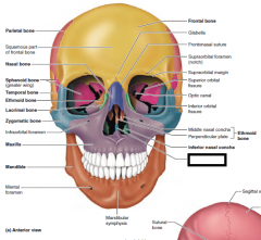
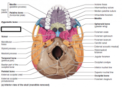
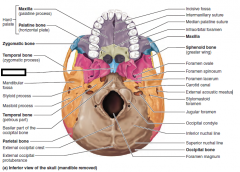
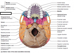
Ethmoidal Bone
|
|
|
Ethmoidal Bone(nose)
|
|
|
frontal sinus
|
|

|
Inferior Nasal Concha
|
|

|
mandible
|
|

|
maxilla
|
|
|
Maxillary sinus
|
|

|
Middle Nasal Concha
|
|
|
Nasal Bone
|
|

|
Optic canal
|
|
|
Parietal bone
|
|

|
sphenoid process
|
|

|
sphenoid sinus
|
|
|
superior orbital
|
|

|
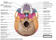
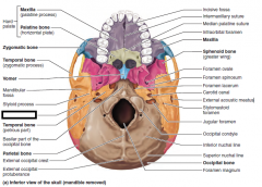
temporal bone
|
|
|
vomer
|
|
|
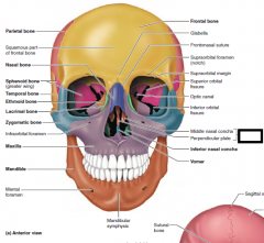
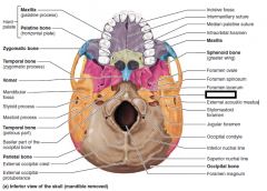
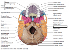
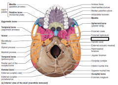
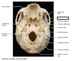
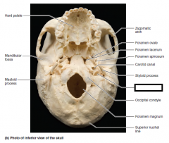
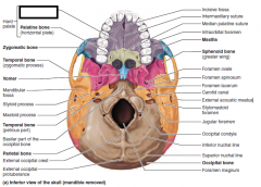
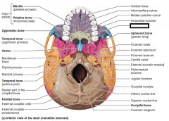
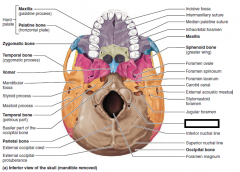
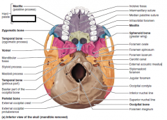
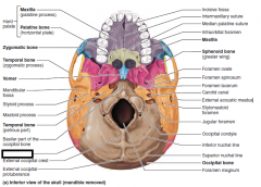
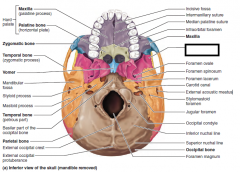
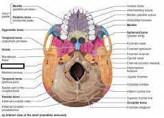
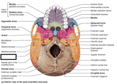
cartoid canal
|
|
|
cartoid canal
|
|
|
ext accoustic meatus
|
|
|
foramen lacerum
|
|
|
foramen magum
|
|
|
foramen ovale
|
|
|
jugular foramen
|
|
|
mastoid process
|
|
|
maxilla
|
|
|
maxilla(inferior)
|
|
|
occipital condyle
|
|
|
palentine bone
|
|
|
parietal bone
|
|
|
sphenoid bone
|
|
|
styloid process
|
|
|
temporal bone(petrous part)
|
|
|
temporal bone(zygomatic process)
|
|
|
vomer
|
|
|
zygomatic bone
|
|
|
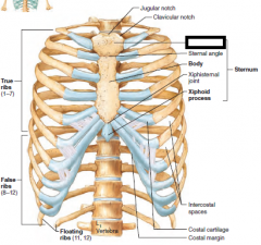
manubrium
|
|
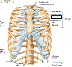
|
sternum body
|
|
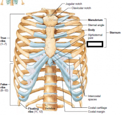
|
sternum xiphoid process
|
|
|
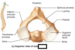
cervical
3 holes |
|
|
C1
|
|
|
C2
|
|
|
lumbar
|
|
|
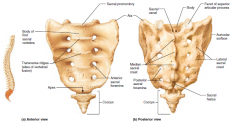
sacrum
|
|
|
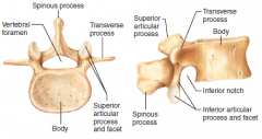
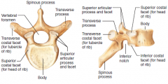
thoracic
|
|
|
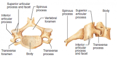
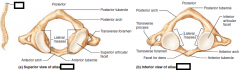
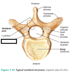
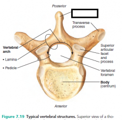
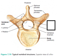
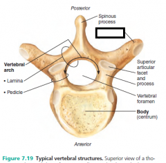
vertebral arch
|
|
|
body
|
|
|
vertebral foramen
|
|
|
lamina
|
|
|
pedicle
|
|
|
spinous process
|
|
|
Superior articular process and facet
there's an inferior |
|
|
transverse process
|
|
|
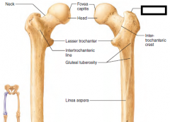
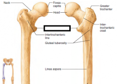
greater trochanter
|
|
|
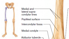
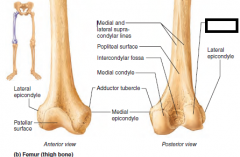
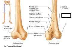
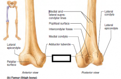
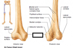
Lateral Condyle
|
|
|
Lateral epicondyle
|
|
|
lesser trochanter
|
|
|
medial condyle
|
|
|
Medial epicondyle
|
|
|
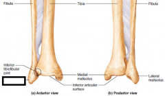
lateral malleolus
|
|
|
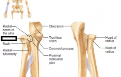
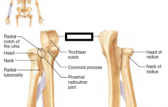
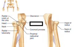
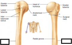
head
|
|
|
neck
|
|
|
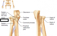
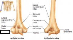
olecranon process
|
|
|
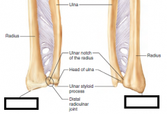
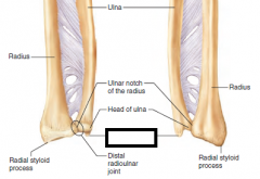
radius
|
|
|
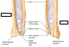
styloid process (radius)
|
|
|
styloid process ulna
|
|
|
Trochlear notch
|
|
|
capitulum
|
|
|
deltoid tuberosity
|
|
|
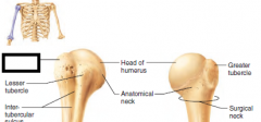
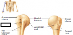
greater tubercle
|
|
|
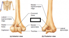
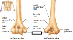
lateral epicondyle
|
|
|
lesser tubercle
|
|
|
medial epicondyle
|
|
|
olecranon fossa
|
|
|
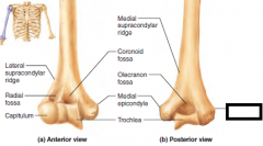
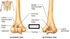
trochlea
|
|
|
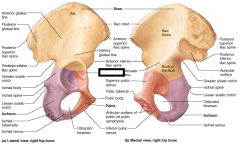
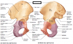
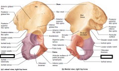
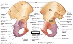
acetabulum
|
|
|
ilium
|
|
|
ischium
|
|
|
pubis
|
|
|
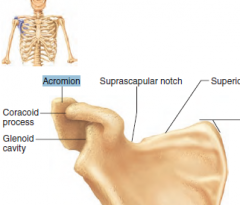
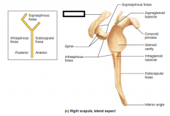
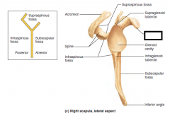
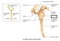
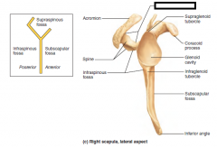
acromion
|
|
|
coracoid process
|
|
|
glenoid cavity
|
|
|
supraspinous fossa
|
|
|
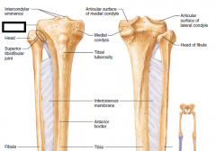
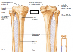
lateral condyle
|
|
|
medial condyle
|
|
|
medial malleolus
|
|
|
tibial tuberosity
|
|
|
adipose
|
|
|
areolar
|
|

|
anaphase
|
|
|
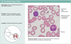
blood
|
|
|
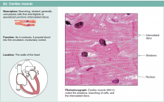
cardiac muscle
|
|
|
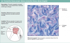
dense irregular
|
|
|
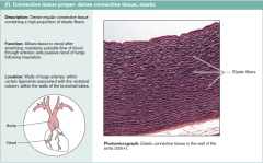
elastic
|
|
|
elastic cartilage
|
|
|
fibrocartilage
|
|
|
hyaline cartilage
|
|
|
interphase
|
|

|
metaphase
|
|
|
nerveous
|
|
|
osseous
|
|
|
prophase
|
|
|
pseudostratified columnar
|
|
|
reticular
|
|
|
simple cuboidal
|
|
|
simple columnar
|
|
|
simple squamos
|
|
|
skeletal muscle
|
|
|
smooth muscle(pink)
|
|
|
stratified squamos
|
|
|
telophase
|
|
|
transitional epithelium
|