Reading...
![]()
Play button
![]()
Play button
![]()
Use LEFT and RIGHT arrow keys to navigate between flashcards;
Use UP and DOWN arrow keys to flip the card;
H to show hint;
A reads text to speech;
147 Cards in this Set
- Front
- Back
|
What is the composition of the blood made out of?
|
--Plasma: fluid matrix, 55%
(90% is water, rest dissolved nutrients) --Formed elements: RBCs, WBCs, platelets, 45% |
|
|
What are some characteristics of erythrocytes?
|
7-8 um in diameter
pale pink biconcave 4.5 to 5 million per cubic millimeter no nucleus when mature |
|
|
What are some characteristics of leukocytes?
|
Nucleated cells
4000 to 10,000 per cubic millimeter fight infections, immunity can move in/out blood vessels (emigration or diapedesis) Two groups: granulocytes and agranular leukocytes |
|
|
What type of leukocytes are granulocytes? (3)
|
Neutrophil
Eosinophil Basophil |
|
|
What type of leukocytes are agranular leukocytes?
|
Lymphocyte
Monocyte |
|
|
How do you know order of abundancy of leukocytes?
|
Never Let Monkeys Eat Bananas
Neutrophil, Lymphocyte, Monocyte, Eosinophil, Basophil Most abundant --> least abundant |
|
|
What is the order of leukocytes in largest to smallest?
|
Monocyte, Neutrophil/eosinophil, basophil, lymphocyte
|
|
|
What are platelets?
|
Cell fragments from larger megakaryocytes
2-4 um darkly staining, irregularly shaped 140,000 to 450,000 per millimeter cubed blood clotting |
|
|
What are neutrophils?
|
Part of "granulocyte" sub category
Most abundant of the white blood cells "polymorphonuclear" leukocyte (3-7 lobed nucleus) pale purple cytoplasm, active phagocytes numbers increase during acute infections |
|
|
What are eosinophils?
|
Part of "granulocyte" sub category
Nucleus is bilobed cytoplasmic granules stain red-orange numbers increase during allergies and parasite infections |
|
|
What are basophils?
|
Part of "granulocyte" sub category
Least abundant leukocyte Large U or S shaped nucleus deep purple cytoplasmic granules with several chemicals Release histamine --Dilates blood vessels Release heparin --Prevents blood clotting |
|
|
What are lymphocytes?
|
Agranular leukocyte (no observable cytoplasmic granules)
Smallest leukocytes Generally spherical nucleus 3 types: B lymphocytes, T lymphocytes, NK cells involved in immunologic responses |
|
|
What are monocytes?
|
largest of leukocytes
kidney-shaped nucleus abundant cytoplasm active phagocyte |
|
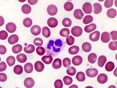
What leukocyte is this?
|
Neutrophil
|
|
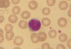
What leukocyte is this?
|
Eosinophil
|
|
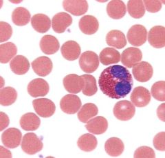
What leukocyte is this?
|
Basophil
|
|
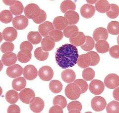
What leukocyte is this?
|
Lymphocyte
|
|
What leukocyte is this?
|
Monocyte
|
|
|
What are platelets?
|
Cell fragments from larger megakaryocytes
2-4 um, 140,000 to 450,000 per milimeter cubed Instrumental in blood clotting |
|
|
What is a differential white blood cell count?
|
Preformed during a physical examination; counting # WBC
Elevated or depressed numbers of white blood cell types are signs of certain diseases |
|
|
What is the central blood-containing space of a blood vessel?
|
Lumen
|
|
|
Name the innermost tunic of a blood vessel. What is the tunic made of?
|
Tunica intima/interna-- single layer smooth endothelium
smooth endothelium=specialized simple squamous epithelial cells In arteries: consists also of thick layer elastic fibers called the "internal elastic membrane" |
|
|
Name the middle tunic of a blood vessel. What is this tunic made of?
|
Tunica media
smooth muscle tissue in framework of loose connective tissue How vessels constrict and dilate |
|
|
Name the outer tunic of a blood vessel? What is this tunic made of?
|
Tunic adventitia/externa--collagen connective tissues
stabilization/anchoring |
|
|
Which tunic is thickest in an artery?
|
Tunica media
|
|
|
Which tunic is thickest in a vein?
|
Tunica adventitia/externa
|
|
|
Which single tunic is present in capillaries?
|
Tunica intima/interna (endothelium)
|
|
|
What are the 3 types of arteries found in the body?
|
Elastic arteries (largest) , muscular arteries (second largest) and arterioles (no tunica externa)
|
|
|
How is blood flow different in the arteries than in the veins?
|
Arteries bring blood away from the heart; veins carry blood toward the heart
|
|
|
What material is most abundant in elastic arteries?
|
Elastic fibers
|
|
|
What anatomical structures help to regulate blood flow through the capillary beds?
|
Arterioles and venules
The precapillary sphincter |
|
|
What three adaptations help return venous blood to the heart?
|
1. Valves
2. large diameter lumen 3. Muscular pumps and respiratory pumps |
|
Which is the artery and which is the vein?
|

Here!
|
|
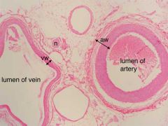
In this picture:
For artery-- What is thick red portion? What is lighter portion? What is innermost portion? |
Tunica media
Tunica adventitia Tunica interna |
|
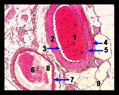
Describe what the tunics look like in a vein.
|
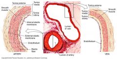
Look at this pic.
|
|
|
What is the ductus venous? What does it become?
|
A shunt that bypasses the liver in the fetal heart.
From the umbilical vein a majority of the blood passes through the ductus and directly into the fetal inferior cava. It closes at birth and becomes ligamentum venosum. |
|
|
What is the foramen ovale? What does it turn into?
|
A hole in the septum separating the left and right atrium of the fetal heart. It allows blood to bypass the collasped fetal lungs.
It closes at birth leaving an indentation called the fossa ovalis. |
|
|
What is the ductus arteriosus? What does it turn into?
|
A bridge between the pulmonary artery and the aorta, allows blood to bypass the pulmonary circuit. It closes at time of birth and leaves the ligamentous arteriosum.
|
|
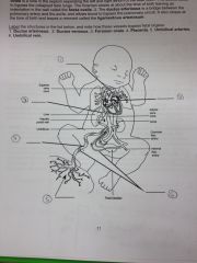
Label the items in this picture of fetal heart circulation.
|

|
|
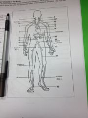
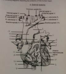
Label the arteries of the head and neck:
--Left common carotid --Right common carotid --external carotid --internal carotid |
--Left common carotid=10A
--Right common carotid=10B --external carotid=1 --internal carotid=9 |
|

Label the ascending aorta and its branches:
--Ascending aorta --Arch of aorta --Brachiocephalic --Subclavian (L) |
--Ascending aorta=14
--Arch of aorta=13 --Brachiocephalic=2 --Subclavian (L)=11 |
|

Label the descending aorta and its branches
--Abdominal aorta --Renal --Superior mesenteric --Common iliac |
--Abdominal aorta=5
--Renal=4 --Superior mesenteric=15 --Common iliac=7 |
|

Label the arteries of the upper limb
--Axillary --Brachial --Radial --Ulnar |
--Axillary=12
--Brachial=3 --Radial=6 --Ulnar=16 |
|

Label the arteries of the lower limb.
--External iliac --Femoral --Anterior tibial --Dorsalis pedis |
External iliac=7
Femoral=18 Anterior tibial=8 Dorsalis pedis=19 |
|
Label the following veins
--Axillary, Brachial, Brachiocephalic, Cephalic, Common iliac, femoral, great saphenous, inferior vena cava, internal iliac, internal jugular, median cubital, radial, renal, subclavian, superior vena cava, ulnar |
--Axillary (4), Brachial (7), Brachiocephalic (3), Cephalic (5), Common iliac(13), femoral (16), great saphenous(15), inferior vena cava(8), internal iliac(14), internal jugular(1), median cubital(9), radial(11), renal(10), subclavian(2), superior vena cava(6), ulnar(12)
|
|
|
Why is blood considered a connective tissue?
|
nonliving fluid matrix (plasma) in which living elements (formed elements) are suspended and fibers typical of a connective tissue matrix become visible as fibrin threads during clotting
|
|
|
How much blood does the normal human have in body?
|
5-5.5 L
|
|
|
What is a hemocytometer and how did we use it?
|
Hemocytometer=counting chamber of known volume
--first we diluted blood with a fluid that prevented blood coagulation and caused the WBC cells to rupture --cells placed in hemocytometer and cells are counted by microscopic inspection, then corrections for dilution and chamber volume are made to obtain the results in cubic millimeter of blood |
|
|
What is polycythemia and how do we detect it?
|
Increase in the number of RBCs
may result from bone marrow cancer or from living at high altitudes where less oxygen is available |
|
|
What is anemia?
|
A decrease in the oxygen carrying capacity of blood
This may result from either a decrease in RBCs (hemorrage or can't make anymore) or decreased hemoglobin content of RBCS |
|
|
What is leukocytosis?
|
An increase in the total number of white blood cells
May indicate an infection, poisoning or hemorrhage |
|
|
What is leukopenia?
|
A decrease in white blood cell counts below 4000/mm^3
Diseases that could cause this are tuberculosis, measles or infectious hepatitis |
|
|
What is leukemia?
|
A malignant disease of the lymphoid organs that is characterized by an increase in abnormal white blood cells with a decrease in red blood cells and platelets
|
|
|
How do we count cells of a hemocytometer and how do we get the total red blood cell counts from there?
|
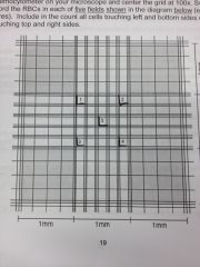
Count cells touching bottom and left of each of the squares above
Add the cells in all five groups and multiple by 10,000 We get this 1000 by multiplying our dillution factor (2000 in this case) x the volume correction factor (50) to get 10,000 |
|
|
What is the hematocrit (and it's nickname)? When is it determined? How?
|
Aka Paced Cell Volume (PCV), generally considered equal to the RBC volume (bc WBC=only 1% total blood volume) and considered as a percentage to plasma
Determined when anemia is suspected because more accurate than total RBC count Centrifuge whole blood so formed elements at bottom and plasma at top, then hold up card to determine percentages |
|
|
What is perhaps the most accurate way of measuring the oxygen-carrying capacity of blood?
|
Determining the hemoglobin content, aka the hemoglobin concentration in g per 100 mL of blood
Use Tallquist hemoglobin scale to determine color of blood Generally ratio between hematocrit and grams of hemogobin per 100 mL is 3:1 |
|
|
What is Mean Corpuscular Volume and how do we calculate it?
|
Average volume of an individual RBC, in fL
MCV=hematocrit/RBC count(per L of blood) |
|
|
What is the normal range for MCV? What are conditions if this is off?
|
80-100 fL (10^-15 L)
normocytic=RBC w/ normal size/volume macrocytic=RBC larger than normal microcytic=RBC lower than normal |
|
|
What is 1 fL? 100 mL? 1mm^3?
|
10^-15 L
1 dL 1 x 10^-6 L |
|
|
What is MCHC?
|
MCHC=mean corpuscular hemoglobin concentration
Avg. concentration of hemoglobin in a given volume of red blood cells |
|
|
What is the average range for MCHC? What are some conditions of it?
|
32-36 gm/dl
normochromic=RBC with normal amt hemoglobin hyperchromic=more hemoglobin than normal hypochromic=less hemoglobin than normal |
|
|
How do we calculate MCHC?
|
MCHC=hemoglobin (g/dl) / hematocrit
|
|
|
What is MCH, how do we calculate it and what are some normal values for it?
|
MCH=hemoglobin (g/L) / RBC count per L
Normal=27-31 picograms (10^-12 g) Used along with MCV, MCHC and MCH to distinguish diff. forms of anemia |
|
|
What is iron deficiency anemia? Vitamin B12 deficiency?
|
iron deficiency = RBCs are microcytic and hypochromic
B12=RBCs are macrocytic and normochromic |
|
|
For blood type A, what are the antigens and antibodies? Who could it donate to and receive from? What would the card look like?
|
Antigens:A
Antibodies: Anti-B Receives from: O, A Donate to: AB, A Card: Anti-A serum would have clumps, anti-B serum wouldn't |
|
|
For blood type B, what are the antigens and antibodies? Who could it donate to and receive from? What would the card look like?
|
Antigens: B
Antibodies: Anti-A Receive from: B, O Donate to: AB, B Card: no clumps with A, clumps with B |
|
|
For blood type AB, what are the antigens and antibodies? Who could it donate to and receive from? What would the card look like?
|
Antigens: A and B
Antibodies: none Donate to: AB Receive from: A,B, O Card: clumps for both A and B spots |
|
|
For blood type O, what are the antigens and antibodies? Who could it donate to and receive from? What would the card look like?
|
Antigens: none
Antibodies: anti-A and anti-B Donates to: A, B, AB Receives from: O only Card: no clumps on A or B spots |
|
|
What is the Rh antigen?
|
Rh + means you have it, Rh - says you don't
If you mix blood with anti-Rh serum, get clumping saying you have it Rh + can receive from Rh + or Rh - Rh - can receive from Rh - and Rh + at first, but then start making antibodies for following transfusions |
|
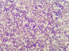
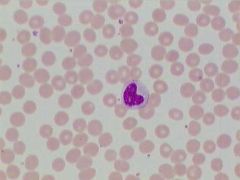
What is this condition?
|
Leukemia, a lymphoid cancer characterized by an increase in abnormal white blood cells with a decrease in red blood cells and platelets
|
|
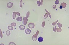
What is this condition?
|
Sickle cell anemia
A genetic disease that results in the production of modified hemoglobin molecules, hemoglobin S that do not pack properly into the red blood cell Causes the red blood cells to take on crescent shapes when deoxygenated and, being fragile, can easily rupture or stick in small blood vessels, diminishing the blood supply |
|
|
What is the flow of arterial blood from the heart to the dorsal surface of the foot (dorsalis pedis)?
|
Left atrium, left ventricle, ascending aorta, descending aorta, abdominal aorta, right common iliac artery, right external iliac artery, femoral artery, popliteal artery, tibial artery, anterior tibial artery, dorsalis pedis
|
|
|
Which large vein delivers blood back to the right side of the heart from the head, neck and upper appendages?
|
Superior vena cava
|
|
|
Which large vein delivers blood back to the right side of the heart from the lower extremities?
|
Inferior vena cava
|
|
|
What blood vessel delivers oxygenated blood from the mother's placenta to the fetus?
|
Umbilical veins
|
|
|
What structure in the fetal heart allows for mixing of blood between the two atria?
|
Foramen ovale
|
|
|
What vessel in the fetus allows blood to bypass the lungs and directly enter the systemic circulation? What is the value of this?
|
Ductus arteriosus: blood does not travel to the collapsed lungs
|
|
|
What are the differences between blood in the right side of the heart versus the left?
|
Left: O2 rich, CO2 poor
Right: O2 poor, CO2 rich |
|
|
In general, where does blood go that is pumped from the right side of the heart?
|
The lungs
|
|
|
In general, where does blood go that is pumped from the left side of the heart?
|
Body cavities, capillaries, tissue
|
|
|
What is the pulmonary circuit?
|
1. Blood leaves heart via pulmonary arteries
2. Lungs 3. Returns to heart via pulmonary veins |
|
|
What is the systemic circuit?
|
1. Blood leaves heart via aorta
2. goes to body 3. goes back to heart via systemic veins |
|
|
Identify three key structures visible on a histological slide of cardiac muscle
|
Intercalated disks
Striations Nuclei (one per cell) |
|
|
What are two types of junctions in an intercalated disk?
|
1. Gap junction
2. desmosomes |
|
|
What is the function of the desmosomes in the intercalated disc?
|
Anchors tissue together
|
|
|
What is the function of gap junctions in the intercalated disc?
|
Allows action potential to proceed thru cells (functional synticium)
|
|
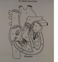
Label this picture of the heart
|
2=aorta
10=aortic semilunar valve 7=bicuspid (mitral) valve 9=chordae tendineae 11=inferior vena cava 14=interventricular septum 6=left atrium 15=left ventricle 16=myocardium 12=papillary muscles 3=pulmonary artery 5=pulmonary semilunar valve 4=right atrium 13=right ventricle 1=superior vena cava 8=tricuspid valve |
|
Label this picture of the heart
|
See above
|
|
|
What term is used to describe the force that blood exerts against the walls of the blood vessels?
|
Blood pressure
|
|
|
In what units is blood pressure expressed?
|
mm Hg
|
|
|
What is laminar flow in a blood vessel?
|
normal blood flow
velocity of blood highest in middle rather than sides of blood vessel minimizes energy loss due to friction |
|
|
What is a pulse?
|
Alternating surges of pressure (expansion and then recoil) in an artery that occur with each contraction and relaxation of the left ventricle
|
|
|
What is systolic pressure and what causes it?
|
Maximum pressure exerted by blood against the artery walls and the peak of ventricular ejection; caused by ventricular systole
|
|
|
What is a typical value for systolic pressure?
|
120 mm Hg
|
|
|
What is diastolic pressure and what causes it?
|
Lowest pressure in artery; caused by ventricular diastole (relaxation)
|
|
|
What is a typical value for diastolic pressure?
|
80 mmHg
|
|
|
What is pulse pressure? What does a narrow range or wide range indicate?
|
Difference between systolic pressure and diastolic pressure; throb you feel when you take a pulse
Narrow (<30 mmHg)=aortic blockage or tachycardia Wide (>40 mmHg)=hypertension |
|
|
What would a normal pulse pressure value be? (Assume a measured pressure of 120/80 mmHg)
|
40 mmHg
|
|
|
How is mean arterial pressure calculated?
|
MAP= diastolic pressure + 1/3 of pulse
|
|
|
What is the significance of MAP?
|
acc. wikipedia: "average arterial pressure during a single cardiac cycle"
ACTUAL pressure that propels blood to tissues (not average because diastole last longer than systole) |
|
|
What do the two sounds correspond to when taking a blood pressure?
|
Systolic pressure (first) and diastolic pressure (second)
|
|
|
What do the semilunar valves prevent backflow of the blood into?
|
The two ventricles
|
|
|
What do the AV valves prevent backflow into?
|
The atriums
|
|
|
Why are the left ventricle walls thicker than the right?
|
Left=needs to pump blood to whole body so needs to be able to supply more force
Right=just needs to pump to lungs |
|
|
Starting from the right atrium, explain the pathway of the blood
|
Right atrium -- tricuspid valve -- right ventricle -- pulmonary semilunar valve -- pulmonary trunk --left and right pulmonary arteries -- capillary beds of lungs --left and right pulmonary veins -- left atrium -- biscuspid mitral valve -- left ventricle --contraction -- aortic semilunar valve -- aorta -- capillaries of tissues --superior and inferior vena cava -- right atrium
|
|
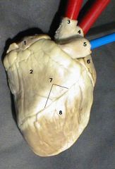
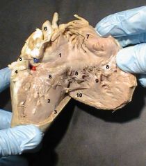
Label this picture of the sheep heart.
|
1. Right Auricle
2. Right Ventricle (thinner/more flexible walls) 3. Brachiocephalic Artery (Oxygenated--first branch aorta) 4. Aortic Arch (Oxygenated) 5. Pulmonary Artery (Deoxygenated) 6. Left Auricle 7. Interventricular Sulcus (has anterior interventricular artery and great cardiac vein) 8. Left Ventricle |
|
|
What forms the outer surface of the heart?
|
Visceral pericardium aka epicardium
One layer of squamous cells |
|
|
How do we know what the pulmonary trunk/artery is?
|
Large vessel between the right and left atria as seen from the anterior side of the heart
|
|
|
How do we know where the aorta is?
|
Large vessel juts beneath (posterior to) pulmonary trunk. First branch of it, the brachiocephalic artery, may be necessary.
|
|
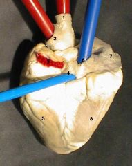
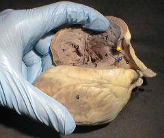
Label
|
1. Brachiocephalic Artery (Oxygenated)
2. Aortic Arch (Oxygenated) 3. Openings for Pulmonary Veins (Oxygenated) 4. Opening for Inferior Vena Cava (Deoxygenated) 5. Left Ventricle 6. Opening for Superior Vena Cava (Deoxygenated) 7. Right Auricle 8. Right Ventricle |
|
|
Where do the pulmonary veins enter?
|
4 of them, the left atrium
seen on posterior (not round) surface |
|
|
Where does the superior vena cava enter?
|
Attached to upper part right atrium; goes into right atrium and out of right ventricle is inferior vena cava
|
|
Label
|
1. Right Atrium
2. Right Ventricle 3. Myocardium (Note Thinness) 4. Opening of Coronary Sinus 5. Inferior Vena Cava 6. Superior Vena Cava 7. Pectinate Muscle 8. Tricuspid Valve 9. Papillary Muscle 10. Moderator Band |
|
|
Where is the pectinate muscle and what is it?
|
Inner comb-like surface of right atrium with ridges (not as many in left atrium)
|
|
|
What is the coronary sinus?
|
Between inferior vena cava and tricuspid valve; where blood of coronary circulation is returned to venous circulation
|
|
|
Where is the tricuspid valve?
|
Between right and left atria
|
|
|
What is the chordae tendinae?
|
Weird stringlike things in ventricles that connect the papillary muscles to the AV valves
|
|
|
What is the trabeculae carneae?
|
Pitted and ridged parts of right inner right and left ventricular wall
|
|
|
What does the moderator band stretch across?
|
Right ventricle
|
|
Label
|
1. Right Ventricle
2. Papillary Muscle 3. Moderator Band 4. Pulmonary Semilunar Valve 5. Pulmonary Artery 6. Left Ventricle |
|
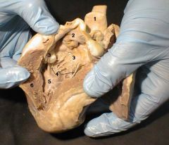
Label
|
1. Aortic Arch
2. Left Atrium 3. Mitral Valve 4. Chordae Tendineae 5. Left Ventricle 6. Myocardium (Note Thickness) |
|

Label!
|
1. Pulmonary Artery
2. Brachiocephalic Artery 3. Aorta 4. Aortic Seminlunar Valve 5. Opening for Coronary Artery 6. Trabeculae Carnae of Left Ventricle 7. Ventricular Myocardium (Note Thickness) |
|
|
What is the "lubb" sound mean?
|
Associated with the closure of the AV valves at the beginning of stole
|
|
|
What is the "dupp" sound mean?
|
Associated as the semilunar valves close at the end of systole
|
|
|
What are abnormal heart sounds called and what do they indicate?
|
Murmurs
Indicate valvular problems |
|
|
What happens in valves that do not close tightly?
|
Closure is followed by a swishing sound due to backflow of the blood (regurgitation)
|
|
|
What does the term pulse mean and what does it often equal?
|
Refers to the alternating surges of pressure (expansion and then recoil) in an artery tht occur with each contraction and relaxation of the left ventricle
Normally equals the heart rate and pulse averages 70 to 76 beats per minute |
|
|
Where is the pulse easily felt?
|
On any superficial artery when the artery is compressed over a bone or a firm tissue
|
|
|
Where is the common carotid artery?
|
At the side of the neck
|
|
|
Where is the temporal artery?
|
Anterior to the ear in the temple region
|
|
|
Where is the facial artery?
|
Clench teeth, palpate the pulse just anterior to the masseter muscle on the mandible (in line with the corner of the mouth)
|
|
|
Where is the brachial artery?
|
In the antecubital fossa, where it bifurcates into the radial and ulnar arteries
|
|
|
Where is the radial artery?
|
At the lateral aspect of the wrist, just above the thumb?
|
|
|
Where is the femoral artery?
|
In the groin
|
|
|
Where is the popliteal artery?
|
At the back of the knee
|
|
|
Where is the posterior tibial artery?
|
Just above the medial malleolus
|
|
|
Where is the dorsalis pedis artery
|
On the dorsum of the foot
|
|
|
What is the apical pulse?
|
The actual counting of the heartbeats; usually faster than the radial because of a slight lag in time as the blood rushes from the heart into the large arteries
|
|
|
What if there is a large difference between the apical and radial pulses?
|
This is a pulse deficit
May indicate cardiac impairment (a weakened heart unable to pump blood into the arterial tree to a normal extent or abnormal heart rhythms) |
|
|
Where do you take the apical pulse?
|
Put sthethoscope over the fifth left intercostal space
|
|
|
What is blood pressure
|
The pressure the blood exerts against any unit area of the blood vessel walls
|
|
|
What is the difference between systolic pressure and diastolic pressure?
|
Systolic=pressure in arteries at the peak of ventricular ejection
Diastolic=pressure during ventricular relaxation |
|
|
What is a sphygmomanometer and how do we use it?
|
Blood pressure cuff
1. Inflate and put stethoscope on brachial artery 2. Release pressure until hear first sound of Korotkoff (systolic pressure) 3. Continue until the sound disappears (diastolic pressure) |
|
|
What is the pulse pressure? What does a narrow range or wide range indicate?
|
=difference between the systolic and diastolic pressures
Narrow range (<30)=aortic blockage or tachycardia Wide range (>40)=hypertension |
|
|
What is mean arterial pressure (MAP)?
|
The TRUE pressure that propels the blood to the tissues, or the actual "working" pressure
MAP=diastolic pressure + (pulse pressure/3) |
|
|
When listening to a HEARTBEAT what is the lupp sound? the dup sound?
|
Lub: beginning of systole, the AV valves closing
Dupp: end of systole, semilunar valves closing |
|
|
What are the papillary muscles?
|
In anatomy, the papillary muscles are muscles located in the ventricles of the heart. They attach to the cusps of the atrioventricular valves (a.k.a. the mitral and tricuspid valves) via the chordae tendinae and contract to prevent inversion or prolapse of these valves.[1][2]
|