Reading...
![]()
Play button
![]()
Play button
![]()
Use LEFT and RIGHT arrow keys to navigate between flashcards;
Use UP and DOWN arrow keys to flip the card;
H to show hint;
A reads text to speech;
49 Cards in this Set
- Front
- Back
|
1. Histology of the red bone marrow. Embryonic/ foetal haematopoiesis. Haematopoietic stem cells and their regulation.
describe stem cells |
Haematopoietic stem cells (HSCs)
- in bone marrow -unique ability to give rise to all of the different mature blood cell types and tissues. - HSCs are self-renewing cells: when they proliferate, at least some of their daughter cells remain as HSCs, so the pool of stem cells does not become depleted.This phenomenon is called asymmetric division. - other daughters of HSCs (myeloid and lymphoid progenitor cells), however can commit to any of the alternative differentiation pathways that lead to the production of one or more specific types of blood cells, but cannot self-renew. -pool of progenitors is heterogeneous and can be divided into two groups, long-term self-renewing HSC and only transiently self-renewing HSC, also called short-terms. This is one of the main vital processes in the body. All precursor cells have some common features: They are larger in diameter than mature red and white blood cells The nuclei have non-condensed chromatin The cytoplasm is rich in free ribosome |
|
|
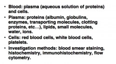
1. blood and hematopoiesis
whats in blood? investigation methods |
|
|
|
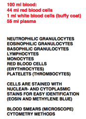
1. 100 ml blood contents
|
|
|
|
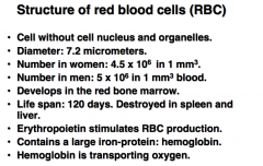
1. rbc structure
|
|
|
|
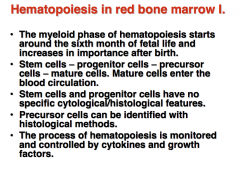
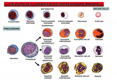
hematopoiesis 1
|
blood cell formation
occurs in RED!! bone marrow of flat bones and proximal ends of long bones 1st trimester- yolk salk 2nd trimester- liver and spleen 3rd trimester- bone marrow |
|
|
hematopoiesis part 2
|

|
|
|
hema in embryo
|
IN THE WALL OF THE YOLK SAC, LIVER, AND SPLEEN
|
|
|
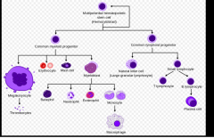
blood cell lineages
|
|
|
|
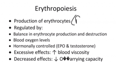
2. The structure and development of erythrocytes and thrombocytes:
-erythropoiesis general |
|
|
|
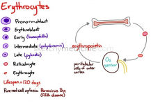
erythrocyte development
|
|
|
|
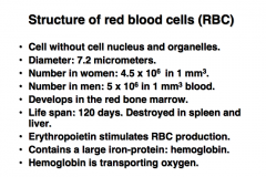
RBC structure
|
|
|
|
thrombocytes
|
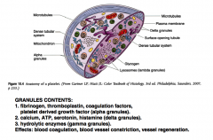
(platelet) structure: three zones of platelet structure, peripheral, structural, and organelle
|
|
|
development of RBC and platelets (lineage) 1
|
|
|
|
lineage 2 (less general)
|
|
|
|
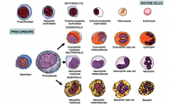
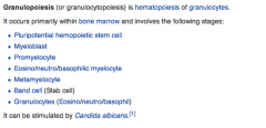
granulocytopoiesis cells invlolved
|
|
|
|
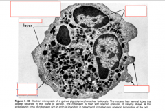
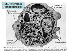
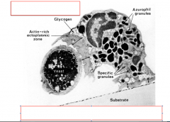
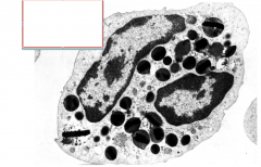
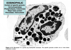
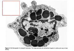
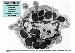
granulocyte sturcture
|
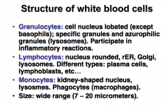
Neutrophils are normally found in the bloodstream and are the most abundant type of phagocyte, constituting 50% to 60% of the total circulating white blood cells
Basophils : least abundant cells in bone marrow and blood (occurring at less than two percent of all cells). Like neutrophils and eosinophils, they have lobed nuclei; however, they have only two lobes, |
|
|
|
|
|
|
|
|

|
|
|
|
|
|
|
|
|
|
|
|
|
|
|
|
|
|
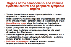
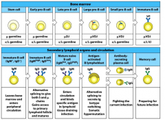
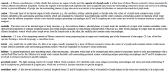
Name Organs of hemopoietic and immune systems: central and peripheral lymphoid organs
|
|
|
|
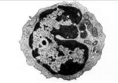
4. Structure of monocytes and the mononuclear phagocytic system. Development
of monocytes. Types, structure and development of lymphocytes. |
-ype of white blood cells (leukocytes).
- largest of all leukocytes. - part of the innate immune system -amoeboid in shape, having clear cytoplasm - bean shaped nuclei that are unilobar, makes them one of the types of "mononuclear leukocytes"(agranulocytes). - 2% to 10% of all leukocytes -They play multiple roles in immune function. (1) replenishing resident macrophages under normal states 2) in response to inflammation signals, monocytes can move quickly (approx. 8–12 hours) to sites of infection in the tissues and divide/differentiate into macrophages and dendritic cells to elicit an immune response. -Half stored in the spleen -identified in stained smears by their large kidney shaped or notched nucleus. These change into macrophages after entering into the tissue spaces. |
|
|
monocyte macrophage development
|
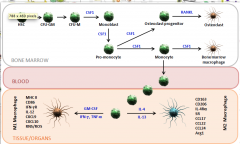
HSC: Hematopoietic stem cell
CFU-M: Colony forming unit-macrophage TNF-α: Tumor necrosis factor alpha IFN-γ: Interferon gamma RNS: Reactive nitrogen species CFU-GM: Colony forming unit-granulocyte/macrophage Arg1: Arginase-1 enzyme CXCL and CCL: Chemokines SR: Scavenger Receptors ROS: Reactive oxygen species IL-: Interleukin- CSF1: Macrophage colony-stimulating factor 1 (M-CSF) RANKL: Receptor activator of NF-κB ligand GM-CSF: Granulocyte macrophage-colony stimulating factor |
|
|
mononuclear phagocytic system description and function
|
![part of the immune system that consists of the phagocytic cells[1] located in reticular connective tissue. The cells are primarily monocytes and macrophages, and they accumulate in lymph nodes and the spleen. The Kupffer cells of the liver and tis...](https://images.cram.com/images/upload-flashcards/45/23/27/5452327_m.png)
part of the immune system that consists of the phagocytic cells[1] located in reticular connective tissue. The cells are primarily monocytes and macrophages, and they accumulate in lymph nodes and the spleen. The Kupffer cells of the liver and tissue histiocytes are also part of the MPS.
|
|
|
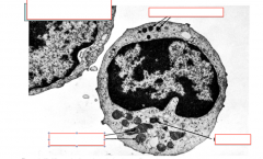
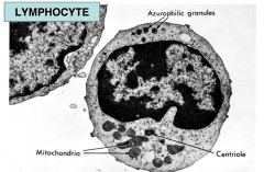
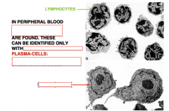
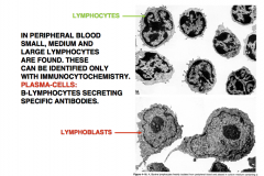
Lymphoctye defined and chart of function and percent
|
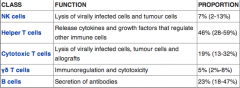
lymphocyte is any of 3 types of white blood cell in a vertebrate's immune system. All 3 are agranulocytes. They include natural killer cells (NK cells) (which function in cell-mediated, cytotoxic innate immunity), T cells (for cell-mediated, cytotoxic adaptive immunity), and B cells (for humoral, antibody-driven adaptive immunity). They are the main type of cell found in lymph, which prompted the name lymphocyte.
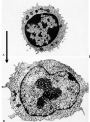
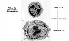
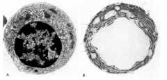
Lymphocytes can be identified by their large nucleus. LYMPHOCYTES CAN BE STIMULATED FOR DIVISION, DEDIFFERENTIATION AND DIFFERENTIATION (DEDIFFERENTIATION = LYMPHOBLASTIC TRANSFORMATION) |
|
|
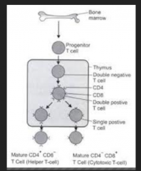
T cell development
|
|
|
|
B cell Development
|
|
|
|
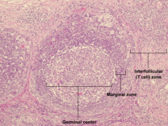
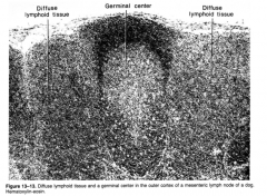
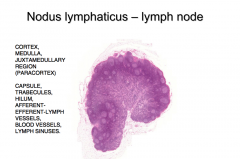
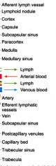
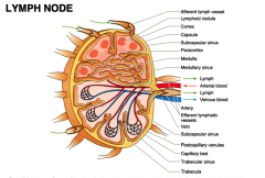
5. Histology of lymphatic follicles and lymph nodes.
- list what to look for |
|
|
|
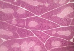
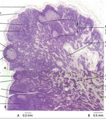
THYMUS
Stained with haematoxylin and eosin 1 - lobules 2 - interlobular connective tissue |
|
|
lymphoid follicle, what is it? function, structure, size. cells present
|
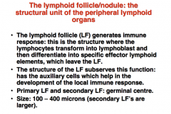
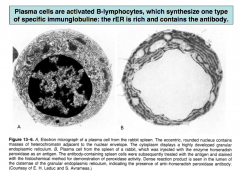
Lymphocytes (B- and T-type).
Lymphoblasts (transformed lymphocytes). Macrophages (phagocytose dead cells). Antigen presenting cells (follicular dendritic cells): take up and digest antigens, then put it into the cell membrane in order to present it to lymphocytes.. Reticular cells and reticular fibers. |
|
|
|
|
|
|
|
|
|
|
|
|
|
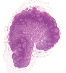
what is it? and parts
|
|
|
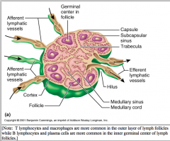
draw a lymph node with these things
|
|
|
|
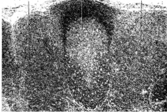
draw another follicle...
|
|
|
|
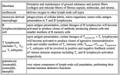
chart of lymphoid cells and functions
|
|
|
|
|
|
|
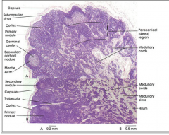
describe parts of lymph node:
capsule of a lymph node cortex medulla trabeculae lobule follicles germinal center medullary cord |
|
|
|
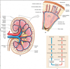
Draw Renal Bloodflow
|
|
|
|
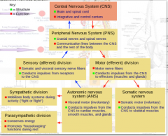
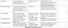
Nerves Nervous system flowchart basic
|
|
|
|
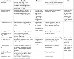
Nevre chart 1
|
|
|
|
Nerve chart 2
|
|
|
|
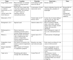
Nerves 3
|
|