Reading...
![]()
Play button
![]()
Play button
![]()
Use LEFT and RIGHT arrow keys to navigate between flashcards;
Use UP and DOWN arrow keys to flip the card;
H to show hint;
A reads text to speech;
45 Cards in this Set
- Front
- Back
|
During the 3rd week of development the intraembryonic mesoderm on each side of the midline differentiates into what 3 mesoderms?
What do these 3 germ layers form? |
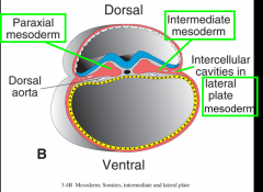
1. paraxial
2. intermediate 3. lateral -form the trilaminar germ disc |
|
|
obj.
Describe the formation of the intraembryonic cavity within lateral plate mesoderm |
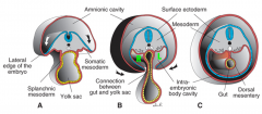
lateral plate mesoderm divides into 2 layers:
-somatic/parietal mesoderm -splanchnic/visceral mesoderm Space btwn these layers = intraembryonic cavity -initially right & left intraembryonic cavities on each side of midline --> merge into single intraembryonic cavitiy (goes from throacic region to pelvic region) |
|
|
obj.
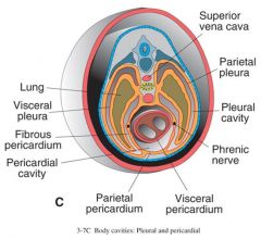
Describe the division of the single intraembryonic cavity into the pericardial, pleural, and peritoneal cavities |
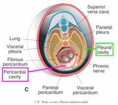
Once the left/right intraembryonic cavity has merged into a single cavity, it will go on to subdivide into three cavities surrounding important structures, later in development
-pericardial cavity (around heart) -plueral cavities (one around each lung) -peritoneal cavity (around abdominal organs) |
|
|
During abdominal development, the temporary primitive umbilical ring is caused by what 2 structures?
What will this area become? |
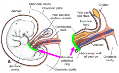
(5 wk embryo on left, 10 wk on right)
1. the vitelline duct (connection btwn midgut & yolk sac) 2. connecting stalk -becomes umbilicus |
|
|
obj.
Discuss development of the serous membranes lining the body cavities |
cells lining the intraembryonic cavity form a thin layer --> serous (mesothelial membrane)
part that develops from parietal mesoderm = parietal layer (lines inside of body wall) part that develops from visceral mesoderm = visceral layer (covers heart, lungs, abdominal organs) |
|
|
obj.
Innervation of serous membranes lining body cavities |
parietal layer of serous membrane:
anterior abdomen- T7-12 & L1 central diaphram- phrenic nerve peripheral diaphram- T7-12 (lower intercostal nerves) pelvic region- obturator nerve visceral layer of serous membrane: sensory innervation from autonomic afferent nerves |
|
|
obj.
function of mesenteries for the peritoneal cavity |
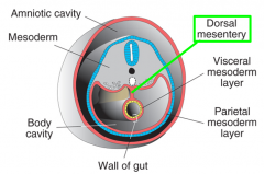
-Parietal & visceral layers of peritoneum (in abdomen) are continuous at dorsal mesentary
-Mesentary is a double layer of peritoneum that provides a pathway for blood vessels, nerves, & lymphatics to reach abdominal organs * |
|
|
The pericardial cavity and the pleural cavity are separated by the development of what membrane?
Why does this membrane develop? |
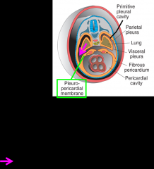
separated by pleuropericardial membranes, which develop as a result of the lungs growing into the body wall on each side, segmenting the intraembryonic cavity
|
|
|
What forms to separate the thoracic and abdominal cavities?
|
respiratory diaphram
|
|
|
obj.
Describe the formation of the respiratory diaphragm |
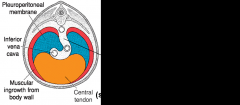
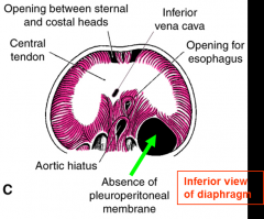
The diaphram develops from the following sources:
-septum transversum--> central tendon -pleuroperitoneal membranes -dorsal mesentery of the esophagus (btwn membrane) -mesoderm (ingrowth) of the body wall |
|
|
obj
Describe the embryological basis for the innervation of the respiratory diaphram |
-septum transversum: C3-5, phrenic nerve
(phrenic supplies all diaphram EXCEPT peripheral sensory) -sensory of peripheral portion (developed from mesoderm of body wall) of diaphram: GSA from lower intercostal nerves |
|
|
What is the septum transversum and how does its position change as the embryo develps?
|
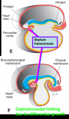
-it is a plate of mesodermal tissue that initially lies opposite cervical somites
(innervated by C3-5) -repositioned caudally by cephalocaudal folding due to differential growth -lies btwn developing heart in thorax & liver in abdomen -forms the central tendon of the diaphram |
|
|
What is differential growth?
|
the vertebral column and associated tissues grow more rapidly than the ventral part of embryo
|
|
|
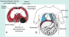
Development of the septum transversum leaves holes btwn thoracic & abdominal cavities, the _________________, on each side of the foregut
How are these holes closed? |
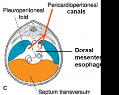
-pericardioperitoneal canals
-closed by development of pleuroperitoneal folds, that fuse w/ septum and dorsal mesentary of esophagus |
|
|
obj.
Discuss the development of congenital diaphragmatic hernias and their clinical importance. |
-the defect (foramen of Bochdalek), most frequently results from failure of one or both pleuroperitoneal membranes to close the pericardioperitoneal canals
* one of the most common malformations of newborns (1:2000), can sometimes be repaired in utero |
|
|
Where do congenital diaphragmatic hernias most commonly occur?
|
on left side in region of the lumbocostal trigone
|
|
|
What is the result of a congenital diaphragmatic hernia?
|
-allows loop of intestine, stomach, spleen, and/or part of liver to enter thoracic cavity
-results in pulmonary hypoplasia* (fatal, unless repaired in utero) |
|
|
obj.
Describe developmental defects of the ventral body wall. |
*result from failure of body folding or failure of fusion of lateral body wall folds in anterior midline
Ectopia cordis- the lateral body folds fail to fuse in the thoracic midline = heart lies outside body Omphalocele- failure of the midgut to return to the body cavity following physiological herniation = viscera covered by amnion (w/i sac), high alpha-fetoprotein levels present Gastroschisis- loops of bowel herniate through weak body wall lateral to umbilicus = NOT covered by amnion, high alpha-fetoprotein levels present in maternal serum & amniotic fluid |
|
|
As a result of cephalocaudel & lateral folding of trilaminar germ disc, most of the __________ is incorporated into the GI tract, which consists of foregut, midgut, & hindgut
|
secondary yolk sac
|
|
|
The respiratory diverticulum/long bud is an outgrowth from the ventral wall of the __________. It's epithelial lining has an _______________ origin, while the rest of it's components are derived from _____________mesoderm.
|
foregut
endodermal origin splanchnic mesoderm |
|
|
Longitudinal trachesophageal ridges fuse to form the _________________________. This separates the esophagus from what?
|
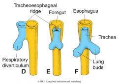
tracheosophageal septum
separates esophagus (foregut) from trachea (long bud) |
|
|
obj.
Describe development of the respiratory system |
-long bud/respiratory diverticulum forms (4 wks)
-long bud separates from foregut when tracheosophageal septum forms -separation differentiates esophagus (dorsally) & trachea (ventral) -laryngeal inlet/orifice maintains communication btwn respiratory primordium & pharynx |
|
|
obj.
Respiratory system, developmental defects that may occur. |
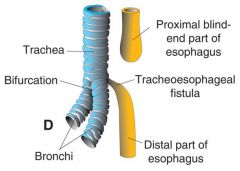
Esophageal Atresia: 90% the upper part of the esophagus ends in a blind pouch w/ lower segment forming a tracheoesophageal fistula
Tracheoesophageal fistula: connection btwn the trachea & esophagus-->pneumonia & polyhydramnios |
|
|
The epithelial lining of the larynx originates from endoderm, in contrast the cartilage & muscles originate from _______________.
|
mesenchyme of the 4th & 6th pharyngeal arches
|
|
|
The intrinsic muscles of the larynx are innervated by ___________
|
vagus nerve (C10)
|
|
|
The long bud forms the trachea, which further divides into 2 bronchial buds. The bronchial buds enlarge to form what?
|
the right and left primary bronchi
|
|
|
Which primary bronchi is more susceptible to inhalation of foreign objects and why?
|
right primary bronchi, it is wider & more vertically oriented
|
|
|
Right primary bronchus form __ secondary (lobar) bronchi & the left form __.
Each lobar bronchus branches into _________________, each of which supplies a bronchopulmonary segment. |
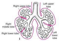
right 3
left 2 branch into tertiary (segmental) bronchi |
|
|
The conducting portion of the bronchial tree ends with ___________________
The respiratory portion of the bronchial tree begin with __________ |
ends w/ terminal bronchioles
begins w/ respiratory bronchioles |
|
|
obj.
Discuss the formation of alveoli |
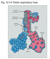
Respiratory bronchioles divide into alveolar ducts
--> alveolar ducts end in alveolar sacs --> alveolar sacs have alveoli (air spaces where gas exchange occurs) |
|
|
Alveoli are lined w/ 2 main cells types of squamous epithelium:
|
1. type 1 pneumocytes
-flattened cells -attenuated w/ cytoplasm -90% of surface lining 2. type 2 pneumocytes -round cells -located in obtuse angles of alveoli -secrete surfactant |
|
|
obj.
Discuss role of surfactant in alveoli |
reduces alveolar surface tension
|
|
|
Stages of Lung Maturation
|
Psuedoglandular period : 5-16 weeks
Canalicular period: 16-26 weeks Terminal sac period: 26 weeks to birth Alveolar period: 8 months to childhood |
|
|
What occurs during the pseudoglandular period?
|
branching continues to form terminal bronchioles
(no resp. bronchioles or alveoli yet) |
|
|
What occurs during the canalicular period?
|
terminal bronchioles divide into 2 or more respiratory bronchioles which each divide into 3-6 alveolar ducts
|
|
|
What event in lung maturation is key to survival and begins at about 26 weeks?
|
terminal sacs (primitive alveoli) form
capillaries form close contact *Terminal sac period |
|
|
what occurs during alveolar period?
|
mature alveoli have well developed epithelial endothelial capillary contacts
|
|
|
Pleural cavities are partitioned from rest of intraembryonic coleom by formation of ________________ and ________________
|
pleuropericardial membranes
(btwn pericardial & pleural cavities) and pleuroperotpneal folds (btwn plueral & peritoneal cavities) |
|
|
Splanchnic mesoderm covering outside of lungs forms (visceral/parietal) pluera, which is innervated by ___________________
|
visceral
GVA fibers from pulmonary plexuses *insensitive to pain |
|
|
Somatic mesoderm covering inside of body walls forms (visceral/parietal) pluera, which is innervated by ___________________
|
parietal
GSA fibers from intercostal nerves *very sensitive to pain |
|
|
Potential space btwn visceral & parietal pluerae is ___________
|
pleural cavity
|
|
|
obj.
Discuss factors necessary for normal lung development. |
necessities:
-fluid in the lungs -fetal breathing movements -adequate amniotic fluid volume (space for growth) |
|
|
At birth, lungs are 1/2 filled w/ fluid, fluid is cleared via 3 routes:
|
1. pressure on fetal thorax
(during delivery, fluid comes out of mouth & nose) 2. resorption into pulmonary capillaries 3. resorption into pulmonary lymphatic vessels |
|
|
obj.
Discuss the cause of respiratory distress syndrome (hyaline membrane disease) |
cause: insufficient synthesis of surfactant by type II alveolar cells (pneumocytes)
leads to: increase in surface tension & collapse of alveoli, common cause of premi death* -can be treated w/ maternal glucocorticoid therapy or artifical surfactant adiministration to infant |
|
|
obj.
Discuss the causes of hypoplastic (underdeveloped) lungs |
cause:
-oligohydramnios, prevents expansion in thoracic cavity -congenital diaphragmatic hernia, most common cause leads to: underdeveloped lungs |