![]()
![]()
![]()
Use LEFT and RIGHT arrow keys to navigate between flashcards;
Use UP and DOWN arrow keys to flip the card;
H to show hint;
A reads text to speech;
59 Cards in this Set
- Front
- Back
|
At what point on the hind limb do the extensors and flexors act exactly the same as the front limb? |
at the tarsus. All of the tendons are going join there and head on down 9 |
|
|
What does the SDF and the DDF look like on a pelvic limb? |
DDF is against bone and the SDF runs on top of it |
|
|
What are the major parts of teh calcanean tendon? |
tendons of gastrocnemius, SDF< al little biceps femoris, a little semitendinosus, a little gracilis |
|
|
What has a HUGE bursa in the lower pelvic limb? |
A huge calcaneal bursa to keep it from fraying |
|
|
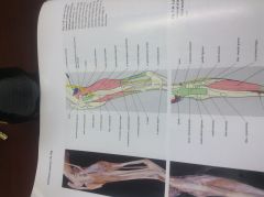
What are the flexor tendons on the thoracic limb? |
FCR, SDF, DDF
The SDF and DDF share a sheeth |
|
|
What are the extensor tendons on the front limb? |
CDE, LDE |
|
|
What is the eqivilant of the extensor digitorum longus in the front limb and what do they all do? |
Common Digital extensor
Extensor process of distal phalanx of digits 2-4 (its going to go on top of the bone) |
|
|
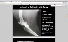
Where is arthtritis in the knee really going to hurt? |
joint capsule because there are so many nerves |
|
|
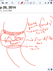
How many sacs do dogs have in the joints of dogs |
3 or 4
2 Femorotibila (under / above each condyle on tib or femur)
1 underneath the patella
There is also one underneath the long digital extensor |
|
|
What does the long digital extensor capsule look like from the front limb? |
The capsule around the biceps brachaie |
|
|
What do all of the joint sacs do with eachother in the stifle? |
communicate |
|
|
What is a joint sac? |
|
|
|
What does everybone below the hip have and can be named easily? |
Every bone below hip has collateral ligaments. Each joint has a medial and a lateral ligament |
|
|
Where are all of the ligaments located in reguards to the joint sacs? |
Outside of them |
|
|
What is the patellar ligament essentially? what is it called? What sections are there |
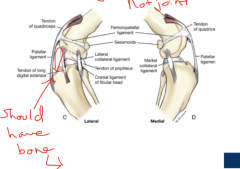
a collateral ligament of the patella bone. It prevens it from going side to side. It is called the femoropatellar ligaments
there is a medial and lateral section |
|
|
Which of the patellar ligaments is most likely to be damaged in a bow legged dog and how? |
When a dog runs it puts too much mechanical stress on the lateral patellar ligament and will cause the ligament to tear and pull the patella out of the trochlea of the femur |
|
|
What are the four sesmoids of the femur? |
1. The patella its its own little sesamoid 2. the three fabella 2 for the gastroc 1 for the popliteal |
|
|
Where do the cruciate ligaments oringinate and how do we name them |
They originate in the intercondylar fossa
The caudal crux originates on caudal portion of tibia
The cranial originates on cranial portion |
|
|
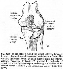
What are the functions of the cruciates? |
as the stifle is flexed the lateral collateral ligaments loosen allowing internal rotation on each other. The cruciates counteract this by twisting together and stopping that from happening. As the leg is extended these cruxiates untwist and have no affect on limiting external rotation
LEARN WHICH IS WHICH FROM THE BACK AND Front |
|
|
What is the function of the intermeniscal ligament? |

attach medial to lateral |
|
|
What is the funciton of the meniscal tibial ligament? |

Attach meniscal to tibia |
|
|
What was the function of the meniscal-tibial ligament |

attach meniscal to tibia |
|
|
What was the function of a meniscal femoral ligament |
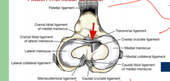
attach meniscal to femur |
|
|
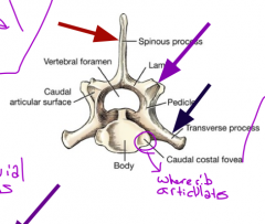
What is the function of the body of the vertebra? |
It helps articulate with the one infront of it.
Finish this at home |
|
|
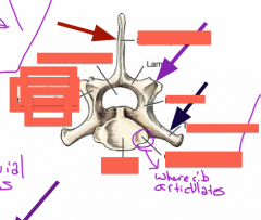
What part of the vertebrae make up the vertical canal |
the stacking vertebrale foramen |
|
|
What are the transverse processes used for |
Muscle attachment, they are always paired, usually lateral |
|
|
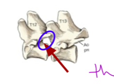
How many articular processes are there on a vertebra and where are they |
caudal and cranial (2) the ribs will articulate here |
|
|
What makes up the vertebral arch |
The two pedicles coming down and meeting with the body. |
|
|
Where do the vertebral notches occur and what is their function? |
They occur at the base of the pedicles where it meets with teh bodyOne cranial and one caudal. When these two meet you get a intervertabiral foramen |
|
|
What forms the intervertablral foramen |
The vertebral notches of a forward vertebrae and the ones of the one directly following it |
|
|
|
|
|
Draw about the invtervertebral foramen |
|
|
|
Where do the ribs articulate with the vertebrae |

the costal fovea |
|
|
What kind of joints occur at the articular process? |
Synovial joints |
|
|
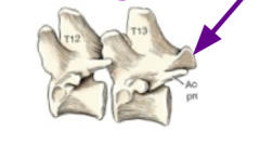
What is characteristic of thoracic vertebrate |
GIANT HEAD
TIny body
There are 13 |
|
|
What is the body of the veretbra |
That solid piece of bone at the base of the vertebra. |
|
|
What is the lamina of the vertebra |
The portion that holds up the spinous process |
|
|
what is the function of the pedicle? |
connects the lamena to the body of vetrebra |
|
|
What does each vertebrae have to hang on to each other |
vertebral processes with articular surfaces (articular fossa) One pair at the top and one pair at the bottom |
|
|
How do the ribs articulate? |
The first 10 ribs articulate in 2 places articulates with two bodies
The last 3 articulate only in 1 place |
|
|
Where does the rib articulate on 1-10> |
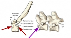
head of rib connects to caudal portion of one vertebrae to the cranial portion of another. The rounded head has two facets the areas of articulation are called the caudal coastal fovea ON THE BODY |
|
|
Where does the rib articulate on last 3 |

Only once. Head connects to On the cranial area of the body |
|
|
What does the tubercle of the rib do? |
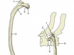
Connect to the transverse process to provide a 2 or 3 articulation |
|
|
What is the costocondral junction? |
Where the cartilage attaches to the rib about midway downish somewhere |
|
|
What is the costal cartilage? |
At the distal end of the rib when you have just cartlige |
|
|
Where does the cost cartilage go? |
1-9 are going to attach only to sternum
CC of 10, 11 and 12 form costal arch
Rib 13 is a pain and just floats |
|
|
How does the costal arch connect with the sterum |
via rib 8s cc, each cc overlaps to connect indirectly to sternum |
|
|
Why is the costal arch so important? |
It indicates the lateral margin of division of thoracic and adbominal cavities |
|
|
Where is the acronion |
around the 6-7 space |
|
|
Where do you want to cut to do a thoracotomy |
Cutaneous trunci Latissimus dorsi Serratus ventralis and dorsalis Intercostal X 2 |
|
|
Where is the superspinous ligament |

(3) Above the thoracic vertebra on the top. Spide of T1 to Cd3 |
|
|
What is the nuchal ligament |
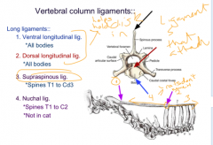
above the spine of T1 to C2. NOT IN CAT (4). Lots of fibrous fiber |
|
|
What is the dorsal longitudinal ligament |
* |
|
|
What is the ventral longtidunal ligament |
Names for where it lays on the body |
|
|
What lies between vertebral |
intervertebral discs, the lognitudnal ligaments help hold in place |
|
|
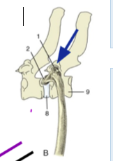
What is between the head of a rib and the vertebra |
a synovial joint |
|
|
What is are the parts of the vertebral disk |
The inner jelly is called the inner nucleus pulposus
The thing that surrounds it is called the outer annulus fibrosus |
|
|
What does the intercapital ligament do? |
It goes between the two rib heads across spinal cord the top of the spinal cord Dorsal to the IV disc but ventral to the dorsal longitudinal ligament |
|
|
What does the yellow ligament do? |
It is between the two arches on the dorsal side. they connect the lamina |