![]()
![]()
![]()
Use LEFT and RIGHT arrow keys to navigate between flashcards;
Use UP and DOWN arrow keys to flip the card;
H to show hint;
A reads text to speech;
19 Cards in this Set
- Front
- Back
|
The anterior compartment of the leg is located anterior to the interosseous membrane. T/F |
True |
|
|
What are the two retinaculums associated with the anterior compartment? |
- superior and inferior extensor retinaculum |
|
|
What forms the retinacula around the ankle joint region? |
deep crural fascia |
|
|
What is the origin of extensor hallucis longus? |
- middle third of the medial surface of the fibular |
|
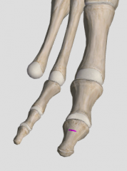
What inserts here? |
extensor hallucis longus - dorsal surface of the base distal phalanx of the big toe |
|
|
What is the innervation and action of EHL? |
- deep fibular nerve (L4, L5) - extension of great toe; support of medial longitudinal arch |
|
|
What muscle has origin underneath EHL? |
fibularis tertius |
|
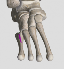
What inserts here? |
fibularis tertius - dorsal surface of the shaft of the 5th metatarsal bone |
|
|
What are the actions of the fibularis tertius? |
extends and everts foot |
|
|
What is the origin of tibialis anterior? |
- lateral tibial condyle - 1/2 lateral surface of tibial shaft - interosseous membrane |
|
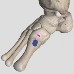
What inserts here? |
tibialis anterior - medial cuneiform - base of first metatarsal |
|
|
What are the actions of TA? |
- strongest extensor and invertor of foot - supports medial longitudinal arch |
|
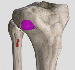
What originates here? |
extensor digitorum longus - lateral tibial condyle - proximal medial surface of fibula |
|
|
What is the insertion of EDL? |
- extensor expansion - 2 bands to distal phalanges - 1 band to middle phalange |
|
|
What structures pass deep to the superior extensor retinacula? |
- tibialis anterior - extensor digitorum longus - anterior tibial artery and veins (2) - deep fibular nerve - fibularis tertius tendon - extensor hallucis longus tendon |
|
|
What are the attachments of the inferior extensor retinaculum? |
- laterally it is attached to the superior and lateral surfaces of calcaneus - medially; superior band attaches to medial malleolus and its inferior band attaches to the plantar aponeurosis |
|
|
Why is the superior extensor retinaculum more likely to experience friction/inflammation? |
- only 1 synovial sheath (TA) - extensor hallucis longus and extensor digitorum longus do not |
|
|
What passes under the inferior extensor retinaculum? |
- TA (in synovial sheath) - EHL (in synovial sheath) - dorsalis pedis artery and veins (2) - EDL (in synovial sheath) - fibularis tertius - deep fibular nerve |
|
|
Under the superior fibular retinaculum FL and FB share a common tendinous sheath. T/F |
True; divided in the inferior fibular retinaculum |