Reading...
![]()
Play button
![]()
Play button
![]()
Use LEFT and RIGHT arrow keys to navigate between flashcards;
Use UP and DOWN arrow keys to flip the card;
H to show hint;
A reads text to speech;
304 Cards in this Set
- Front
- Back

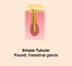
What type of glandular epithelium is this and where is it found?
|
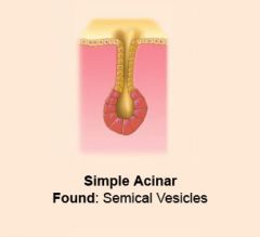
|
|

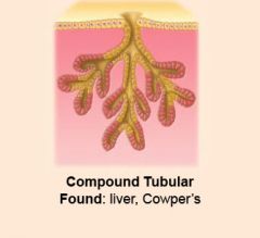
What type of glandular epithelium is this and where is it found?
|
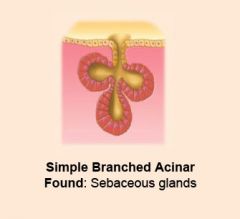
|
|

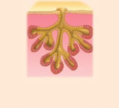
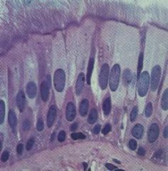
What type of glandular epithelium is this and where is it found?
|

|
|
What type of glandular epithelium is this and where is it found?
|
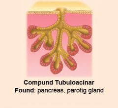
|
|

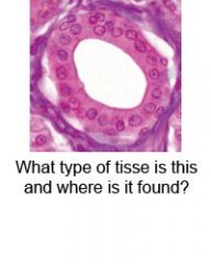
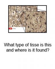
What type of epithelium is this and where is it found?
|
|
|

What type of glandular epithelium is this and where is it found?
|
|
|
|
What are glycolipids?
|
Glycolipids are carbohydrate-attached lipids.
Their role is to provide energy and also serve as markers for cellular recognition. |
|
|
Definition of cervical
|
Narrowing area
|
|
|
Cholesterol
|
Classified as lipid
Most abundant steroid in animal tissue Located in cell membranes. Used for synthesis of steroid hormones & bile salts. |
|
|
Mediastinum
|
Central portion of thoracic cavity between the lungs.
Extends from sternum to vertebral column and form neck to diaphragm. Contains: heart, thymus, esophagus, trachea and several large blood vessels. |
|
|
Monosaccharides
|
A simple sugar such as glucose, fructose or deoxyribose that has a single ring
|
|
|
Antecubital
|
Where arm bends.
|
|
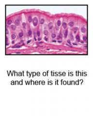
Identify this type of tissue and tell where it might be found.
|

Pseudostratified Ciliated Columnar Epithelium
* 1 layer of ciliated columnar cells packed tightly together. * Appear multilayered * Functions: protection, secretion & movement of mucus. * Found: lines respiratory & reproductive tracts. |
|
|
Amino acids
|
An amino acid is a molecule containing both amine and carboxyl functional groups.
The monomer is used to synthesis polypeptides and proteins. |
|
|
Popliteal
|
Area right behind knee.
|
|

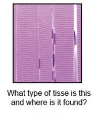
Identify the tissue type and tell where it might be found.
|

* Single, flat layer of cells.
* Functions: Filters, permits diffusion. * Found: lining body cavities, covers viscera, lines capillaries and blood vessels (endothelium), forms air sacs (alveoli) in lungs. |
|

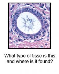
Identify this tissue type and where it might be found.
|

Stratified Cuboidal Epithelium
Found: Larger sweat glands. |
|
|
What is Otic?
|
Related to, or in the region of the ear.
|
|
|
Orbit
|
The bony, pyramidal-shaped cavity of the skull the holds the eyeball.
|
|
|
What is a lipoprotein?
|
Large class of conjugated proteins composed of a complex of protein and lipid (LDLs - good, HDL - not good)
|
|
|
Mental
|
Chin area.
|
|
|
Steroid
|
A large nonpolar region consisting of 4 rings of carbon and a hydrocarbon tail.
|
|
|
Buccal
|
Of or relating to or toward the cheek.
|
|
|
Glycerol
|
A chemical compound also commonly called glycerin or glycerine. It is a colorless, odorless, viscous liquid that is widely used in pharmaceutical formulations.
|
|
|
Cranial
|
The part of the skull that encloses the brain.
|
|
|
Lactic acid
|
An acid produced as a result of anaerobic respiration in muscles and red blood cells ie when glycogen is used as an energy source for respiration rather than oxygen.
An acid produced in the muscles during intense exercise that causes pain and soreness. |
|
|
Cutaneous membrane
|
Skin- squamous stratified epithelial tissue
|
|
|
What are the two regions of skin?
|
The epidermis and the dermis.
|
|
|
Serous membrane
|
* A smooth membrane consisting of a thin layer of cells which excrete serous fluid.
* Serous membranes line and enclose several body cavities, known as serous cavities, where they secrete a lubricating fluid which reduces friction from muscle movement. |
|
|
Matrix components:
|
collagen fibers
elastic fibers reticular fibers |
|
|
Collagen fibers:
|
Main protein of connective tissue in animals and the most abundant protein in mammals,
|
|
|
Elastic fibers:
|
Bundles of proteins (elastin) found in extracellular matrix of connective tissue and produced by fibroblasts and smooth muscle cells in arteries.
These fibers can stretch up to 1.5 times their length, and snap back to their original length when relaxed. |
|
|
Reticular fibers:
|
- made of the same molecules as collagen but thinner, they form an internal mesh-like network within organs. This produces an endoskeleton or stroma for soft organs such as the spleen, liver, etc.
|
|
|
What is ground substance?
|
A viscous, clear substance with a high water content which occupies the space between the cells and fibers.
|
|
|
What is endoderm?
|
A primary germ layer of the developing embryo.
*Gives rise to the gastrointestinal tract, urinary bladder, urethra, and respiratory tract. |
|
|
What is mesoderm?
|
The middle primary germ layer that gives rise to connective tissues, blood and blood vessels, and muscles.
|
|
|
What is ectoderm?
|
The primary germ layer that gives rise to the nervous system & epidermis of skin and its derivatives.
|
|
|
What is lactose?
|
a sugar comprising one glucose molecule linked to a galactose molecule.
|
|
|
What is acromion?
|
The outermost point of the spine of the shoulder blade.
|
|
|
What is mesenchyme?
|
That part of the mesoderm of an embryo that develops into connective tissue, bone, cartilage etc.
|
|
|
What is glucose?
|
A monosaccharide sugar that has several forms; an important source of physiological energy.
|
|
|
Axillary
|
Armpit
|
|
|
Connective tissue
|
One of the most abundant and widely distributed tissues in the body.
|
|
|
Nucleid acid
|
DNA & RNA
|
|
|
What are the 4 types of body tissues?
|
Epithelium, connective, muscle and nervous tissue.
|
|
|
What is the function of epithelium?
|
Covers body surfaces & lines hollow organs, body cavities and ducts. It also forms glands.
|
|
|
What is the function of connective tissue?
|
Protects and supports the body and its organs.
Various types of connective tissue bind organs together, store energy reserves as fat, and help provide immunity to disease-causing organisms. |
|
|
What is the function of muscle tissue?
|
Generates the physical force needed to make the body structures move.
|
|
|
What is the function of nervous tissue?
|
Detects changes in a variety of conditions inside & outside the body & responds by generating nerve impulses that help maintain homeostasis.
|
|
|
Tight junctions
|
Weblike strands of transmembranal proteins that fuse the outer surfaces of adjacent plasma membranes together.
They slow the passage of substances between cells, prevent the contents of these organs from leaking into blood or surrounding tissues. * The portion of the cell exposed to the lumen is called its apical surface. * The rest of the cell (i.e., its sides and base) make up the basolateral surface. |
|
|
Adherens junctions.
|
Adherens junctions provide strong mechanical attachments between adjacent cells.
* They hold cardiac muscle cells tightly together as the heart expands and contracts. * They hold epithelial cells together. * They seem to be responsible for contact inhibition. * Some adherens junctions are present in narrow bands connecting adjacent cells. * Others are present in discrete patches holding the cells together. |
|
|
Desmosomes
|
Localized patches that hold two cells tightly together. They are common in epithelia (e.g., the skin). Desmosomes are attached to intermediate filaments of keratin in the cytoplasm.
|
|
|
Hemidesmosomes
|
These are similar to desmosomes but attach epithelial cells to the basal lamina ("basement membrane") instead of to each other.
|
|
|
What is brachium?
|
A branching or armlike part of an animal.
|
|
|
Antebrachium
|
The forearm is the structure on the upper limb, between the elbow and the wrist.
|
|
|
Mucous membrane (mucosa)
|
epithliem on loose connective tissue. Lines body cavities open to the exterior.
|
|
|
Serous membrane (serosa)
|
Serous = watery.
Lines a body cavity that does not open directly to the exterior and covers organs that lie within the cavity. |
|
|
Synovial membranes
|
Syn = together, referring here to a place where bones come together, line the cavities of freely movable joints.
Secretes synovial fluid. |
|
|
Histones
|
The chief protein components of chromatin.
|
|
|
Carpals/Tarsals
|
Carpals = wrists
Tarsals = ankles |
|
|
Adipose connective tissue
|
Loose connective tissue
Its main role is to store energy in the form of fat, although it also cushions and insulates the body. |
|
|
Chromatin
|
The complex combination of DNA, RNA, and protein that makes up chromosomes.
|
|
|
Pectoral
|
Either of two large muscles of the chest.
|
|
|
Areolar connective tissue
|
Loosely organized fibers, abundant blood vessels, and significant empty space. Its fiber run in random directions and are mostly collagenous, but elastic and reticular fibers are also present.
|
|
|
Endomembrane system
|
Composed of the different membranes that are suspended in the cytoplasm within a eukaryotic cell.
These membranes divide the cell into functional and structural compartments, or organelles. |
|
|
Membrane channels
|
A family of biological membrane proteins which allow the passive movement of ions (ion channels), water (aquaporins) or other solutes to passively pass through the membrane down their electrochemical gradient.
|
|
|
Gated channels
|
What opens and closes the channels.
|
|
|
Diffusion
|
The process in which there is movement of a substance from an area of high concentration of that substance to an area of lower.
|
|
|
Osmosis
|
Passage of water through a membrane in response to concentration differences.
|
|
|
Filtration
|
The process whereby fluids pass through a filter or a filtering medium.
|
|
|
Facilitated diffusion
|
A process of diffusion, a form of passive transport facilitated by transport proteins.
|
|
|
Symporter
|
A transmembrane transporter protein that moves two substances, oftan Na+ & another substance, in the same direction across a plasma membrane.
Also called cotransporter. |
|
|
Uniporter
|
An integral membrane protein that is involved in facilitated diffusion.
They can be either a channel or a carrier protein. |
|
|
Antiporter
|
Inner membrane transport proteins that expel one compound that is present in the cytoplasm in exchange for importing a different compound into the cell.
|
|
|
Active transport
|
Transport of a substance (as a protein or drug) across a cell membrane against the concentration gradient; requires an expenditure of energy.
|
|
|
Endocytosis
|
Cells absorb material.
|
|
|
Exocytosis
|
Cells excretes substance.
|
|
|
Pinocytosis
|
"cell-drinking"
|
|
|
Phagocytosis
|
Engulf and digest microorganisms and cellular debris.
|
|
|
Ligand-gated channels
|
Ionotropic receptors, this group of channels open in response to specific ligand molecules binding to the extracellular domain of the receptor protein.
|
|
|
G-protein
|
Short for guanine nucleotide-binding proteins.
|
|
|
Voltage-gated channels
|
A class of transmembrane ion channels that are activated by changes in electrical potential difference near the channel.
|
|
|
Desmosome
|
A structural unit that functions in the adhesion of cells to form tissue.
|
|
|
Hemidesmosome
|
While desmosomes link two cells together,
hemidesmosomes attach one cell to the extracellular matrix. |
|
|
Intercalating disc
|
Holds cardiac muscle fibers together.
|
|
|
Aquaporins
|
Pproteins embedded in the cell membrane that regulate the flow of water.
They are "the plumbing system for cells." |
|
|
Integrins
|
Family of transmembranal glycoproteins in plasma membranes that function in cell adhesion; they are present in hemidesmosomes, which anchor cells to a s basement membrane.
|
|
|
Adherens junction
|
(Zonula adherens) are protein complexes that occur at cell-cell junctions in epithelial tissues, usually more basal than tight junctions.
|
|
|
Levels of organization
|
chemical level > Cell > Tissue > Organ > Organ system > Organism
|
|
|
Cytoskeleton
|
Network of filaments, microfilaments and microtubules in the cytoplasm.
|
|
|
Mitosis:
|
Orderly division of the nucleus of a cell that ensure that each new daughter nucleus has the same number and kind of chromosomes.
|
|
|
Interphase:
|
This is the longest period of the complete cell cycle during which DNA replicates, the centrioles divide, and proteins are actively produced.
|
|
|
Prophase:
|
Nucleolus fades and chromatin (replicated DNA and associated proteins) condenses into chromosomes.
|
|
|
Metaphase:
|
Tension applied by the spindle fibers aligns all chromosomes in one plane at the center of the cell.
|
|
|
Anaphase:
|
Spindle fibers shorten, the kinetochores separate, and the chromatids (daughter chromosomes) are pulled apart and begin moving to the cell poles.
|
|
|
Telophase:
|
The daughter chromosomes arrive at the poles and the spindle fibers that have pulled them apart disappear.
|
|
|
Anatomical position
|
The position of the human body, standing erect, with the face directed anteriorly, the upper limbs at the sides and the palms turned anteriorly.
|
|
|
Actin
|
A protein found in muscle that together with myosin functions in muscle contraction.
|
|
|
Collagen
|
Main protein of connective tissue in animals and the most abundant protein in mammals,[1] making up about 25% to 35% of the whole-body protein content.
|
|
|
Myosin
|
Protein that uses ATP to drive movements along actin filaments and facilitate muscle contraction.
|
|
|
Keratin
|
Make skin, hair and nailes tough and waterproof.
|
|
|
Purine bases
|
Organic compounds structurally related to purine,
particularly adenine and guanine (A & G). |
|
|
Pyrimidine bases
|
DNA = Thymine and cytosine are the
RNA = Uracil |
|
|
Coronal/Frontal
|
Separates the body into Anterior and Posterior parts.
|
|
|
Transverse/horizontal/cross-sectional plane
|
Separates the body into Superior and Inferior parts.
|
|
|
Midsaggital/parasaggital
|
Separates body into Right and Left parts.
|
|
|
Saggital/longitudinal plane
|
Any plane parallel to the median plane.
|
|
|
Oblique plane
|
Diagonal cut.
|
|
|
Anterior/posterior
|
Front/back
|
|
|
Dorsal/ventral
|
Backside/belly side.
|
|
|
Cephalic/caudal
|
head/away from the head.
|
|
|
Superior/inferior
|
above/below
|
|
|
Proximal/distal
|
proximal = nearer the trunk or attached end
distal = farther from the trunk or point of attachment |
|
|
Medial/lateral
|
medial = towards the midline
lateral = away from the midline |
|
|
Ipsilateral/Contralateral
|
Ipsilateral = same side
Contralateral = opposite side |
|
|
Sternal
|
"Chest" or breastbone.
Is a long flat bone (or, in some models, set of three bones) located in the center of the thorax (chest). |
|
|
Fibroblast
|
Large, flat cells with branching processes.
Present in several connective tissues. Most numerous. |
|
|
Mitochondria
|
The principal energy source of the cell and convert nutrients into energy as well as doing many other specialized tasks.
|
|
|
Umbilical
|
Pertaining to the central region of the abdomen.
|
|
|
Reticular connective tissue
|
Thin, delicate bundles & coating of glycoprotein.
Provides support in wall of blood cells and form network around cells in some tissues. |
|
|
Consists of fine interlacing reticular fibers & reticular cells.
Forms the stroma of the liver, spleen and lymph nodes. Binds together smooth muscle cells. |
Reticular connective tissue
|
|
|
Elastic fibers
|
Yellow. Less strong, more flexible. Contains elastin.
Medium size fiber that branch & join together to form a network w/in a tissue. |
|
|
Collagen fiber
|
Colla = glue.
Very strong and resistant to pulling forces, but are not stiff. |
|
|
Chloroplast
|
Organelles found in plant cells containing chlorophyll and other pigments; in plants that carry out photosynthesis.
|
|
|
Mast cells
|
Produce histamine and heparin.
Abundant alongside blood vessels that supply connective tissue. |
|
|
Plasma cells
|
Develop from a type of WBC called B lymphocyte.
Secrete antibodies, proteins that attack or neutralize foreign substances in body. |
|
|
Histocytes
|
A macrophage that is found in connective tissue.
|
|
|
Macrophage
|
macro = large
phages = eater Develop from monocytes, a type of WBC. Engulf bacteria & cellular debris by phagocytosis. |
|
|
Types of cells present in connective tissue:
|
Fibroblasts
Macrophages Plasma cells Mast cells |
|
|
ATP
|
Adenosine Triphosphate
Occurs in muscle tissue; the major source of energy for cellular reactions. |
|
|
NAD
|
Nicotinamide adenine dinucleotide
A derivative of vitamin B |
|
|
FAD
|
Flavin adenine dinucleotide, a derivative of vitamin B2.
|
|
|
Lumbar
|
Refers to the lower region of the back.
Near the part of the back between the ribs and the hipbones; "lumbar vertebrae"/. |
|
|
Heparin
|
A chemical released by basophils and mast cells that causes nearby tissues to become swollen and inflamed.
|
|
|
Histamine
|
Substance released from mast cells during an allergic reaction.
|
|
|
Antibodies
|
Protein produced by plasma cells in response to a specific antigen.
The antibody combines w/that antigen to neutralize, inhibt or destroy it. Also called immunoglobulin. |
|
|
Cytoplasm
|
The contents of a cell except for the nucleus. It contains the organelles.
|
|
|
Hypochondriac
|
Upper lateral region on each side of the body and below the thorax; beneath the ribs.
|
|
|
Chondroitin sulfate
|
Part of a large protein molecule that gives cartilage elasticity.
|
|
|
Ribosome
|
Organelle involved in the production of proteins by translating messenger RNA
|
|
|
Inguinal
|
Of or relating to or near the groin .
|
|
|
Smooth ER
|
Synthesis of lipids and steroids.
Is connected to the nuclear envelope. |
|
|
Pubic
|
Relating or near the pubis; "pubic bones".
|
|
|
Hyaluronic acid
|
Slippery substance that binds cells together, lubricates joints and helps maintain shape of eyeballs.
|
|
|
Rough ER
|
Contains ribosomes which function in the manufacture of membrane-bound proteins.
|
|
|
Sacral
|
Of or relating to or near the sacrum.
|
|
|
Hyaline cartilage
|
Covers surface of bones forming synovial joint; also called articular cartilage.
"Gristle" |
|
|
Golgi body
|
Modifies proteins delivered from the RER.
Involved in the transport of lipids around the cell, and the creation of lysosomes. |
|
|
Scapular
|
Latin scapula = shoulder.
|
|
|
Elastic cartilage
|
Yellow cartilage is a type of cartilage present in the outer ear, larynx, and epiglottis.
|
|
|
Centrioles
|
Involved in mitosis.
Composed of nine triplet microtubules and form the asters. |
|
|
Characteristics of cartilage
|
Dense network of collagen fibers & elastic fibers firmly embedded in chondroitin sulfate (a gel-like component of ground substance.)
|
|
|
Basal bodies
|
Also called basal granule or kinetosome.
An organelle formed from a centriole. Found at the base of cilium or flagellum and serves as a nucleation site for the growth of the axoneme microtubules. |
|
|
Femoral
|
Of or relating to or near the femur or thigh.
|
|
|
Characteristics of epithelium
|
1. Thin, toughly packed cells resting on a basement membrane (lamina propria).
2. Avascular 3. Named to: a) # of layers: simple (1 layer) or stratified (multiple layers) b) Cell type on top: Squamous, cuboidal, columnar. |
|
|
Flagellum
|
A whip-like appendage which is the organ of locomotion of a motile cell.
|
|
|
Popliteal
|
The area behind the knee joint.
|
|
|
Characteristics of nervous tissue
|
Neurons - (nerve cells) conduct electrical signals.
Neuroglia - (support cells) provide support, nourishment for neurons. |
|
|
Characteristics of muscle
|
* excitability - responds to stimuli (e.g., nervous impulses)
* contractility - able to shorten in length * extensibility - stretches when pulled * elasticity - tends to return to original shape & length after contraction or extension |
|
|
Cilia
|
Hairlike projection from the surface of a cell; provides locomotion in free-swimming unicellular organisms.
|
|
|
Patellar
|
Relating to the patella or kneecap; "patellar tendon".
|
|
|
Plasma membrane
|
A thin membrane (a double layer of lipids) enclosing the cytoplasm of a cell.
|
|
|
Plantar
|
The bottom surface of the foot.
|
|
|
Reticular connective tissue
|
Composed of interlacing fibers of collagen called reticular fibers. This tissue forms supporting structures for many organs, such as the spleen.
|
|
|
Nucleus
|
Control center
Contains DNA and RNA and responsible for growth and reproduction. |
|
|
Calcaneal
|
Relating to the heel bone or heel.
|
|
|
Adipose connective tissue
|
Specialized to contain triglycerides as a large centrally located droplet.
Reduces heat loss through skin, serves as energy reserve. |
|
|
Nucleolus
|
Contain RNA and are involved in protein synthesis.
|
|
|
Pollux
|
Thumb
|
|
|
Fibrocartilage
|
Chondrocytes are scattered among clearly visible bundles of collagen fibers w/in the matrix of this type cartilage.
Lacks a perichondrium. |
|
|
Nuclear membrane
|
A double lipid bilayer that encloses the genetic material in eukaryotic cells.
|
|
|
Hallux
|
Big toe
|
|
|
Articular cartilage
|
Also called hyaline cartilage.
Contains a resilent gel as its ground substance and appears in the body as bluish-white, shiny substance. Gives support and maintains shape. |
|
|
Nuclear pores
|
An opening in the nuclear envelope that allows the passage of small molecules such as salts, small proteins, and RNA molecules.
|
|
|
Digit
|
One of several most proximal parts of a limb, present in many vertebrates.
|
|
|
Lacunae
|
Small spaces containing osteocytes in bone or chondrocyte in cartilage.
|
|
|
Centrosome
|
The microtubule organizing centre that forms the mitotic spindle in dividing cells.
Contains the centrioles and serves to organize the microtubules. |
|
|
Olecranal
|
Elbow
|
|
|
Chondrocyte
|
Greek chondros cartilage + kytos cell, are the only cells found in cartilage.
|
|
|
Lysosome
|
Lysosomes are organelles containing digestive enzymes (acid hydrolases).
|
|
|
Membrane channels
|
A family of biological membrane proteins which allow the passive movement of ions (ion channels), water (aquaporins) or other solutes to passively pass through the membrane down their electrochemical gradient.
|
|

|
Simple Squamous
Found:Lines body cavities, covers viscera, lines capillaries and blood vessels, forms air sacs (alveoli) in lungs. Function: Filtration, diffusion, osmosis and secretion in serous membranes. |
|
|
Simple Cuboidal Epithelium
Found:Covers ovaries, lines kidney tubules, ducts. Function: Secretion & absorption |
|

|
Nonciliated simple columnar epithelium
Found: Uterus, tubes, tubes of the GI Tract. Function: Secretion & absorption |
|
|
Ciliated simple columnar epithelium
Found: lines portions of upper respiratory tract, uterine tubes, uterus. Function: Moves mucus and other substances by ciliary action. |
|
|
Stratified Cuboidal Epithelium
Found: Ducts of adult sweat glands & esophageal glands & part of male urethra. Function: Protection & limited secretion & absorption. |
|

|
Stratified Columnar Epithelium
Found: Lines male urethra, covers epiglottis Function: Secretion and protection. |
|

|
Areolar (loose) Connective Tissue
Found: Under skin, epithelial tissues, between muscles. Function: Bind, support. |
|

|
Adipose (fat) Connective Tissue
Found: Under skin, around kidneys & heart Function: Protection, insulation. |
|
|
Reticular Connective Tissue
Found: Walls of solid organs (liver, spleen), Stroma. Function: Form supportive network. |
|
|
Dense Regular Connective Tissue
Found: Fascia (sheets covering muscles), tendons (cords connecting muscles to bone), ligaments (cords connecting bones to bones). Function: provides strong attachment between various structures. |
|

|
Dense Irregular Connective Tissue
Found: Dermis, membranes covering bones, cartilage. Function: Provides strength. |
|

|
Hyaline Cartilage
Found: Joint surfaces, C-rings of the trachea, soft portion of the nose, fetal skeleton. Functions: flexible support; serves as template for bone. |
|

|
Fibrocartilage Connective Tissue
Found: At the symphysis of pubis, between vertebrae & at knee. Function: Strongest cartilage; acts as shock absorber. |
|

|
Elastic Cartilage Connective Tissue
Found: Ear pinnae, portions of larynx, epiglottis. Function: Provides flexible support. |
|
|
Compact Bone (Osseous) Conntective Tissue
Found: Both compact & spongy bone tissue make up the varous parts of bones of the body. Function: Support, protection, storage; houses blood-forming tissue. Serves as levers that act together w/muscle tissue to enable movement. |
|

|
Blood (Vascular) Connective Tissue
|
|
|
Skeletal Muscle Tissue
Found: Attached to bones. Function: Voluntary movments. Contains: Strands of actin & myosin (strations). |
|

|
Cardiac Muscle Tissue
Found: In heart. Function: Involuntary heart movements. Contains: uninucleated, specialized cell junctions (intercalated discs), regularly arranged strands of actin and myosin. |
|

|
Smooth Muscle Tissue
Found: Walls of hollow internal organs. Function: Involuntary movements of internal organs. Contains: uninucleated, no obvious striations. |
|
|
Peroxisomes
|
Site of production and degradation of hydrogen peroxide, a toxic byproduct of cell metabolism.
|
|
|
Gluteal
|
Rear end
|
|
|
Vesicle
|
A small, intracellular, membrane-enclosed sac that stores or transports substances within a cell.
|
|
|
Tendon/ligament
|
A tough band of fibrous connective tissue that usually connects muscle to bone[1] and is capable of withstanding tension.
|
|
|
Vacuole
|
A membrane-bound, fluid-filled sac inside plant and animal cells.
|
|
|
Occipital
|
The back part of the head.
|
|
|
Pubic symphysis
|
The front part of the pelvis.
|
|
|
Microfilament
|
The thinnest filaments of the cytoskeleton found in the cytoplasm of all eukaryotic cells.
|
|
|
Parietal
|
On the top and side of the skull.
|
|
|
Osseous Connective Tissue
|
Osseous tissue forms the rigid part of the bone organs that make up the skeletal system.
|
|
|
Visceral
|
Pertaining to the organs, or the coverings of the organs.
|
|
|
Calcium Carbonate
|
It is a common substance found in rock in all parts of the world, and is the main component of shells of marine organisms, snails, pearls, and eggshells.
|
|
|
Microtubule
|
Long, unbranched hollow tubes composed of the protein tubulin.
|
|
|
Temporal
|
Latin tempus = time; hence, the temporal area of the scalp, where grey hair first appears, marking the progress of ageing.
|
|
|
Hydroxyapatite
|
The mature, hard, somewhat crystalline mineral compounds in bone tissue.
|
|
|
Microvilli
|
Minute projections on the surface of epithelial cells of the villi in the small intestine.
They increase the surface area of the intestine to improve absorption of digested nutrients. |
|
|
Lamellae
|
Concentric rings of hard, calcified matrix found in compact bone.
|
|
|
Resting membrane potential
|
Relatively static membrane potential of quiescent cells is called resting membrane potential (or resting voltage).
|
|
|
Osteon
|
The fundamental functional unit of much compact bone.
|
|
|
The Na+/K+ Pump
|
Found in the membranes of many types of cells. In particular, it plays a very important role in nerve cell membranes.
|
|
|
Comact/Cancellous, Cortical Bone
|
Hard, strong outer shell of bone.
|
|
|
Integral (intrinsic) proteins
|
Bound within the plasma membrane of a cell.
|
|
|
Epithelial Tissue
|
Epithelial tissue covers the whole surface of the body.
|
|
|
Trabecular/Spongy Bone
|
Cancellous bone, synonymous with trabecular bone or spongy bone, is one of the two types of osseous tissue which form bones.
|
|
|
Peripheral (extrinsic) proteins
|
Do not penetrate the membrane itself.
Usually bound to integral proteins by non-covalent bonds, and can therefore be extracted from the membrane without destroying it (using some kind of chemical wash). |
|
|
Transitional epithelium
|
Multiple layers of epithelial cells which can contract and expand.
These cells, part of the epithelium, are usually found in the urinary tract, especially around the urinary bladder. |
|
|
Hemopoiesis
|
The formation of blood cells in the living body (especially in the bone marrow).
|
|
|
Osteoblast/osteocyte
|
(From the Greek words for "bone" and "germ" or embryonic)
Cells which aid in the growth and development of teeth and bones. |
|
|
Chondroblast/chondrocyte
|
(Greek chondros cartilage + kytos cell) are the only cells found in cartilage.
|
|
|
Haversian system Central canal
|
Any of the anastomosing channels of the haversian system in compact bone, containing blood and lymph vessels and nerves.
|
|
|
Volkman's canal Perforating canal
|
Canals communicating with the haversian canals, for passage of blood vessels through bone.
|
|
|
Canaliculi
|
Also known as lacrimal ducts, these tube-like structures carry the tears from the eyes to the lacrimal sac.
|
|
|
Cytoskeleton
|
The internal framework of a cell, composed largely of actin filaments and microtubules.
|
|
|
Keratin
|
A tough, nonwater-soluble protein found in the nails, hair, and the outermost layer of skin. Human hair is made up largely of keratin.
|
|
|
Caudal/Cephalic
|
Located on, in, or near the head.
|
|
|
Pericyte
|
One of the elongated, contractile cells found wrapped about precapillary arterioles outside the basement membrane.
|
|
|
Perichondrium
|
The dense irregular fibrous membrane of connective tissue covering the surface of cartilage except at the endings of joints.
|
|
|
Osteoclast
|
Large multinuclear cell associated with absorption and removal of bone.
|
|
|
Epidermis
|
The nonvascular outer protective layer of the skin, covering the dermis.
|
|
|
Dermis
|
Below epidermis.
Consists of a bed of vascular connective tissue, and containing the nerves and organs of sensation, the hair roots, and sebaceous and sweat glands. |
|
|
Subcutaneous layer
|
A continuous sheet of areolar connective tissue and adipose tissue between the dermis of the skin and the deep fascia of the muscles.
Also called the superficial fascia. |
|
|
Merkel's disc
|
Fine touch and pressure receptor connected to large sensory cells.
|
|
|
Pacinian (lamellated) corpuscle
|
Any of numerous small oval bodies that are sensitive to pressure.
Found in the skin of the fingers and elsewhere, are formed of concentric layers of connective tissue. |
|
|
Apocrine gland
|
A type of large, branched, specialized sweat gland, after puberty producing a viscous secretion that is acted on by bacteria to produce a characteristic acrid odor.
|
|
|
Eccrine gland
|
One of the ordinary, or simple, sweat glands, which is of the merocrine type.
|
|
|
Sebaceous gland
|
One of the holocrine glands in the dermis that secrete sebum.
|
|
|
Ceruminous gland
|
Cerumen-secreting glands in the skin of the external auditory canal.
|
|
|
Gastric gland
|
The secreting glands of the stomach, including the fundic, cardiac, and pyloric glands.
|
|
|
Goblet cell
|
An epithelial cell that secretes mucous.
|
|
|
sudoriferous gland
|
Any of the glands in the skin that secrete perspiration.
|
|
|
Mammary gland
|
Milk-secreting organ of female mammals.
|
|
|
Merocrine
|
Remains in tact throughout process of formation & discharge of secretory product, as in salivary & pancreatic glands.
|
|
|
Holocrine
|
A type of gland in which entire secretory cells, along with their accumulated secretions, make up the secretory product of the gland, as in the sebaceous (oil) glands.
|
|
|
Papillary region
|
The uppermost layer of the dermis, intertwined with the rete ridges of the epidermis.
Composed of fine and loosely arranged collagen fibers. |
|
|
Reticular region
|
The lower layer of the dermis, composed of thick, densely packed collagen fibers, and the primary location of dermal elastic fibers.
|
|
|
Stratum spinosum
|
Cells moving toward surface.
It lies on superior to the stratum basale. |
|
|
Stratum Granulosum
|
A layer of the epidermis found between the stratum corneum (and possibly stratum lucidum) and stratum spinosum.
|
|
|
Stratum Lucidum
|
A layer of the epidermis found only in palmoplantar skin (the thicker skin of the palms and soles), between the stratum granulosum and stratum corneum layers.
It is composed of three to five layers of dead, flattened keratinocytes |
|
|
Stratum Corneum
|
The outermost layer of the epidermis, composed of large, flat, polyhedral, plate-like envelopes filled with keratin, which is made up of dead cells that have migrated up from the stratum granulosum.
|
|
|
Keratinocyte
|
The primary cell types found in the epidermis, the outer layer of skin.
|
|
|
Melanocyte
|
A cell in the skin that produces the pigment melanin.
|
|
|
Langerhan's cell
|
Dendritic cells in the epidermis, containing large granules called Birbeck granules.
|
|
|
Pheomelanin
|
A red-yellow form of pigment characteristically found in fair-skinned, red-headed people.
|
|
|
Eumelanin
|
The most abundant type of human melanin, found in brown and black skin and hair.
|
|
|
Hemoglobin
|
A type of protein in the red blood cells that carries oxygen to the tissues of the body.
|
|
|
Arrector Pili
|
Tiny muscle fibers attached to each hair follicle, which contract to make the hairs stand on end.
|
|
|
Tryptophan
|
One of the 20 standard amino acids, as well as an essential amino acid in the human diet. It is encoded in the standard genetic code as the codon UGG.
|
|
|
Tyrosine
|
An amino acid found in most proteins; a precursor of several hormones
|
|
|
Cysteine
|
A sulfur-containing amino acid found in many proteins. Valuable as a source of sulfur in metabolism.
|
|
|
Carotene
|
An orange-yellow pigment usually found in vegetables.
A precursor of vitamin A. |
|
|
Albinism
|
A rare, inherited disorder characterized by a total or partial lack of melanin (skin pigment) in the skin.
|
|
|
Striated Muscle
|
Skeletal muscle: a muscle that is connected at either or both ends to a bone and so move parts of the skeleton.
|
|
|
Smooth/Visceral Muscle
|
Involuntary non-striated muscle.
Found: within the tunica media layer of large and small arteries and veins, the bladder, uterus, male and female reproductive tracts, gastrointestinal tract, respiratory tract, the ciliary muscle, and iris of the eye. |
|
|
Eukaryotic
|
True nucleus, organized nucleus.
|
|
|
Prokaryotic
|
Cells that lack membrane-bound nuclei.
|
|
|
Neuron
|
A cell that is specialized to conduct nerve impulses.
Consists of: cell body, dendrites and an axon. |
|
|
Neuroglia
|
Also known as simply "glia".
(Greek for "glue"). Non-neuronal cells that provide support and nutrition, maintain homeostasis, form myelin, and participate in signal transmission in the nervous system. |
|
|
Multipolar
|
A type of neuron that possesses a single (usually long) axon and many dendrites.
|
|
|
Bipolar
|
Type of neuron which has two extensions. Bipolar cells are specialized sensory neurons for the transmission of special senses.
|
|
|
Pseudounipolar
|
This neuron contains a long dendrite and a short axon that connects to the spinal cord.
|
|
|
Nail Matrix
|
the only living part of the nail. It is situated behind and underneath the Nail Fold and produces protein keratin which makes up the Nail Plate. If the matrix is ever damaged in any way, it would affect the Nail Plate growth.
|
|
|
Hair Matrix
|
Produces the actual hair shaft as well as the inner and outer root sheaths.
|
|
|
Fingerprint
|
A friction ridge is a raised portion of the epidermis.
|
|
|
Types of burns
|
From heat, electricty, corrosion chemicals.
Partial thickness 1 - 1st degree: only epi, painful (sunburn) Partial thickness 2 - 2nd degree - epi & part of dermis, painful blister. Full thickness 3 - 3rd degree - epi, dermis & subcutaneous. No pain, eschar. |
|
|
Sjogren's Syndrome
|
autoimmune, especially of exocrine glands.
|
|
|
Third degree burn
|
The whole Epidermis and Dermis region is destroyed. Regeneration is not possible only skin graphing.
|
|
|
Mucous Membranes
|
epithliem on loose connective tissue. Lines body cavities open to the exterior.
|
|
|
Second Degree Burns
|
Injury of the Epidermis and the Upper Dermis Region-regeneration can also occur
|
|
|
Stratum Basale
|
Closest to the Dermis- provides nourishment to the Epidermis via diffusion
|
|
|
Skin color
|
Melanin- pigment produced my melanocytes
|
|
|
Third Degree Burn
|
The whole Epidermis and Dermis region is destroyed. Regeneration is not possible only skin graphing.
|
|
|
Basic Skin Function
|
Keeps water and molecules in the body.
Insulates and protects organs from mechanical and chemical damage. |
|
|
Stratum Corneum
|
dead layer of skin cells
|
|
|
First Degree Burns
|
Epidermis is damaged and regenration can occur.
|
|
|
Cutaneous Glands
|
release secretions to the skin layer via ducts.
|
|
|
Sebaceous Glands
|
Oil Glands- empty into hair folicules and keep skin sof and prevent brittled hair.
|
|
|
Stratum Lucidum
|
Cells are continuing to die, the skin is hairless and thick (palms,feet) where no hair can grow
|
|
|
Apocrine Glands
|
Sweat Glands that empty into a hair follicue- contains fatty acids and protein.
|
|
|
Reticular Layer
|
Deepest skin layer- contains blood vessels and deep pressure receptors.
|
|
|
Eccrine Glands
|
Sweat Glands- produce sweat all over the body
|
|
|
Stratum Granulosum
|
cells are beginning to die
|
|
|
Serous Membranes
|
simple squamous on areolar tissue- lines body cavities closed to the interior.
|
|
|
Serous Membrane
|
02 layers of Visceral serous fluid in beween the membrane layers. Closed to the interior.
|
|
|
Papillary Layer
|
Upper Dermal Region- furnishs nutrients to the Dermis
|