![]()
![]()
![]()
Use LEFT and RIGHT arrow keys to navigate between flashcards;
Use UP and DOWN arrow keys to flip the card;
H to show hint;
A reads text to speech;
140 Cards in this Set
- Front
- Back
|
List the four types of tissues from least to greatest amount of extracellular matrix. |
Nervous, epithelial, muscle, connective. |
|
|
The two types of epithelia and their different functions. |
Covering: layered, covers external surface or internal cavities. Glandular: surround secretion-producing cells. |
|
|
Two types of epithelia, classified by structure. |
Simple: one layer of cells. Stratified: more than one. |
|
|
What are the three different types of linings, and what do they cover? |
Epithelium: surface facing external environment or cavities exposed to external environment. Endothelium: blood and lymph vessels. Mesothelium: cavities not exposed to exterior environment. |
|
|
Three different cavities covered by mesothelium, the name of each type of mesothelium, and what each cavity holds: |
Pleural: pleura, holds lungs.
Pericardial: pericardium, holds heart.
Peritoneal: peritoneum, holds abdominopelvic viscera (internal organs of abdomimal cavity, and parts of urinary and reproductive tracts). |
|
|
What does embryo epithelium start out as, and what three fates can it take on thereafter? |
Starts out as a simple sheet. Three possible fates: 1. Remain as simple epithelium. 2. Become stratified epithelium. 3. Form a gland. |
|
|
What is one major difference between exocrine and endocrine glands that we learned about in class? |
Exocrine may have a lumen. Endocrine do not. |
|
|
What one major step must occur in the formation of endocrine glands? |
Cells connecting the newly formed gland to the external cell layer must disappear. |
|
|
Are epithelia vascularized? |
Nope |
|
|
Are epithelia innervated? |
Yep |
|
|
All the possible functions of epithelia: |
Physical protection Controlled permeability Provide sensation Make secretions |
|
|
Two types of epithelial apical extremities we learned about and where each is found. |
Microvilli (small intestine, kidney) Cilia (respiratory epithelia, fallopian tubes) |
|
|
What is the core of microvilli composed of? |
Actin microfilaments |
|
|
What is the unit membrane of microvilli covered by? |
Glycocalyx, aka the cell coat. |
|
|
What is the core internal structure of cilia? |
Axoneme: 9(2) peripheral + 2 central microtubules. |
|
|
What do microvilli do? |
Increase surface area for absorption. |
|
|
What do cilia do? |
Beat synchronously, which moves fluid all along the surface of the epithelial sheet. |
|
|
The three types of cell-cell attachments, and examples of each: |
Occluding: permeability barrier (tight junctions). Anchoring: mechanical strength (desmosomes, hemidesmosomes). Communicating: ionic/molecular movement between connected cells (gap junctions). |
|
|
What are gap junctions made of? |
Connexon proteins. |
|
|
Purpose of anchoring cells to each other. |
Mechanical strength and stability. |
|
|
Structure of connexon protein. |
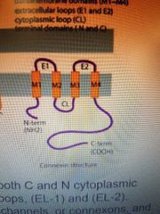
|
|
|
What are the types of simple epithelia (by shape), and where can each be found? |
Squamous (alveoli, endothelium, mesothelium). Cuboidal (certain glands, ducts). Columnar (stomach, small intestine (with microvilli), fallopian tubes (with cilia)). Pseudostratified (respiratory tract) |
|
|
The name of the transmembrane subunits of the connexon protein. |
Connexins |
|
|
The reason why pseudostratified epithelia are known as such. |
The cells are not perfectly aligned and appear to be in two layers, especially considering the fact that goblet cells are dispersed in between. |
|
|
What are the different types of stratified epithelia, and where can they be found? |
Squamous (skin, wet surfaces) Cuboidal/columnar (large excretory ducts of sweat and salivary glands, eylid covering) Transitional (surfaces that must be able to stretch and recoil, like the bladder) |
|
|
What are the two types of stratified squamous epithelia, what makes them special, and where can they be found? |
1. Keratinized. Top layer filled with keratin, has no nucleus or organelles, and protects against dehydration. Can be found in skin. 2. Nonkeratinized. Top layer retain nuclei and has less keratin. Lines internal surfaces like the esophagus, or the cornea. |
|
|
Another name for unucellular gland, and where can it be found? |
Goblet cell, exposed surface of epithelial sheet (stomach lining, respiratory epithelium). |
|
|
Where do endocrine glands secrete their secretions? |
Surrounding interstitial fluid, at which point it goes into nearby blood vessels. |
|
|
Two functional regions in exocrine glands. |
Acinus: the secretory portion. Duct: the conducting portion. |
|
|
Types of exocrine glands by duct structure. |
Simple (unbranched) Compound (branched) |
|
|
Tyles of exocrine glands by secretory unit shape. |
Tubular (tube) Acinar (alveolar) Tubuloacinar (mix between the two) |
|
|
Types of glands by secretion type |
Mucuous Serous Seromucous |
|
|
Types of exocrine glands by mode of secretion |
Merocrine (release secretory product by exocytosis). Apocrine (some cytoplasm released with the product). Holocrine (cell disintegrates into duct). |
|
|
What organ secretes content both by exocrine and endocrine method? |
Pancreas |
|
|
Connective tissue is highly vascularized and is supplies with nerves. What is the exception to that? |
Cartilage and tendon |
|
|
What is the function of ground substance in connective tissue? |
Support and bind cells, store water, allow exchange between blood and cells. |
|
|
What are the five types of mature connective tissue? |
Loose connective tissue Dense connective tissue Cartilage Bone tissue Liquid connective tissue |
|
|
What are the types of loose connective tissue, and where can they be found? |
Areolar (skin) Adipose (fat) Reticular (stroma of spleen, liver, and lymph nodes) |
|
|
What are the types of dense connective tissue, and where can they be found? |
Dense regular (skeletal muscle) Dense irregular (where forces are exerted in many directions, like dermis of skin and heart) Elastic (lung tissue, arteries) |
|
|
What are the types of cartilage and where can they be found? |
Hyaline (fetal precursor to bone) Fibrocartilage (invertebral discs) Elastic cartilage (auricle of ear) |
|
|
What are chondrocytes and where can they be found? |
Cartilage cells, found in spaces called lucunae. |
|
|
What is the pericondrium, and what is it composed of? |
It is a covering of dense irregular connective tissue, surrounding the cartilage. Composed of two layers: outer fibrous layer, inner cellular layer |
|
|
What is the structure in which an osteocyte can be found, and what is the name of the structure that in turn contains that one? |
Central (haversian) canal, contained within an osteon. |
|
|
What are the two main groups of WBCs that we learned about, and what types does each group contain? |
Granulocytes (Neutrophil, eosinophil, basophil) Agranulocytes (monocyte, lymphocyte) |
|
|
Functions of bone |
Support Protection Movement assistance Mineral homeostasis Blood cell production Triglyceride storage |
|
|
List these components of the humerus bone from proximal to distal
(Epiphysis, periosteum, articular cartilage, diaphysis, metaphysis, medullary cavity) |
Articular cartilage, proximal epiphysis, metaphysis, diaphysis (which includes endosteum, medullary cavity), metaphysis, distal epiphysis, articular cartilage. Periosteum surrounds the outside of the bone. |
|
|
What is the most abundant mineral salt? |
Calcium phosphate |
|
|
Which cells are responsible for calcification? |
Osteoblasts |
|
|
Pathway of osteocyte production. |
Osteogenic cells --> osteoblasts --> osteocytes |
|
|
Which cells regulate blood calcium levels by degrading bone matrix? |
Osteoclasts |
|
|
These cells are in the embryonic mesenchyme, and eventually they differentiate into osteoblasts. |
Osteoprogenitors |
|
|
What is the composition of the organic component of bone matrix? |
Type 1 collagen fibers Proteoglycans Glycoproteins |
|
|
Osteocytes are enclosed within (1), and communicate with each other via (2), through which they extend (3). |
1: lacunae 2: gap junctions 3: canaliculi |
|
|
Osteoclasts are derived from fusion of (1), are found in (2), and maximize their surface area via (3). |
1: multiple monocytes from blood. 2: resorption bays/Howship lacunae 3: ruffled border |
|
|
The dense outer fibrous layer of compact bone (1), and its two sub-layers (2), (3). The (3) layer contains these two types of cells (4), (5). |
1: Periosteum 2: Fibrous 3: Cellular 4: Mesenchymal stem cells 5: Osteoprogenitor cells |
|
|
What binds bone matrix to the periosteum? |
Sharpey's fibers |
|
|
This layer of bone covers large internal marrow cavities (1), it has a thin layer composed of these three types of cells: (2),(3),(4). |
1: Endosteum 2: Osteoblasts 3: Osteoclasts 4: Osteoprogenitor cells |
|
|
What is the endosteum responsible for? |
Bone growth and repair, nutrition of osseous tissue. |
|
|
Basic structural units of compact bone. |
Osteons or Haversian system |
|
What are the Ls? |
Lamellae |
|
|
Which structures are used for lamellae to be arranged in spongy bone? What is the other use for those structures? |
Trabeculae, and they also support and protect red bone marrow. |
|
|
Two types of blood vessels in bone that we learned about, and what each does. |
Periosteal arteries: supply periosteum and compact bone. Epiphyseal veins: carry blood away from long bones |
|
|
This portion of bone is rich in sensory nerves. |
Periosteum |
|
|
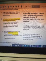
What is the endochondral ossification process (in detail)? |
|
|
|
Growth of bone in length involves these two events: |
1. Growth of cartilage on epiphyseal plate 2. Replacement of cartilage by bone tissue in epiphyseal plate. |
|
|
This process on the diaphyseal surface increases the long bone diameter. |
Intramembranous ossification |
|
|
This process grows the marrow cavity in long bone. |
Osteoclast activity |
|
|
Describe the process of bone repair in detail. |
Blood clot removed by macrophages. Fracture hematoma (procallous tissue) formed. Periosteum establishes continuity over the tissue. Procallous invaded by blood vessels and osteoblasts. Hematoma replaced by trabeculae. Bone remodeled into compact and spongy. Fully functional vasculature established. |
|
|
What two hormones regulate calcium levels in blood and how does each do it? |
Parathyroid hormone: target osteoblasts to secrete osteoclast stimulating factor, increasing Calcium levels. Calcitonin: inhibits osteoclast activity, decreasing Calcium levels. |
|
|
Out of the three muscle types covered, which ones are striated, and which are voluntary? |
Skeletal: striated voluntary. Cardiac: striated involuntary. Smooth: nonstriated involuntary. |
|
|
One unique feature in cardiac muscle tissue. |
Intercalated discs. |
|
|
Describe the organization of a skeletal muscle going from largest to smallest unit and the linings in-between. |
Skeletal muscle is surrounded by epimysium, and is composed muscle fascicles.
A fascicle is surrounded by perimysium, and is composed of multiple muscle fibers (individual muscle cells).
A muscle fiber is surrounded by endomysium, then sarcolemma, and is made up of many myofibrils (filamentous proteins). Myofibrils are made up of myofilaments. |
|
|
Blood vessels and nerves together form these in muscle. |
Neurovascular bundles. |
|
|
The nuclei of skeletal muscle are positioned in this manner. |
Periphery of the fiber. |
|
|
The name of a skeletal muscle fiber cell membrane. |
Sarcolemma |
|
|
The name of muscle fiber cytoplasm. |
Sarcoplasm |
|
|
Extensions of sarcolemma perpendicular to cell surface. |
T-tubules |
|
|
These chambers in myofibers act as calcium storage. |
Sarcoplasmic reticulum |
|
|
Expansions of sarcoplasmic reticulum on either side of T-tubule. |
Terminal cisterna |
|
|
The name for a t-tubule and adjacent terminal cisternae. |
Triad |
|
|
Contractile units of striated muscle consisting of regular arrays of thin and thick filaments (making striations). |
Sarcomeres |
|
|
What are thin filaments in sarcomeres? |
Actin, tropomyosin, troponin. |
|
|
Describe the process of muscle contraction at the level of the sarcomere. |
Calcium binds to TnC unit of troponin. Troponin has a conformational shift. Myosin binding site on actin exposed, so myosin head binds to actin. ATP breaks down into ADP, energy released, myosin head moves. The above process happens all at once for many myosin heads, resulting in thin filaments sliding over thick ones. |
|
|
This is what covers the active site of G actin in sarcomeres (1). And this is what holds it in place (2). |
1: Tropomyosin 2: Troponin |
|
|
What are the monomers of sarcomere actin called, and what is the polymer called? |
G actin is the monomer. F-actin is the filament. |
|
|
What are thick filaments in sarcomeres? |
200-300 myosin molecules bundled together. |
|
|
What are the different areas in a sarcomere? |
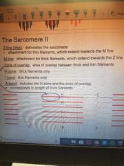
|
|
|
These units transmit electrical impulses to muscle (1), and this neurotransmitter is released as a result (2). |
1: Neuromuscular Junction 2: Acetylcholine |
|
|
What is acetylcholine's mechanism of action? |
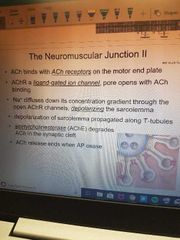
|
|
|
What important principle ensures that muscle contraction is presicely coordinated? |
All-or-none principle (a motor unit, which is connected to multiple NMJs, will activate all of them or none). |
|
|
What do cardiac muscle intercalated discs contain? |
Desmosomes to hold fibres together, and gap junctions. |
|
|
This anchors actin filaments to terminal sarcomeres in cardiac muscle. |
Fascia adherens |
|
|
Explain how the different components of an ECG wave synchronize with electrical activity in the heart. |
P: Depolarization of atria in response to SA node trigerring. PR: Delay of AV node to allow filling of ventricles. QRS: Depolarization of ventricles, main pumping contractions triggered. ST: Start of ventricular repolarization. T: Ventricular repolarization. |
|
|
These cells set the basic rate of contraction in the heart. |
Pacemaker cells. |
|
|
What factor is stored in factors aggregated at the nuclear pore in cardiomyocytes? What is its job? |
ANF (Atrial Natriuretic Factor) It acrs on kidneys to cause sodium and water loss. |
|
|
What is one major structutal difference that smooth muscle cells have when compared to skeletal or cardiac? |
They are nonstriated, and this is due to them having an irregular arrangement of myofilaments. |
|
|
What does grey matter contain? |
Neuron cell bodies, dendrites, unmyelinated axons, axon terminals, neuroglia. |
|
|
What does white matter contain? |
Myelinated axons of many neurons. |
|
|
What type of matter contains neuron cell bodies? |
Grey |
|
|
In the brain, which type of matter is on the outside and which is on the inside? |
Grey on the outside, white on the inside. |
|
|
In the spinal cord, which type of matter is on the outside and which is on the inside? |
White on the outside, grey on the inside. |
|
|
What are the two ways neurons can communicate? |
Electrically: change in transmembrane potential. Chemically: synaptic transmission. |
|
|
This is the part of the neuron where sodium channel packing is the densest, and action potential starts here. |
Axon hillock |
|
|
These substances in Neuronal somas contain rough ER and free ribosomes for protein synthesis. |
Nissl bodies |
|
|
What are the two types of summation that can trigger an action potential? |
Spatial: multiple neurons together simultaneously excite the post-synaptic neuron to reach the threshold. Temporal: a neuron sends excitatory signals, one after another, until the post-synaptic neuron reaches threshold . |
|
|
What are the five types of neurons (by structural classification), and what are the characteristics of each type? Where are they found? |
Multipolar: several dendrites and one axon. Found in brain and spinal cord. Bipolar: one (main) dendrite, and one axon. Found in retina of eye, inner ear, and in olfactory area of the brain. Unipolar: dendrites and axon fused together, examples include sensory neurons. Purkinje: highly branched dendrites. Found in cerebellum. Pyramidal: pyramidal shape. Found in cerebral cortex. |
|
|
What are the three types of neurons according to functional classification? What are their characteristics? |
Sensory (afferents, unipolar): sensory receptors at dendrites. Motor (efferents, multipolar): convey action potentials away from CNS to effectors. Interneurons (between sensory and motor): they process information from sensory neurons and determine whether to elicit a response. |
|
|
Each thick filament is surrounded by how many thin filaments? |
6 |
|
|
What are the functions of the lymphatic system? |
Drain excess interstitial fluid, transport dietary lipid, carry out immune responses. |
|
|
What are the primary and secondary lymphatic structures? |
Primary: thymus, red bone marrow. Secondary: Tonsils, cervical lymph nodes, axillary lymph nodes, inguinal lymph nodes, spleen, MALT (small intestine). |
|
|
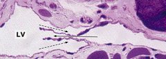
What features of lymphatic vessels make them different from blood vessels? |
They are closed at one end, have thinner walls, and more valves, slightly larger diameter than blood capillaries. |
|
|
These organs are encapsulated with masses and B and T cells. |
Lymph nodes. |
|
|
What is chyle? |
Lymph with lipids. |
|
|
What happens in lymphatic capillaries when interstitial fluid accumulates? |
Anchoring filaments pull openings wider. |
|
|
True or false: interstitial fluid can flow out of lymphatic capillaries. |
False, the capillaries only allow fluid to flow inwards. |
|
|
Where are lymphatic capillaries not found? |
Avascular tissues, the CNS, portions of spleen, and bone marrow. |
|
What is represented by the solid and dotted arrows? |
The solid arrow represents the direction of lymph flow, the dotted arrow represents the back flow prevented by valves. |
|
|
These leukocytes are found in both blood and lymph circulation. What are they responsible for (in a general sense)? |
Lymphocytes, responsible for specific immunity. |
|
|
What do lymphatic vessels unite to form? |
Lymph trunks. |
|
|
What are the principal lymph trunks? |
Lumbar, intestinal, bronchomediastinal, subclavian, jugular. |
|
|
Where does lymph pass from lymph trunks? |
Two main channels: thoracic, and right lymphatic ducts. |
|
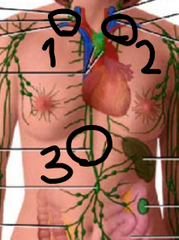
Identify the lymphatic ducts at positions 1, 2, and 3, respectively. |
1: Right lymphatic duct. 2: Thoracic duct. 3: Thoracic duct (cont'd). |
|
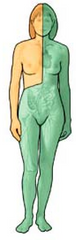
Which portion is drained by which lymphatic duct? |
Orange: right lymphatic duct. Green: thoracic duct. |
|
|
Where does lymph go from its two main channels? |
Bloodstream. |
|
|
Which two pumps that aid in venous return also aid in lymph flow? |
Skeletal muscle pump, and the respiratory pump. |
|
|
Where is the thymus located? |
On top of the heart. |
|
|
What happens to the thymus as an individual ages? |
It atrophies. |
|
|
What are the two parts of the thymus, and what is each responsible for? |
Outer cortex: positive selection of T cells, and macrophages to clear out dead and dying cells. Medulla: more mature T cells present, as well as more epithelial cells, dendritic cells, and macrophages. |
|
|
What are the two parts of a lymph node, and what is each responsible for? |
Stroma: the supportive connective tissue. Parenchyma: the functional part. |
|
|
What are the parts of a lymph node parenchyma? |
Outer cortex, inner cortex, and medulla. |
|
|
What is the exact function of lymph nodes? |
Filter lymph (trap foreign substances, destroy using macrophages or lymphocytes). |
|
|
Describe the pathway of lymph starting at the point when it is about to enter the node, up until when it leaves. |
Enters node through afferent lymphatic vessels, go into sinuses (irregular channels), into the medulla, then into efferent lymphatic vessels, out of node. |
|
|
What is the spleen parenchyma made up of? |
White pulp (lymphatic tissue), and red pulp (blood-filled venous sinuses and splenic cords). |
|
|
Which portion of the spleen produces blood cells during fetal life? |
Red pulp. |
|
|
Which part of the lymphatic system is not surrounded by a capsule? |
Lymphatic nodules/follicles. |
|
|
Where are lymphatic follices scattered? |
Lamina propria of mucous membranes lining GI, urinary, reproductive, and respiratory tracts. |
|
|
What is the MALT? |
Mucosa-associated lymphatic tissue - diffuse lymphatic tissue. |
|
|
Where are larger lymphatic follicles located? |
Tonsils, Peyer's patches, appendix. |
|
|
Where are Peyer's patches located? |
Wall of the ileum. |
|
|
Where is the appendix located? |
Cecum. |