![]()
![]()
![]()
Use LEFT and RIGHT arrow keys to navigate between flashcards;
Use UP and DOWN arrow keys to flip the card;
H to show hint;
A reads text to speech;
333 Cards in this Set
- Front
- Back
|
Which neuroglia are present in the CNS? What is the main function of each? |
Astrocytes: maintain blood-brain barrier. Oligodendrocytes: produce myelin sheath. Microglia: immune system in nervous system. Ependymal cells: secretion and passage of cerebrospinal fluid. |
|
|
What are the neuroglia of the PNS, and what are the functions of each? |
Schwann cells: myelin sheath. Satellite cells: exchange of material betwern neurons and interstitial fluid. |
|
|
These cells provide inner covering around the brain. |
Cells of pia matter |
|
|
What are the functions of astrocytes? |
Structural support Spatial buffering Nutrient transfer Blood-brain barrier Calcium wave Neuronal migration/guidance Providing ECM molecules |
|
|
What is the function of the Calcium wave in the CNS? |
A signal of damage to the brain, such as ischemia. |
|
|
What contributes to the blood-brain barrier? |
Astrocyte endfeet surrounding the capillaries Tight junctions in capillaries blocking pericellular passage of molecules. Basal laminae |
|
|
What is the difference between oligodendrocytes and schwann cells? |
Oligodendrocytes wrap myelin sheath around several axons. Schwann cells wrap just one. |
|
|
What is the special name of conduction used in most of our nerves? What makes it special? |
Salutatory conduction. In it the action potential appears to jump from one node of Ranvier to the next, as action potential cannot be triggered in portions of axon wrapped in myelin sheath. |
|
|
These portions of axons are not wrapped by myelin sheath. |
Nodes of Ranvier |
|
|
Explain the organization of peripheral nerves from macro to the micro level. |
Epineurium (entire nerve) Perineurium (each fascicle) Endoneurium (each axon) |
|
|
What protein is present in axons at the end of sheath in high density? |
Contactin-associated protein (Caspr) |
|
|
Channels for which ions are present in nodes of Ranvier? In sheath? |
Sodium in nodes of Ranvier, potassium in sheath. |
|
|
What triggers the release of neurotransmitters via synaptic vesicles in synaptic end bulbs? |
Calcium ion influx |
|
|
How many neurons are betweeb CNS and tissue in somatic motor system? In autonomic motor system? |
1 in somatic, 2 in autonomic. |
|
|
What is the preganglionic neurotransmitter in the autonomic nervous system? |
Acetylcholine |
|
|
What is the postganglionic neurotransmitter in the sympathetic nervous system? |
Norepinephrine, or acetylcholine |
|
|
What is the postganglionic neurotransmitter in the parasympathetic nervous system? |
Acetylcholine |
|
|
List some effects of sympathetic nervous signalling in the body. |
Pupil dilation, opening of airways, heart rate increase, digestion inhibition, skin blood vessel constriction, skeletal muscle blood vessel dilation. |
|
|
List some effects of parasympathetic nervous signalling in the body. |
Pupil constriction, airway constriction, heart rate decrease, digestion promotion, gut blood vessel dilation. |
|
|
Which segments of the spinal column send out sympathetic signals? |
Thoracic, and lumbar (T1-L2). |
|
|
Which segments of the spinal column send out parasympathetic signals? |
Cranial, and sacral. |
|
|
For an organ or tissue that normally receives signals from both sympathetic and parasympathetic nervous systems, what would happen if signalling from one system stopped? |
Signalling from the other would ensue unopposed. |
|
|
What is an example of sympathetic and parasympathetic nervous systems behaving cooperatively? |
Sympathetic contracting internal bladder sphincter allowing the bladder to fill up, then parasympathetic contracting the bladder to empty the contents. |
|
|
What are examples of organs or tissues receiving only sympathetic input? |
Blood vessels in skin, sweat glands, hair follicles, radial muscles of the eye. |
|
|
What are examples of organs or tissues receiving only parasympathetic input? |
Lacrimal glands, sphincter muscles of the eye. |
|
|
Out of sympathetic and parasympathetic preganglionics, which ones favour widespread activity, and which favour localized? |
Sympathetic favour widespread, parasympathetic favour localized. |
|
|
What are the sympathetic ganglia, from anterior to posterior (1)? What is another name for sympathetic ganglia (2)? |
1: Superior cervical ganglion, middle cervical ganglion, inferior cervical ganglion, celiac ganglion, superior mesenteric ganglion, inferior mesenteric ganglion. 2: Collateral ganglia. |
|
|
What are the three major sympathetic output pathways? |
Via sympathetic chain ganglia, via collateral ganglia, or via direct innervation. |
|
|
For sympathetic output via chain ganglia, what do the postganglionics do? |
One of the following: either rejoin spinal nerves to innervate blood vessels, sweat glands, hair follicles of skin, or pass into nerves innervating thoracic organs. |
|
|
For sympathetic output via collateral ganglia, what do the postganglionics do? |
Either innervate organs in the head, or innervate organs in the abdomen or pelvis (via splanchnic nerves). |
|
|
For sympathetic output via direct innervation, what do the postganglionics do? |
There are no postagnglionics, hence the "direct innervation". Splanchnic nerve directly innervates the adrenal gland, triggering the release of epinephrine into the bloodstream. |
|
|
How come sympathetic innervation is "all-or-none"? |
Preganglionics branch out and those branches travel up and down the sympathetic chain, spreading the signal |
|
|
The other name for cranial nerve III, the name of the ganglion that it reaches, the tissue its signal reaches, and the effect the signal has. |
Oculomotor, ciliary ganglion, sphincter muscle of the iris, pupil constriction. |
|
|
The other name for cranial nerve VII, the name of the ganglion that it reaches, the tissue its signal reaches, and the effect the signal has. |
Facial, sub-mandibular ganglion, salivary gland in jaw, production of saliva. |
|
|
The other name for cranial nerve IX, the name of the ganglion that it reaches, the tissue its signal reaches, and the effect the signal has. |
Glossopharyngeal, otic ganglion, salivary gland below ear, production of saliva. |
|
|
The other name for cranial nerve X, the name of the ganglion that it reaches, the tissue its signal reaches, and the effect the signal has. |
Vagus; thoracic ganglia and abdominal ganglia; visceral organs including lungs, heart, and stomach; promoting urination, defecation, digestion. |
|
|
What specific nerves do Pelvic Splanchnic Nerves consist of, what structure do they contribute to, what organs do they innervate, and what effect do they have? |
Sacral nerves 2-4; nerve plexus; lower digestive tract, bladder, and reproductive organs; urination, defecation, reproductive function. |
|
|
From where to where is information regarding emotion and behaviour relayed? |
From limbic system to hypothalamus. |
|
|
What is responsible for controlling the autonomic nervous system? What else is it responsible for? |
The hypothalamus, and it is also responsible for maitenance of homeostasis. |
|
|
Most descending pathways have a diffuse arrangement in the spinal cord. What are is the sympathetic exception to that? If not diffuse, what arrangement is it in? |

The descending sympathetic track for sweating and breathing. |
|
|
Most descending pathways have a diffuse arrangement in the spinal cord. What is the parasympathetic exception to that? If not diffuse, what arrangement is it in? |

Paraependymal tracts on either side of the central canal that contain reciprocal connections between the hypothalamus and preganglionics of the ANS. |
|
|
On the dorso-ventral axis, where in the spinal cord are sympathetic preganglionics located? |
Intermediolateral column |
|
|
On the dorso-ventral axis, where in the spinal cord are parasympathetic preganglionics located? |
Intermediomedial column |
|
|
Another name for the outflow of sympathetic preganglionic neurons in lateral horns of T1-T12, L1-2. |
White rami communcantes |
|
|
Another name for the outflow of sympathetic postganglionic neurons in lateral horns of T1-T12, L1-2. |
Gray rami communicantes |
|
|
How do myelinated and unmyelinated fibers differ in terms of their conduction velocity? |
Myelinated: saltatory conduction Unmyelinated: continuous conduction |
|
|
Which ion channels are located in and around nodes of ranvier? |

Sodium channels in the actual node. Contactin-associate protein (caspr) right by the edge facing the node. Potassium channels next to caspr. |
|
|
What is the neurotransmitter in the somatic nervous system? |
Acetylcholine |
|
|
What is the postganglionic sympathetic neurotransmitter? |
Norepinephrine or sometimes acetylcholine. |
|
|
What neurotransmitter activates the adrenal medulla? |
Acetylcholine |
|
|
What neurotransmitter activates sweat glands. |
Acetylcholine |
|
|
What is the major parasympathetic postganglionic neruotransmitter? |
Acetylcholine |
|
|
What is another name for the autonomic nervous system? |
Visceral motor system |
|
|
In terms of the big picture, what is the autonomic nervous system responsible for? |
Maintaining homeostasis in the body. |
|
|
Describe how baroreceptor reflexes work. |
There are pressure-sensitive receptors in internal carotid arteries and other large arteries in neck and chest. If blood pressure falls, baroreceptors are stretched less, there is a slower rate of impulses to cardiovascular center in the spinal cord (CV), CV decreases parasympathetic (inhibitory) stimulation, and therefore sympathetic stimulation increases. |
|
|
Which reflex helps regulate blood pressure in brain? |
Carotid sinus reflex |
|
|
Which reflex helps regulate systemic blood pressure? |
Aortic reflex |
|
|
Describe how chemoreceptor reflexes work. |
These receptors are located close to baroreceptors of carotid sinus and aortic arch. They detect hypoxia, hypercapnia, and acidosis, and upon detecting any of the three they send signals to cardiovascular center in the spine (CV). CV increases sympathetic stimulation to arteries and veins, resulting in vasoconstriction, and an increase in blood pressure.
These receptors also provide input to respiratory center to adjust breathing rate. |
|
|
What are the peripheral chemoreceptors, and what are their subtypes? |
Sensory neurons that track O2 and CO2 pressures, and pH. The subtypes are carotid body and aortic body. |
|
|
List the compartments throught which blood flows in order during circulation, starting with venae cava. |
Supererio/inferior vena cava, right atrium, right ventricle, pulmonary trunk/arteries, capillary beds of lungs, pulmonary veins, left atrium, left ventricle, aorta, arteries, arterioles, capillaries, venules, veins. |
|
|
What are the three main groups of arteries and what does each group supply? |
Ascending aorta: cardiac muscle (via the coronary arteries). Arch of aorta: head, neck, shoulders, upper limbs. Descending aorta: everything below the heart. |
|
|
What are the three main veins bringing blood into the right atrium, and where do they bring it from? |
Superior vena cava: everything above the heart.
Inferior vena cava: everything below the heart.
Coronary sinus: from cardiac muscle. |
|
|
This connects two capillary beds. |
A portal vein. |
|
|
What is a portal system? |
Capillary beds + intervening vein. |
|
|
Where are the first capillary beds, and what do they drain into? |
In digestive organs, drain into hepatic portal vein. |
|
|
The hepatic portal vein branches in the liver, forming what? |
Hepatic sinusoids |
|
|
What does blood drain into from hepatic veins? |
IVC |
|
|
What are the three layers of blood vessels and what are they composed of (go from inner-most). |
Tunica interna: endothelium. Tunic media: smooth muscle. Tunica externa: collagen, sometimes elastic lamina. |
|
|
What modulates bloodflow through the release of nitric oxide? |
Endothelium |
|
|
What is the vasa vasora? |
"Vessel of the vessels", it supplies the external layers of large blood vessels with blood. |
|
|
What is the tunica media of a muscular artery made of primarily? |
Smooth muscle |
|
|
What is the tunica media of an elastic artery made of primarily? |
Layers of smooth muscle intercalated by elastic laminas. |
|
|
What is the adventitia? |
Another name for tunica externa. |
|
|
Specific features of large elastic arteries. |
Large, closest to heart, internal and external elastic laminae present, tunica media has higher proportion of elastin. |
|
|
Specific features of muscular arteries. |
Intermediate in size, distribute blood to body organs, internal and external elastic laminae present, tunica media thicker than usual with a higher proportion of smooth muscle fibers. Controlled by sympathetic nervous system. |
|
|
Specific features of arterioles. |
No internal or external elastic laminae, thin smooth muscle layer, vascular tone controlled by sympathetic nervous system, thin tunic external compared to usual. |
|
|
What does sympathetic stimulation of venous smooth muscle cause? |
Venoconstriction, which then causes the movement of blood out of this blood reservoir into arteriolar/capillary systems. |
|
|
Specific features of veins. |
Tunica intima and media less developed than those of arteries. |
|
|
Specific features of venules. |
Little or no tunica media, thick tunica externa. |
|
|
Specific features of medium-sized veins. |
Thin tunica media, few smooth muscle fibers, tunica externa thickest. |
|
|
Specific features of large veins. |
Largest diameter, tunica externa thickest. |
|
|
What is the purpose of the venous valve, and what is it made of? |
The venous valve helps venous blood overcome gravity by preventing backflow. It is made of a folding of tunica intima. |
|
|
What forces venous blood back into the heart from the lower half of the body (specifically the legs)? |
Skeletal muscle pump. |
|
|
Which veins accompany systemic arteries, and what do they drain? |
Deep veins drain deep structures of limbs, head, thorax, abdomen, and pelvis. |
|
|
Which veins have no accompanying arteries? How deep are they tissue level-wise, and what do they drain? |
Superficial veins, which are subcutaneous, drain superficial tissues. |
|
|
What do capillaries consist of (material-wise)? |
Endothelium and BM rolled into a tube. |
|
|
What are the three types of capillaries? |
Continuous, fenestrated, and discontinuous sinusoids. |
|
|
What are the defining features of continuous capillaries? |
Complete endothelial lining with tight junctions and desmosomes, they are the least permeable and the most common, and they have transcytosis occurring. |
|
|
What are the defining features of fenestrated capillaries? |
They have pores/fenestrae in the endothelial cell wall, which permit rapid exchange of peptides and small proteins. |
|
|
Give examples of where fenestrated capillaries are present. |
Any of the following: endocrine tissue, small intestine, choroid plexus, kidney. |
|
|
What are the defining features of discontinuous sinusoids? |
They have pores larger than what is found in fenestrated capillaries, a larger diameter of vessels, with a thinner or discontinuous basement membrane. They also allow for rapid exchange for solutes larger than what is allowed in fenestrated capillaries. |
|
|
Give examples of where discontinuous sinusoids are present. |
Any of the following: liver, bone marrow, spleen, lymph nodes. |
|
|
What is the smooth muscle controlling the entrance to a capillary, and how is it controlled? |
The precapillary sphincter relaxes with increased CO2 levels, and constrict with decreased. |
|
|
What do capillary collaterals form? What is the structure's purpose? |
Arterial anastomoses. They are there to take on the blood flow from arterioles to venules in case the capillaries are closed off (which can happen in a situation of low CO2 levels). |
|
|
What action keeps blood flowing during ventricular relaxation? |
Recoil of elastic arteries. |
|
|
What major arteries make up the Circle of Willis? |
Internal carotid artery anterior cerebral artery, posterior cerebral artery. |
|
|
What are the functions of the lymphatic system? |
Drain excess interstitial fluid, transport dietary lipid, carry out immune responses. |
|
|
What are the primary and secondary lymphatic structures? |
Primary: thymus, red bone marrow.Secondary: Tonsils, cervical lymph nodes, axillary lymph nodes, inguinal lymph nodes, spleen, MALT (small intestine). |
|
|
What features of lymphatic vessels make them different from blood vessels? |
They are closed at one end, have thinner walls, and more valves, slightly larger diameter than blood capillaries. |
|
|
These organs are encapsulated with masses and B and T cells. |
Lymph nodes. |
|
|
What is chyle? |
Lymph with lipids. |
|
|
What happens in lymphatic capillaries when interstitial fluid accumulates? |
Anchoring filaments pull openings wider. |
|
|
True or false: interstitial fluid can flow out of lymphatic capillaries. |
False, the capillaries only allow fluid to flow inwards. |
|
|
Where are lymphatic capillaries not found? |
Avascular tissues, the CNS, portions of spleen, and bone marrow. |
|
|
These leukocytes are found in both blood and lymph circulation. What are they responsible for (in a general sense)? |
Lymphocytes, responsible for specific immunity. |
|
|
What do lymphatic vessels unite to form? |
Lymph trunks. |
|
|
What are the principal lymph trunks? |
Lumbar, intestinal, bronchomediastinal, subclavian, jugular. |
|
|
Where does lymph pass from lymph trunks? |
Two main channels: thoracic, and right lymphatic ducts. |
|
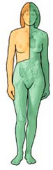
Which portion is drained by which lymphatic duct? |
Orange: right lymphatic duct.Green: thoracic duct. |
|
|
Where does lymph go from its two main channels? |
Bloodstream. |
|
|
Which two pumps that aid in venous return also aid in lymph flow? |
Skeletal muscle pump, and the respiratory pump. |
|
|
Where is the thymus located? |
On top of the heart. |
|
|
What happens to the thymus as an individual ages? |
It atrophies. |
|
|
What are the two parts of the thymus, and what is each responsible for? |
Outer cortex: positive selection of T cells, and macrophages to clear out dead and dying cells.Medulla: more mature T cells present, as well as more epithelial cells, dendritic cells, and macrophages. |
|
|
What are the two parts of a lymph node, and what is each responsible for? |
Stroma: the supportive connective tissue.Parenchyma: the functional part. |
|
|
What are the parts of a lymph node parenchyma? |
Outer cortex, inner cortex, and medulla. |
|
|
What is the exact function of lymph nodes? |
Filter lymph (trap foreign substances, destroy using macrophages or lymphocytes). |
|
|
Describe the pathway of lymph starting at the point when it is about to enter the node, up until when it leaves. |
Enters node through afferent lymphatic vessels, go into sinuses (irregular channels), into the medulla, then into efferent lymphatic vessels, out of node. |
|
|
What is the spleen parenchyma made up of? |
White pulp (lymphatic tissue), and red pulp (blood-filled venous sinuses and splenic cords). |
|
|
Which portion of the spleen produces blood cells during fetal life? |
Red pulp. |
|
|
Which part of the lymphatic system is not surrounded by a capsule? |
Lymphatic nodules/follicles. |
|
|
Where are lymphatic follices scattered? |
Lamina propria of mucous membranes lining GI, urinary, reproductive, and respiratory tracts. |
|
|
What is the MALT? |
Mucosa-associated lymphatic tissue - diffuse lymphatic tissue. |
|
|
Where are larger lymphatic follicles located? |
Tonsils, Peyer's patches, appendix. |
|
|
Where are Peyer's patches located? |
Wall of the ileum. |
|
|
Where is the appendix located? |
Cecum. |
|
|
What are the protective coverings of the brain? |
The cranium, and the cranial meninges: dura mater, arachnoid mater, and pia mater. |
|
|
What are the three extensions of the dura mater, and what parts of the brain does each separate? |
The falx cerebri separate the two cerebral hemispheres. The falx cerebrilli separate the two cerebellar hemispheres. The tentorium cerebelli separate the cerebrum from the cerebellum. |
|
|
What are brain ventricles? |
Cavities in the brain filled with CSF. |
|
|
Describe the location of ventricles within a brain, as taught in class. |
An interventricular foramen on each side connects a lateral ventricle to the third ventricles, and the aqueduct of the midbrain connects the third ventricle to the fourth. |
|
|
Where are the lateral ventricles located? |
Cerebral hemispheres. |
|
|
Where is the third ventricle located? |
Diencephalon. |
|
|
Where is the cerebral aqueduct located? |
Midbrain. |
|
|
Where is the fourth ventricle located? |
Brain stem and the cerebellum. |
|
|
Which artery/arteries supply brain with blood? |
Internal carotid and vertebral. |
|
|
Which vein(s) return blood from the brain? |
Internal jugular. |
|
|
Where does the CSF circulate? |
Ventricles, spinal cord's central canal, and subarachnoid space. |
|
|
What is the purpose of CSF? |
Absorb shock, protect brain and spinal cord, help transport nutrients and wastes from the blood and the nervous tissue. |
|
|
These cells line the brain ventricles. |
Ependymal cells. |
|
|
These are the networks of capillaries in the walls of the ventricles. |
Choroid plexuses. |
|
|
Where does CSF get its fluid from? |
Plasma from choroid plexuses, which is drawn through ependymal cells into the CSF. |
|
|
Where are choroid plexuses located? |
Lateral, third, and fourth ventricles. |
|
|
Describe the flow of CSF from the furthest choroid plexus to the blood vessels, noting any other points of CSF entry. |
Lateral ventricle choroid plexus --> lateral ventricles --(through interventricular foramina)--> third ventricle (CSF can also come in this choroid plexus) --(through aqueduct of the midbrain aka the cerebral aqueduct)--> fourth ventricle (CSF can also come in this choroid plexus) --> subarachnoid space or central canal --(arachnoid villi)--> venous blood. |
|
|
What does the brain stem consist of? |
Midbrain, pons, and medulla oblongata. |
|
|
This is what forms the central core of the brainstem. |
Reticular formation. |
|
|
Which important processes is the reticular formation involved in? |
Reticulospinal tracts (voluntary movements and posture), autonomic nervous system control centre, consciousness. |
|
|
What are the three anatomical columns of the RF? |
Median, medial, and lateral. |
|
|
What are the RF nuclei in the midbrain responsible for? |
Eye movements, consciousness, ARAS (sleep-wake cycle). |
|
|
What are the RF nuclei in the pons responsible for? |
Eye movements, consciousness, ARAS (sleep-wake cycle), blinking reflex, autonomic control. |
|
|
What are the RF nuclei in the medulla responsible for? |
Swallowing and couching, gagging and vomiting, respiration and circulation, balance and posture, pain, and autonomic control. |
|
|
What can lesions to the medulla result in (symptom-wise)? |
Respiratory distress, strange breathing behaviors (going back-and-forth between hyperventilation and deep breathing), yawning, sneezing, hiccuping. |
|
|
The cardiovascular center in the brain: what part of the brain is it in, what does it receive input from, and where does it send output to? |
It is in the medulla oblongata. It receives input from higher brain centers, proprioceptors, baroreceptors, and chemoreceptors. It provides output to the autonomic nervous system. |
|
|
What are the features of diffuse brain systems? |
Each has a small number of neurons (10-15K); the axon of each neuron is really long, has many branches, and may influence more than 100K widely-spaced post-synaptic neurons, the neurons release neurotransmitters into the extracellular space. |
|
|
What are the four diffuse brain systems? |
Norepinephrine, serotonin, dopamine, and acetylcholine. |
|
|
Norepinephrine system: 1: Where are the neurons for it? 2: What is it involved in? 3: What occurs in the case of a lesion? |
1: Locus Coeruleus. 2: Attention, arousal, sleep/wake cycle, mood disorder, pain, anxiety, learning and memory. 3: No sleep. |
|
|
Serotonergic system: 1: Where are the neurons for it? 2: What is it involved in? |
1: Raphe nuclei. 2: Mood disorders such as depression, eating disorders, sleep-wake cycle, arousal, anxiety, aggressive behaviour. |
|
|
Both serotonin and norepinephrine depletion are correlated with the onset of which mood disorder? |
Depression. |
|
|
This class of drugs increases brain serotonin levels (1). An example taught in lecture of this class is (2). |
1: SSRIs, 2: Prozac. |
|
|
These classes of drugs maintain or increase brain norepinephrine levels. |
MAO inhibitors and tricyclic drugs. |
|
|
Very little is known about the function of this brain diffuse system. |
Acetylcholine system. |
|
|
What nuclei does the acetylcholine system contain? |
PMTC, and Basal Nucleus of Meynert. |
|
|
Alzheimer's Disease is characterized by the degeneration of which neurons? |
Meynert and PMTC. |
|
|
What is a good Alzheimer's treatment? |
Acetylcholinesterase inhibitors. |
|
|
Where are the important neurons of the dopamine system? |
Substantia nigra, and ventral tegmental area (VTA) in midbrain. |
|
|
The diffuse brain system responsible for the reward pathway (1), and the location of neurons specifically involved in that (2). |
1: Dopamine system, 2: VTA. |
|
|
What causes Parkinson's Disease? What are the symptoms? |
Cause: degeneration of midbrain dopaminergic neurons in substantia nigra, but not the ventral tegmental area. Symptoms: muscular rigidity, involuntary tremor, akinesia (inability to initiate movement). |
|
|
What is a good treatment for Parkinson's, and how does it work? |
L-dopa is a precursor of dopamine, and it increases dopamine production in remaining substantia nigra neurons. |
|
|
What is the crucial part of the dopaminergic reward pathway? |
Projections from the VTA releasing dopamine to the Nucleus Accumbens and prefrontal cortex in response to rewarding or pleasurable stimuli. |
|
|
What are the cranial nerves (number and name)? |
I: Olfactory. II: Optic. III: Oculomotor. IV: Trochlear. V: Trigeminal. VI: Abducens. VII: Facial. VIII: Vestibulocochlear. IX: Glossopharyngeal. X: Vagus. XI: Accessory. XII: Hypoglossal. |
|
|
What is cranial nerve I responsible for? |
Sense of smell. |
|
|
What is cranial nerve II responsible for? |
Vision. |
|
|
What is cranial nerve III responsible for? |
Movements of eyeball and upper eyelid. |
|
|
What is cranial nerve IV responsible for? |
Movement of eyeball. |
|
|
The smallest of the cranial nerves. |
Trochlear nerve (IV). |
|
|
The largest cranial nerve. |
Trigeminal nerve (V). |
|
|
What is cranial nerve V responsible for? |
Sensation of touch, pain, and temperature. Chewing. |
|
|
What are the three branches of cranial nerve V? |
Opthalmic, maxillary, and mandibular. |
|
|
What is the origin of cranial nerve I? |
Olfactory cells converging. |
|
|
What is the origin of cranial nerve II? |
Ganglion cells in retina of each eye joining. |
|
|
What is the origin of cranial nerve III? |
Midbrain. |
|
|
What is the origin of cranial nerve IV? |
Midbrain. |
|
|
What is the origin of cranial nerve V? |
Mixed. |
|
|
What is cranial nerve VI responsible for? |
Abduction of eyeball (lateral rotation). |
|
|
What is the origin of cranial nerve VI? |
Pons. |
|
|
What is the origin of cranial nerve VII? |
Sensory portion in taste buds of anterior two-thirds of tongue, motor portion from pons. |
|
|
What is cranial nerve VII responsible for? |
Taste and facial expression. |
|
|
What are the branches of cranial nerve VIII, and what is each responsible for? |
Vestibular branch is for equilibrium, cochlear branch is for hearing. |
|
|
What is the origin of cranial nerve VIII? |
Inner ear. |
|
|
What is cranial nerve IX responsible for? |
Taste and saliva release. |
|
|
What is the origin of cranial nerve IX? |
Sensory portion in taste buds of posterior one-third of the tongue, motor portion in medulla. |
|
|
What is cranial nerve X responsible for? |
Proprioception, stretching, swallowing, vocalization. |
|
|
What is the origin of cranial nerve X? |
Mixed, motor portion comes from medulla. |
|
|
What is cranial nerve XI responsible for? |
Head movements. |
|
|
What are the subunits of cranial nerve XI? |
Cranial accessory and spinal accessory. |
|
|
What is cranial nerve XII responsible for? |
Speech and swallowing. |
|
|
What is the origin of cranial nerve XII? |
Medulla oblongata. |
|
|
Which of the cranial nerves carry parasympathetic signals? |
III, VII, IX, X. |
|
What plane is this? |
Frontal or (when referring to sections passing through the skull) coronal. |
|
What plane is this? |
Sagittal. |
|
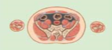
What plane is this? |
Transverse, horizontal, or cross-sectional. |
|
|
This plane separates the anterior and posterior portions of the body, it is oriented parallel to the long axis. Which one is it? |
Frontal or (when referring to sections passing through the skull) coronal. |
|
|
This plane separates the left and right portions of the body, it is oriented parallel to the long axis. Which one is it? |
Sagittal. |
|
|
This plane is oriented perpendicular to the long axis. Which one is it? |
Transverse, horizontal, or cross-sectional. |
|
|
Directional term that means "toward the head" (1), and its antonym (2). |
1: Carnial/cephalic, 2: Caudal. |
|
|
Directional term that means "toward the front" (1), and its antonym (2). |
1: Anterior/Ventral, 2: Posterior/Dorsal. |
|
|
Directional term used to mean "above" (1), and its antonym (2). |
1: Superior, 2: Inferior. |
|
|
Directional term used to mean "toward the midline" (1), and its antonym (2). |
1: Medial, 2: Lateral. |
|
|
Directional term used to mean "closer to an attached base" (1), and its antonym (2). |
1: Proximal, 2: Distal. |
|
|
Directional term used to mean "closer to the body surface" (1), and its antonym (2). |
1: Superficial, 2: Deep. |
|
|
Which directional term is absolute? |
Median. |
|
|
Directional term used to mean "front of the head" (1), and its antonym (2). |
1: Rostral, 2: Caudal. |
|
|
What are the the types of articulations classified by functionality? |
Synathroses (no movement), amphiathroses (little movement), diathroses/synovial joints (free movement). |
|
|
What are the types of synathroses? |
Fibrous (joined by dense irregular CT), cartilaginous (cartilage), and bony fusions (two bones becoming one). |
|
|
Give examples of fibrous synathroses. |
Suture, and gomphosis. |
|
|
Give an example of a cartilaginous synathrosis. |
Synchondrosis. |
|
|
Give an example of a bony fusion. |
Synostosis. |
|
|
What are the types of amphiathroses? |
Fibrous (joined by ligament or band of CT), and cartilaginous (cartilage). |
|
|
Give an example of a fibrous amphiathrosis. |
Syndesmosis. |
|
|
Give an example of a cartilaginous amphiathrosis. |
Symphysis. |
|
|
What are the core components of diathroses? |
Joint cavity, fibrous joint capsule, synovial membrane, articular cartilage. |
|
|
What are the synovial joint accessory structures? |
Articular discs/menisci, fat pads, tendons, bursae. |
|
|
What is the result of bursae inflammation? |
Bursitis. |
|
|
What movements can joints perform? |
Gliding, angular movement, rotation, special. |
|
|
A painful disorder of supporting structures (bone, muscles, tendons, ligaments) of the body. |
Rheumatism. |
|
|
A form of rheumatism in which the joints are painful, swollen, and stiff. |
Arthritis. |
|
|
An autoimmune disease in which cartilage and joint linings are attacked. What is it characterized by? |
Rheumatoid arthritis, characterized by inflammation of the joint, causing swelling, pain, and loss of function. |
|
|
The three subdivisions of the thoracic cavity. |
Pleural cavities (x2), mediastinum, pericardial cavity. |
|
|
The muscular sheet that separates the thoracic cavity from the abdominopelvic cavity. |
Diaphragm. |
|
|
The abdominopelvic cavity can be subdivided into these two spaces: |
Abdominal/peritoneal cavity, and pelvic cavity. |
|
|
They line walls of body cavities and their contents. |
Serous membranes. |
|
|
Serous membranes can be divided into these two portions. |
Parietal (line walls of cavity), and visceral (line contained spaces). |
|
|
Aside from function, are the parietal and visceral portions of serous membrane different in any way? If so, how. |
They are not. Histologically they are identical, and structurally they are continuous with each other. |
|
|
This is located between the two portions of serous membrane (1). It contains this (2). |
1: Serous cavity, 2: transudate. |
|
|
What is the mediastinum? |
It is the compartment between the two pleural cavities, and it contains the heart (in the pericardial subcavity), its vessels, trachea, esophagus, phrenic nerves, thoracic duct, thymus, and lymph nodes. |
|
|
What are the boundaries of the mediastinum? |
Superior thoracic aperture, diaphragm, R&L pleurae, sternum, bodies of T1-T12. |
|
|
What are the subdivisions of the mediastinum? |
Superior, and inferior. |
|
|
What are the boundaries of the superior mediastinum? |
Superior thoracic aperture, plane of sternal angle, manubrium, T1-T4. |
|
|
What are the contents of the superior mediastinum? |
Superior vena cava, arch of aorta and branches, trachea, esophagus, vagus and phrenic nerves. |
|
|
What are the subdivisions of the inferior mediastinum? |
Anterior, middle, posterior. |
|
|
Where is the anterior mediastinum relative to the pericardial sac and the sternum? |
Anterior to the pericardial sac, posterior to the sternum. |
|
|
What are the contents of the anterior mediastinum? |
Thymus or remnants, blood vesels, loose connective tissue, and lymph nodes. |
|
|
What is the middle mediastinum? What does it contain? |
Another name for pericardial sac, so it contains the heart and great vessels. |
|
|
Where is the posterior mediastinum relative to the pericardial sac and vertebral column? |
Posterior to pericardial sac, anterior to vertebral column. |
|
|
What does the posterior mediastinum contain? |
Trachea, primary bronchi, esophagus, azygous and hemiazygous veins, thoracic duct, descending aorta. |
|
|
What are the vertebral regions, and how many pieces are in each? |
Cervical (7), thoracic (12), lumbar (5), sacral (5), coccygeal. |
|
|
Where are the primary spinal curvatures, when do they develop, and why? |
Thoracic and sacral curves, develop prenatally, to accommodate viscera. |
|
|
Where are the secondary spinal curvatures, when do they develop, and why? |
Cervical and lumbar curves, develop postnatally, to hold head up (cervical), and maintain balance in posture (lumbar). |
|
|
What are the parts of a typical vertebra? |
Anterior vertebral body (bears weight), posterior vertebral arch, vertebral foramen (houses the spinal cord), spinous process, tranverse processes, superior articular processes, inferior articular processes. |
|
|
What is the purpose of the vertebral foramen? |
To hold the spinal cord. |
|
|
What are the two main changing trends in the spinal cord you see when you look in the rostrocaudal direction? Why? |
Size of vertebral body increases (because the load increases), and diameter of vertebral foramen/canal decreases (because the spinal cord diameter decreases. |
|
|
What are the intervertebral articulations? |
Vertebral canal, intervertebral foramina, zygapophyseal joints, intervertebral discs, intervertebral ligaments. |
|
|
What is the purpose of the intervertebral foramina? |
Accommodate spinal nerves. |
|
|
What are the intervertebral discs made up of? |
Fibrocartilagenous annulus fibrosus, and nucleus pulposus. |
|
|
What are the different pairs of ribs, and how are they numbered? |
True ribs (1-7), false ribs (8-10), floating ribs (11,12). |
|
|
What makes true ribs different from false ribs? |
True ribs articulate directly with sternum via costal cartilages, while false ribs join sternum via costal cartilages of the rib above. |
|
|
What are the two major openings of the rib cage? |
Superior and inferior thoracic apertures. |
|
|
What nerves control the intercostal muscles? |
Ventral primary rami. |
|
|
Where does the arterial supply to the thoracic wall come from? |
Intercostal arteries. |
|
|
Where does the thoracic wall drain blood? |
Intercostal veins. |
|
|
How many spinal nerve pairs are there, and what is the breakdown by type? |
31 in total: 8 cervical, 12 thoracic, 5 lumbar, 5 sacral, 1 coccygeal. |
|
|
What are the major spinal nerve branches we have to know? |
Meningeal branches, ventral ramus, dorsal ramus, rami communicantes (gray and white). |
|
|
What are the important nerves of brachial plexus? |
Musculocutaneous, axillary, radial, median, ulnar. |
|
|
Which spinal nerve pairs are related to the phrenic nerve? |
C3, C4, C5. |
|
|
Which rami form lumber plexus? |
L1-L4 (anterior). |
|
|
Which rami form sacral plexus? |
L4-L5 and S1-S4 (anterior). |
|
|
The sacral plexus gives rise to the longest nerve in the body. What is it? |
Sciatic. |
|
|
In which segment of the spine does the spinal cord end in adults? |
L1 |
|
|
What are the intercostal nerves made of? |
Ventral rami of thoracic spinal nerves. |
|
|
Which rami form the brachial plexus? |
C5-T1 |
|
|
Which subsections does the sciatic nerve divide into? |
Common fibular, and tibial. |
|
|
An individual strip of skin supplied by an individual spinal nerve. |
Dermatome. |
|
|
What is the purpose of the pericardium? |
Protect and anchor the heart, prevent overfilling, and provide friction-free environment. |
|
|
What are the layers of the heart wall? |
Epicardium (visceral layer of serous pericardium), myocardium (cardiac muscle), endocardium (endothelium lining the heart chambers). |
|
|
What separates the two heart atria? |
Interatrial septum. |
|
|
What separates the two heart ventricles? |
Interventricular septum. |
|
|
The surface groove dividing the heart ventricles. |
Anterior interventricular sulcus. |
|
|
The surface groove dividing heart atria from ventricles. |
Coronary sulcus. |
|
|
True or false: the coronary sinus feeds into the vena cava. |
False: the coronary sinus feeds straight into the right atrium. |
|
|
What is the main cardiac vein that feeds into the right atrium? |
Coronary sinus. |
|
|
What separates the right and left atria of the heart? What is the fetal version of that? |
Foramen ovalis, in fetal circulation it is foramen ovale which is not completely closed. |
|
|
How does foramen ovale become foramen ovalis. |
After birth the lungs are developed for independent respiration, and their increased pressure pushes on the foramen ovale, closing it and turning it into the foramen ovalis. |
|
|
This holds the aorta and pulmonary trunk together. What is the fetal version of it? |
Ligamentum arteriosum, in fetus known as ductus arteriosus (pulmonary trunk and aorta have blood mixing). |
|
|
What are the types of heart valves, and what valve is in each type? |
Atrioventricular: tricuspid and bicuspid valves. Semilunar: pulmonary and aortic valves. |
|
|
The tricuspid valve is located between which chambers of the heart? |
Right atrium and right ventricle. |
|
|
The pulmonary valve is located between which chambers of the heart? |
Right ventricles and pulmonary arteries. |
|
|
The mitral/bicuspid valve is located between which chambers of the heart? |
Left atrium and left ventricle. |
|
|
The aortic valve is located between which chambers of the heart? |
Left ventricle and aorta. |
|
|
What do anastomoses do? |
Provide alternate routes/collateral circuits in coronary circulation, allowing heart muscle to receive sufficient oxygen if an artery is partially blocked. |
|
|
When does blood perfuse cardiac tissue? |
During the diastolic phase when the myocardium is relaxed. |
|
|
Trace the cardiac conduction system. |
SA node in right atrial wall, through atria via gap junctions, AV node, AV bundle/Bundle of His, right and left bundle branches, Purkinje fibers. |
|
|
What are the 2 hear sounds that can be heard? |
Lubb (AV valves close), dupp (SL valves close). |
|
|
What are the two components of the respiratory system? |
Conducting portion (from nose up to and including terminal bronchioles), and respiratory portion (from respiratory bronchioles up to and including alveoli). |
|
|
What is the lining of the conducting portion of the respiratory system? What are its two layers? |
Respiratory mucosa, consisting of respiratory epithelium and underlying lamina propria (loose CT). |
|
|
What subdivides the nasal cavity? |
Superior, middle, and inferior nasal conchae. |
|
|
Air-filled spaces lined by mucous membrane. |
Paranasal sinuses. |
|
|
What is the nasal mucosa bound to? |
Periosteum and perichondrium of supporting structures. |
|
|
Where do axons from olfactory epithelium pass to? |
Through cribriform plate as cranial nerve I to synapse on nerves in olfactory bulbs. |
|
|
What are the three subdivisions of the pharynx? |
Nasopharynx, oropharynx, and laryngopharynx. |
|
|
The nasopharynx contains the opening of what? |
Auditory tubes. |
|
|
Where is the laryngopharynx compared to the larynx? |
Posterior. |
|
|
What is the laryngeal inlet, and what is the other name for it? |
It is the opening into the larynx, another name for it is the aditus. |
|
|
How is food prevented from getting into the larynx during swallowing? |
The bolus folds the epiglottis, blocking passage into larynx. |
|
|
What are the three unpaired cartilages in the trachea? |
Thyroid cartilage, cricoid cartilage, and epiglottis. |
|
|
What are the three surfaces of the lungs? |
Costal, mediastinal, diaphragmatic. |
|
|
This in the left lung is what accommodates the heart. |
Cardiac notch. |
|
|
This is what separates lung lobes from each other. |
Fissures. |
|
|
What is the root of the lung? |
Primary bronchi, pulmonary arteries and veins, nerves, and lymphatic vessels. |
|
|
This is the region containing the root of the lung. |
Hilus of the lung. |
|
|
This is what joins tracheal rings at the anterior end. |
Annular ligaments. |
|
|
This is what covers the posterior aspect of the trachea. |
Trachealis (a smooth muscle). |
|
|
This is where the trachea bifurcates into the primary bronchi. |
Carina. |
|
|
Describe the bronchial tree: how many primary, secondary, and tertiary bronchi are there? |
Primary: one per lung, secondary: one per lobe, tertiary: one per bronchopulmonary segment. |
|
|
What constitutes the blood supply of the diaphragm? |
Superior and inferior phrenic arteries and veins. |
|
|
What is the nerve supply of the diaphragm? |
Phrenic nerves. |
|
|
How does the position of ribs influence the volume of the thorax? |
Elevation of ribs increases lateral and AP dimensions of the thorax. Depression of ribs does the opposite. |
|
|
These join adjacent ribs. |
Intercostal muscles. |
|
|
What are the three layers of intercostal muscles? |
External, internal, innermost. |
|
|
What happens to the diaphragm during inhalation? |
Flattening. |
|
|
When is exhalation active? |
Forced expiration. |
|
|
What are the two important players in neural control of respiration? |
Inspiratory centre, expiratory centre. |
|
|
Branching of bronchioles ends here. |
Terminal bronchioles. |
|
|
What are the components of a bronchus? |
Mucosa, cartilage. |
|
|
In a cross-section picture of trachea, what layers do you see from the surface layer-downwards? |
Mucosa, submucosa, hyaline cartilage, adventitia. |
|
|
What are the components of a bronchiole? |
Mucosa, lamina propria. |
|
|
These cells in the small bronchiole have no cilia, and possess secretory granules in their apex that release a protein similar to surfactant. |
Clara cells. |
|
|
What are the components of terminal and respiratory bronchioles? |
Mucosa, smooth muscle, elastic CT under epithelia. |
|
|
The surface epithelial cells of the alveoli. |
Pneumocytes. |
|
|
What are the three main cell types found in alveoli? What is the function of each? |
Type I pneumocytes (simple squamous blood-air barrier), type II pneumocytes (surfactant), alveolar macrophages (protection from pathogens). |
|
|
What are the lobes of the right lung? |
Superior, middle, inferior. |
|
|
What are the lobes of the left lung? |
Superior, inferior. |
|
|
The muscle fibers of the diaphragm insert into this. |
Central tendon. |
|
|
This major vein passes through the diaphragm. |
Inferior vena cava. |
|
|
Which muscles pull during exhalation? During inhalation? |
Internal intercostal during exhalation, external intercostal during inhalation. |