![]()
![]()
![]()
Use LEFT and RIGHT arrow keys to navigate between flashcards;
Use UP and DOWN arrow keys to flip the card;
H to show hint;
A reads text to speech;
180 Cards in this Set
- Front
- Back
|
The spine consists of seven ___ cervical vertebrae, |
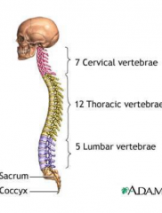
7 Cervical |
|
|
The spine consists of ___ moveable vertebrae
|
24
|
|
|
Size of the vetrebral body ___ as we go down. |
increases
|
|
|
The spine is curved to support ___
|
weight
|
|
|
There are __- normal curves that are formed by the vertebral column.
|
two
|
|
|
The curves function like connected arches adding additional ___ and ___.
|
flexibility
shock absorption |
|
|
The additional flexibility (shock absorbance) is dynamic and maintained by ___ muscle groups
–___ flexors resist hyperlordosis in LS –Long ___ resist hyperkyphosis in TS |
antagonistic
Abdominal back extensors |
|
|
Primary curves: also known as ___ curves are found in the ___ and ___ regions.
|
kyphotic
thoracic sacral |
|
|
___ curves are concave anteriorly due mainly to a decreased ___ anteriorly and an increased height ___ of the vertebral bodies.
|
Kyphotic
height posteriorly |
|
|
The kyphoses develop during ___ when the fetus is in a C-shaped position (fetal position) and are maintained throughout life
|
gestation
|
|
|
Secondary curves: also known as ___ curves are found in the cervical and lumbar regions.
|
lordotic
|
|
|
Lordotic curves are concave ___ and are maintained due to height differences in the intervertebral discs.
|
posteriorly
|
|
|
Cervical ___ develops when the infant begins to hold the head up (___ months)
|
lordosis
~4 |
|
|
Lumbar ___ develops when the infant begins to stand erect and walk (___ months)
|
lordosis
10-18 |
|
|
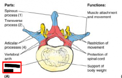
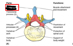
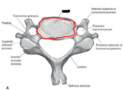
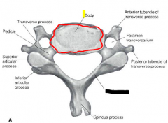
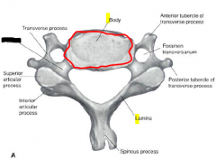
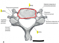
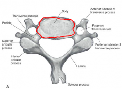
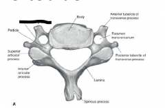
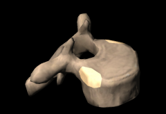
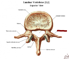
Bone markings that are common to cervical, thoracic, and lumbar vertebrae:
Body - Thick anterior portion that is designed for ___ bearing. The size of the vertebral bodies ___ as you descend the spinal column. In situ, the superior and inferior surfaces of the body are covered with ___ cartilage and are known as the vertebral end plates. |
weight
increase hyaline |
|
|
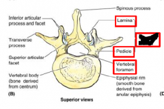
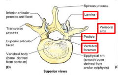
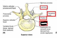
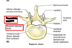
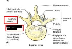
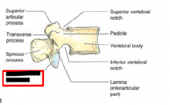
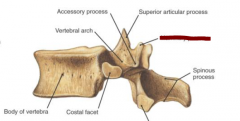
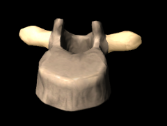
Bone markings that are common to cervical, thoracic, and lumbar vertebrae:
___ (little feet) - Two short, thick processes located on the posterior body. They will project posteriorly |
Pedicle
|
|
|
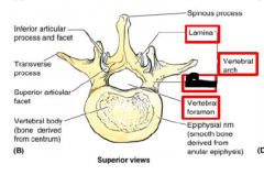
Bone markings that are common to cervical, thoracic, and lumbar vertebrae:
___ - Two flat processes that come off the pedicles and meet in the midline. |
Lamina
|
|
|
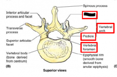
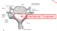
Bone markings that are common to cervical, thoracic, and lumbar vertebrae:
The pedicles and laminae along with the spinous process form the ___. Along with the body they form the ___. |
vertebral arch
vertebral foramen |
|
|
Vertebral foramina of multiple levels form the ___.
|
vertebral canal
|
|
|
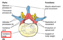
Vertebral Body |
|
|

Lamina |
|
|

Vertebral Arch |
|
|
Pedicle |
|
|

Vertebral Foramen |
|
|
___ processes that extend off of the vertebral arch: |
Seven |
|
|
Seven processes that extend off of the vertebral arch: |
Spinous Process |
|
|
Seven processes that extend off of the vertebral arch:
2)___ - extend laterally to either side of the vertebra at the junction of the pedicle and lamina. |
Transverse Processes
|
|
|
Seven processes that extend off of the vertebral arch cont: |
Fill in***** |
|
|
Spinous Process
|
|
|
Transverse Process
|
|
|
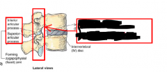
Superior Articular Facet
|
|
|
Inferior Articular Facet
|
|
|
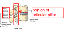
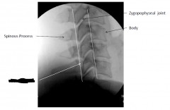
portion of articular pillar |
|

|
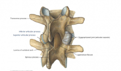
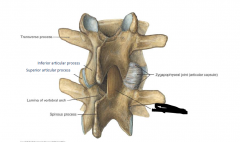
Zygoapophyseal Joint |
|
|

ligamentum Flavum |
|
|
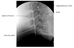
Articular Pillar |
|
|
C1, C2 and C7 are ___ C3-C6 are __- |
atypical typical |
|
|

Body |
|
|

Lamina |
|
|

Pedicle |
|
|
Spinous process |
|
|

Vertebral arch |
|
|

Transverse process |
|

|
Vertebral foramen |
|
|
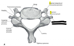
Typical Cervical Vertebrae C3-C6:
Spinous Process - C2-C6 have a ___ spinous process (cleaved in 2) |
bifid |
|
|
Typical Cervical Vertebrae C3-C6:
Vertebral (spinal) foramen - large and ___ shaped |
triangle
|
|
|
Typical Cervical Vertebrae C3-C6:
In the cervical spine C1-C7 have foramen of the ___ process (foramen transversarium). |
transverse
|
|

|
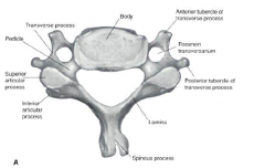
foramen transversarium (foramen of the transverse process)
|
|
|
The superior facets face ___, ___, & ___. |
posteriorly, superiorly, and medial. |
|
|
The __- processes in C/S consist of anterior (costal process) and posterior (true transverse process) parts joined together by the intertubercular lamella. |
transverse |
|

|
Anterior Tubercle of Transverse Process
|
|
|
posterior Tubercle of Transverse Process
|
|
|
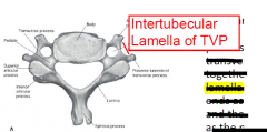
Intertubercular lamella of Transverse Process |
|
|
The anterior tubercle at C6 is also known as the ___ because the common ___ artery can be compressed against the tubercle at this level. |
carotid tubercle
carotid |
|
|
The foramen of the transverse process is located in the TP B/L ___.
|
C1-C7
|
|
|
The vertebral a. enters the foramen of the TP at the ___ level and runs up through C1 where it enters the ___. |
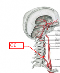
C6 |
|
|
Running with the vertebral a. is a ___ of nerves
|
sympathetic plexus
|
|
|
The vertebral v enters the foramen of the TP at the ___ level and continues through C7 and then drains into the ___.
|
C1
subclavian v |
|
|
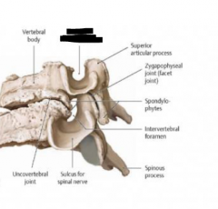
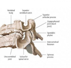
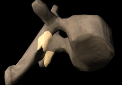
On the superior surface of each cervical vertebra the ___ form a lateral and slightly posterior raised rim |
uncinate processes
|
|
|
Uncinate processes help to limit ___ and to prevent IVD protrusion. |
lateral flexion
|
|

|
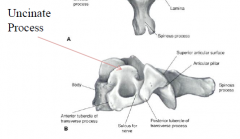
Unicate Process |
|
|
The uncinate processes may form a ___ (uncovertebral joints/joints of von Luschka) with the vertebra above, C3-C6. |
synovial joint |
|
|
The joint formed by uncinate processes is a common site of ___ formation
|
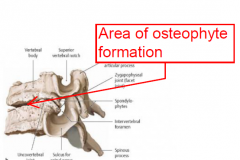
osteophyte |
|
|
Superior Vertebral Notch |
|
|
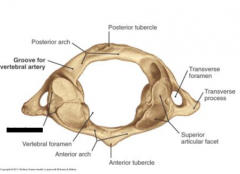
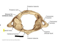
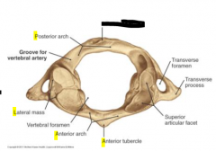
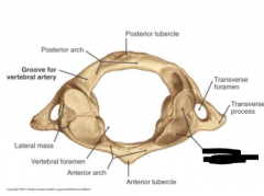
C1 does not have a vertebral body, but has two ___ connected by anterior and posterior arches |
lateral masses |
|
|
Each lateral mass of C1 has a superior and inferior ___ and a TP.
|
articular process
|
|
|
The superior articular facets are shaped like a ___ and articulate with the occipital condyles.
|
peanut
|
|
|
The inferior articular c1 facets are regularly shaped ___ and articulate with C2.
|
oval
|
|
|
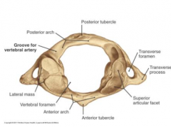
Lateral Mass of C1 |
|
|

Anterior Arch of C1 |
|
|

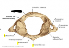
Anterior Tubercle of C1 |
|
|

Posterior Arch of C1 |
|
|

Posterior Tubercle of C1 |
|
|

Superior Articular Facet of C1 |
|
|
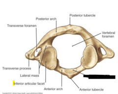
Inferior Articular Facet of C1
|
|
|
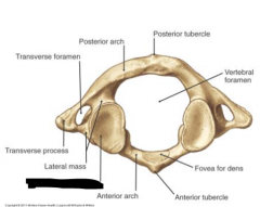
Facet for Dens of C1 |
|
|
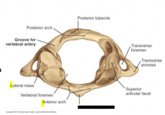
Anterior Arch of C1 |
smaller
|
|
|
Anterior Arch of C1
Roughly in the middle of the arch on the anterior surface is the ___. This tubercle is the attachment site for the anterior longitudinal ligament, and the longus colli muscles. |
anterior tubercle |
|
|
Anterior Arch of C1
On the posterior surface of the arch is found a smooth surface, the facet for the ___. It is covered in hyaline cartilage and forms a diarthrodial, synovial pivot joint with the ___ (dens). This joint allows the majority of rotation in the C/S. |
dens |
|
|
Posterior Arch of C1
The ___ of the two arches, the posterior arch also has a posterior elevation called the ___, which serves as an attachment for the ligamentum nuchae and the origin of rectus capitis posterior minor mm. |
larger |
|
|
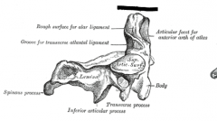
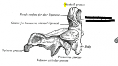
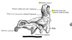
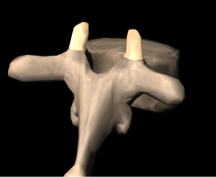
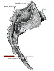
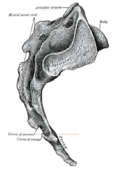
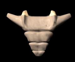
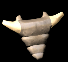
The distinguishing characteristic of C2 is the tooth-like process that protrudes superiorly off the body, the ___
|
dens/odontoid process.
|
|
|
The dens has a ___ lined articular facet anteriorly, and a ___ posteriorly for the transverse portion of the cruciform l. (transverse ligament of atlas).
|
hyaline |
|
|

Odontoid Process/ Dens of C2 |
|
|
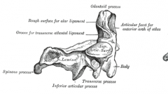
Articular facet for anterior arch of C1 |
|
|

Inferior Articular Process of C2 |
|
|
The superior articular processes of C2 are on the ___ (not the pediculolaminar junction) and appear more as a smoothed out region than an actual process. |
pedicles |
|
|
Like the inferior articular facets of C1, the superior articular facets of C2 are smooth and ___ shaped.
|
oval
|
|
|
C7 is also called the ___ because of its prominent spinous process. The SP usually projects directly posterior and is not bifid.
|
vertebra prominens
|
|
|
The foramen of the TP is usually a little smaller and only the ___ passes through. NO VERTEBRAL A. OR SYMPATHETIC PLEXUS!
|
vertebral v.
|
|
|
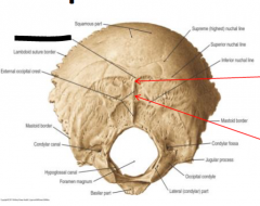
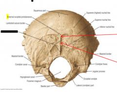
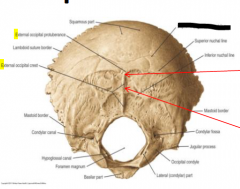
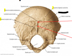
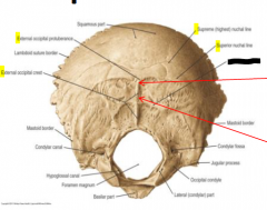
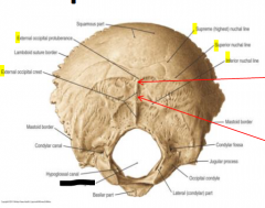
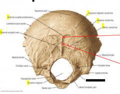
The most prominent bump of the Occiput is known as the ___
|
external occipital protuberance (EOP) / Inion |
|
|
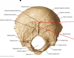
External Occipital Protuberance
|
|
|

External Occipital Crest |
|
|

Supreme Nuchal Line |
|
|

Superior Nuchal Line |
|
|

Inferior Nuchal Line |
|
|

Foramen Magnum |
|
|

Occipital Condyle |
|
|
Continuing inferiorly from the EOP is the ___ |
external occipital crest (EOC)
|
|
|
The EOP is the attachment site for the ___
|
trapezius muscle
|
|
|
There may be as many as ___ horizontal lines on the posterior occiput. |
three (3)
|
|
|
The ___ nuchal line is not always present, but may be located superior to the EOP.
|
highest/supreme
|
|
|
The ___ nuchal line is almost always present and runs horizontally on either side of the EOP. This line is the attachment for the ___, ___, and ___ mm.
|
superior
trapezius splenius capitis SCM |
|
|
The ___ line extends laterally from the EOC about halfway between the EOP and the foramen magnum. Attachment site for ___ post major/minor mm. |
inferior nuchal
rectus capitis |
|
|
Inferiorly on the occipital bone are two (2) ___. The condyles are convex and are located on either side of the foramen magnum. The condyles articulate with the superior articular processes of C1 to form the ___ joints. |
occipital condyles
atlanto-occipital |
|
|
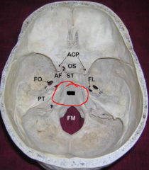
Internally, extending anteriorly from the foramen magnum is the basilar portion of the occipital bone. It articulates with the basilar portion of the sphenoid bone and together they are called the ___.
|
clivus (basi-occiput)
|
|
|
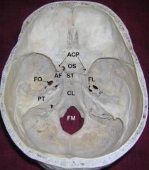
clivus (basi-occiput) of occipital bone |
|
|
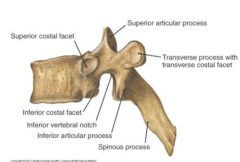
The body of T/S is more ___ shaped and they increase in size as they descend the spine. Also, the bodies are usually a little flattened anteriorly on the left d/t the pulsations of descending aorta. |
heart
|
|
|
Most T/S bodies have four ___, two superior and two inferior, that articulate with the ribs. |
costal demifacets
|
|
|
The ___ costal demifacets of T/S will articulate with the rib of the same number |
superior
|
|
|
The pedicles attach high on the vertebral body so there is usually no ___ associated with the T/S, but there is an extra large ___. |
superior notch
inferior notch |
|
|
Each TP has an articulation for the rib of the same number called the ___ of the TP.
•The TP of the 4th vertebra articulates with the 4th rib. |
transverse costal facet or costal facet
|
|
|
The T/S costal demifacets are half facets located at the superior and inferior borders of the body. A pair of ___ (inferior and superior) articulate with a single rib
|
costal demifacets
|
|
|
___ have both superior and inferior costal demifacets. Other T/S vertebrae are abnormal: |
T2-T9
|
|
|
The ___ articular facets of thorax face posteriorly, superiorly, and laterally. (PLS)
|
superior
|
|
|

T/S Superior Articular Process |
|
|
T/S Inferior Vertebral Notch |
|
|
T/S Mamillary Process |
|
|
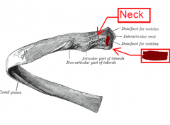
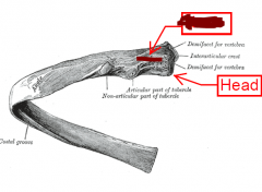
Typical ribs are ribs 3-9, and each has a ___, ___, ___ and a ___.
|
head, neck, tubercle and a shaft. |
|
|
The head of each rib will articulate with two vertebral bodies and the IVD between. It, therefore, has a superior and inferior articular facet.
–The inferior articular facet will articulate with the ___ demifacet of the SAME NUMBER vertebra. |
superior |
|
|
Between the two facets is the ___ of the head. This projects between the two vertebral bodies.
|
crest |
|
|

Head of Rib |
|
|
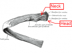
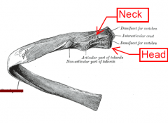
Neck of Rib |
|
|
Costal Groove of Rib
|
|
|
The neck of the rib is located between the head and the tubercle and is an attachment for the ___ and the ___ |
superior costotransverse l.
costotransverse l. |
|
|
The tubercle of the rib is usually located on the posteroinferior margin of the rib. The tubercle articulates with the transverse costal facet of the TP of the ___ number. |
same |
|
|
The non-articular portion of the tubercle is the attachment site for the ___
|
lateral costotransverse l.
|
|
|
The shaft of the rib runs anteriorly and inferiorly. It attaches to the costal cartilage anteriorly and then to the ___.
|
sternum
|
|
|
On the anterior inferior surface of the shaft is the ___. This groove shelters the intercostal vein, artery, and nerve.
|
costal groove |
|
|
The first rib is very flat and short. It runs almost completely horizontal. There are markings for muscle attachments and for vascular depressions. It usually only articulates with ___
|
T1.
|
|
|
The second rib is almost typical, but it does have a large ___ that serves as an attachment for the serratus anterior muscle.
|
tubercle
|
|
|
The 10th rib only has a single ___ because it usually only articulates with T10. As such there is also no crest on the head.
|
facet
|
|
|
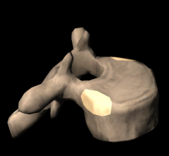
The lumbar vertebral bodies are rather large and are ___ shaped.
They are also wide left to right, and they are thicker anteriorly (helps maintain lumber ___). |
floating( fix)
|
|
|
?? |
kidney |
|
|
L/S There is a small superior vertebral notch, but a very prominent ___ vertebral notch. |
inferior
|
|
|
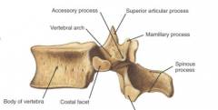
Each L/S TP has an ___ process. |
accessory
accessory |
|
|
The L/S superior articular facets face ___ & ___
|
posterior and medial
|
|
|
Projecting posteriorly off of the superior articular process is the ___ process
|
mamillary |
|
|
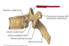
Thoracic Superior Costal Facet/ Costal Demifacet
|
|
|
Thoracic Inferior Costal Facet/ Costal Demifacet
|
|
|
Thoracic Transverse Process
|
|
|
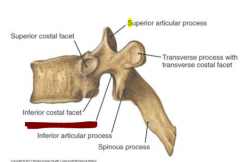
Thoracic Superior Articular Process
|
|
|
Thoracic Inferior Articular Process
|
|
|
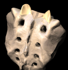
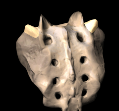
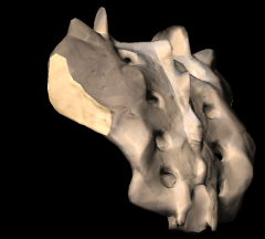
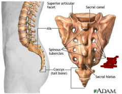
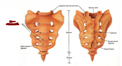
Sacrum: Superior Articular Process
|
|
|
Sacral Ala
|
|
|
Auriticular Surface
|
|
|
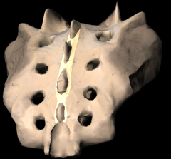
Median Sacral Crest
|
|
|
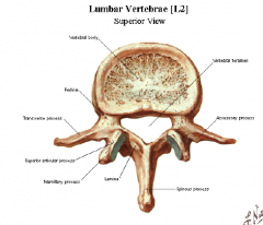
Lumbar Accessory Process |
|
|
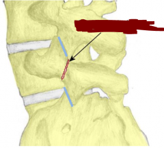
The area between the superior articular process and the inferior articular process is known as the ___. |
pars interarticularis |
|
|
The Pars interarticularis is commonly fractured, which is known as ___ |
spondylolysis
|
|
|
Spondylolysis is associated with ___, which is an anterior displacement of the body, pedicles, TPs, and superior articular processes. |
spondylolisthesis
|
|
|
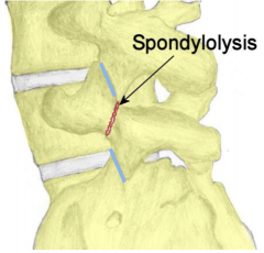
spondylolysis |
|
|

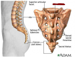
Dorsal Sacral Foramen |
|
|
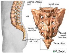
Sacral Canal |
|
|

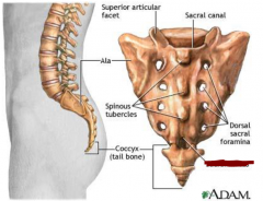
Sacral Hiatus |
|
|
The sacrum consists of five fused vertebrae. The base is located ___ and the apex is located ___.
|
superiorly
inferiorly |
|
|
Unique Characteristics of the Sacral Base
There is a superior articular ___ & ___, which articulate with the inferior articular process of L5. |
process and facet
|
|
|
Unique Characteristics of the Sacral Base
The anterosuperior margin of S1 is known as the ___ |
sacral promontory
|
|
|
Unique Characteristics of the Sacral Base
Left and right sacral ___, also known as lateral sacral masses. On each ala are found the ___ and the ___, which help form the sacroiliac joints. |
alae (ala = wing) |
|
|
Unique Characteristics of the Sacral Base
The vertebral canal continues through the sacrum, but is now known as the ___ |
sacral canal.
|
|
|
Cornu(a) of Sacrum |
|
|
There are ___ vertical ridges on the posterior surface of Sacrum.
|

five (5) |
|
|
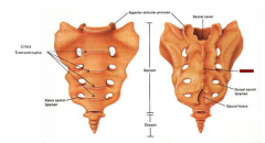
Sacrum:
___ sacral crest is in the midline and is homologous to spinous processes |

Median |
|
|
Sacrum:
the ___ sacral crests, which are homologous to transverse processes |

lateral |
|
|
Sacrum:
Lateral to the median crest are two ___ sacral crests, which are homologous to ___ |
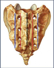
intermediate
articular processes |
|
|
On the ventral and dorsal surfaces there are four pairs of sacral ___
|
foramina
|
|
|
The Sacrum |
posterior
|
|
|
The Sacrum |
anterior |
|
|
The Sacrum |

hiatus |
|
|
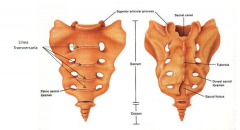
Sacrum: Linea Transversaria |
|
|
Sacrum: Sacral Tubercle |
|
|
Sacral Hiatus
The sacral hiatus is a naturally occurring ___ (S5 lamina don’t fuse) located at the inferior end of the median sacral crest. |

spina bifida |
|
|
Sacral Hiatus
The left and right margins of the hiatus, called the ___, are formed by the inferior tubercles of the intermediate sacral crests. |
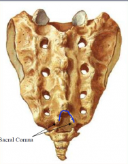
sacral cornua |
|
|
Sacral Hiatus |

S5 & coccygeal nerve roots, |
|
|
The male sacrum is usually ___ right to left and ___ superior to inferior. |

narrower |
|
|
The female sacrum is usually ___ right to left, oriented in a more horizontal plane, and is more concave. |
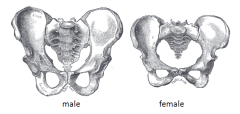
wider |
|
|
The coccyx Greek for ___ (it supposedly looks like a bird’s beak) is made of 4 fused vertebrae.
|
cuckoo
|
|
|
Coccyx |
transverse processes |
|
|
The coccyx serves as an attachment site for |

|
|
|
Coccygeal Cornu(a)
|
|
|
Transverse processs of Coccyx
|