Reading...
![]()
Play button
![]()
Play button
![]()
Use LEFT and RIGHT arrow keys to navigate between flashcards;
Use UP and DOWN arrow keys to flip the card;
H to show hint;
A reads text to speech;
3 Cards in this Set
- Front
- Back
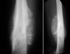
A 24-year-old man "twists" his knee in a softball accident. He denies any preceding complaints and is very active and healthy. Figures A and B include radiographs and histology of the lesion. These findings are most consistent with what diagnosis?
1. Parosteal osteosarcoma 2. Periosteal osteosarcoma 3. Osteochondroma 4. Dedifferentiated chondrosarcoma 5. Synovial Sarcoma |
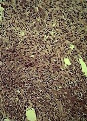
This patient's history is very benign, but the imaging and histology suggest a high grade osteosarcoma. Based on the images, periosteal osteosarcoma is the correct choice. Periosteal osteosarcomas occur on the surface of the bone, most commonly on the femur and tibia and they typically grow in a "sunburst" fashion creating enlarging, linear matrix ossification. In contrast, parosteal osteosarcomas show a "stuck-on" appearance, and frequently show lower-grade histology (illustration A). Osteochondromas have a characteristic continuation with the medullary canal and do not appear aggressive either on xray or pathology. Dedifferentiated chondrosarcomas develop from previous cartilage tumors and as such have a characteristic appearance on pathology. Synovial sarcoma is a soft tissue sarcoma which can calcify and occur around joints, but has a spindle cell pathologic appearance. The cited references describe these key differences which can all occur around joints, show matrix, but have specific differences.
Ans2 |
|
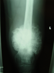
A 47-year-old man presents with increasing left knee pain and swelling. There is no history of trauma and he is otherwise healthy. Representative radiograph, MRI, and histology are shown in Figures A through D. What is the most likely diagnosis?
1. Chondrosarcoma 2. Ewing's sarcoma 3. Periosteal osteosarcoma 4. Giant cell tumor 5. Fibrous dysplasia |
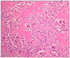
Periosteal osteosarcoma is an extremely rare intermediate grade surface osteosarcoma that usually occurs in patients 15 to 25 years of age. Treatment is the same as intramedullary osteosarcoma and consists of neoadjuvant chemotherapy, surgical excision with reconstruction, and adjuvant chemotherapy.
Hillmann et al. evaluated the functional outcomes of patients with distal femoral or proximal tibial tumors reconstructed with either endoprostheses or rotationplasties. They found that while no statistical difference exists between the two cohorts, patients with rotationplasties tended to have a greater level of activity, more normal gait, and were more confident in their limbs than the endoprostheses cohort. The cosmetic concerns regarding rotationplasties tended to be the largest problem with this method of surgical reconstruction. Manoso et al. review their experience treating osteosarcoma in patients over the age of 40. They found that while historical references suggest no benefit to multimodal chemotherapy, their cohort treated with chemotherapy had a longer overall survival. As such, they now recommend multimodal chemotherapy to patients with osteosarcoma over the age of 40. Ans3 |
|

20 yo F with knee pain and swelling. There is no history of trauma and he is otherwise healthy. Bone Scan is hot.
1-Dx & KIF, 2-Tx & R/Tm 3-C & P) |

1-Dx & KIF(key image finding)
2-Tx & Rehab/Tm 3-Complication & Prevention 1-Dx & KIV=paraosteoal osteosarcoma=intermediate grade surface osteosarcoma that usually occurs in patients 15 to 25 years of age. KIV=histology reveals areas of chondroblastic matrix & "sunburst" or "hair on end" periosteal reaction 2-Tx & R/Tm-neoadjuvant chemotherapy/multi-agent chemotherapy, surgical excision with reconstruction, and adjuvant chemotherapy. Rehab/tm=preoperative chemotherapy given for 8-12 weeks followed by maintenance chemotherapy for 6-12 months after surgical resection 3-C & P= C-20-35% chance of pulmonary metastasis->Chest CT scan, required for staging evaluates for the presence of pulmonary metastasis P-expression of multi-drug resistance (MDR) gene portends very poor prognosis |