![]()
![]()
![]()
Use LEFT and RIGHT arrow keys to navigate between flashcards;
Use UP and DOWN arrow keys to flip the card;
H to show hint;
A reads text to speech;
23 Cards in this Set
- Front
- Back
|
Describe skeletal muscle |
-long and cylindrical fibres (myocytes) which are striated(alternating light and dark bands) -multi nucleated with nuclei residing at the margins -voluntary type of muscle i.e. somatic arm of the nervous system -typically attached to bones via tendons -function: movement, maintaining posture, production of heat andprotection |
|
|
Describe cardiac muscle |
-made of branched striated muscle fibres -nuclei are central – typically only one or two -located in the heart wall -function is to contract the heart to pump blood -contraction is involuntary – regulated by hormones and thesympathetic and parasympathetic arms of the autonomic nervous system -ends of striated myocytes are joined together by thickened regions ofmembrane called intercalated disks |
|
|
Describe smooth muscle |
-non-striated myocytes – hence the term smooth -contraction is involuntary – i.e. regulated by hormones and thesympathetic and parasympathetic arms of the autonomic nervous system -typically found in hollow organs (e.g. vessels, GI tract, respiratorysystem) -myocytes in smooth muscle are thin strands with a single centralnuclei -smooth muscle myocytes are joined together by connexins at gapjunction, which allow them to contract in unison. Some parts of thebody contain smooth muscle which does not have gap junctions – suchas the iris of the eye – which allows the myocytes to contractindividually. -smooth muscle has various functions though all related to movement –e.g. peristalis in the digestive system, constriction of vessels andairways and contraction of the urinary bladder |
|
|
|
|
|
Describe the structure and function of the junctions found within the intercalated discs of cardiac myocytes |
- intercalated diskscontain desmosomes – adhesive intercellular junctions – whichprovide support during contractions. Binding at desmosomes ismediated by cadherins which bind intermediate fllaments to the cellmembrane - intercalated diskscontain gap junctions which are composed of connexins. They permitthe free flow of ions needed changes in membrane potential i.e.contraction. Allow quick depolarisation for successive excitation. - intercalated diskscontain fascia adherens: similar to desmosomes in that they areinvolved in providing support during contraction, and that they alsobind with cadherins – linking the cell membrane to the actincytoskeleton |
|
|
What are the functions of skeletal muscle? |
- movement - stabilising body i.e. posture - storing and moving substances e.g. skeletal muscle pump - heat production i.e. shivering - |
|
|
|
|
|
|
|
|
|
|
|
|
|
|
What are the three types of muscle proteins. Give 2 examples of each |
- contractile proteins: actin and myosin - regulatory proteins: troponin and tropomyosin. - structural proteins: titin and alpha actinin |
|
|
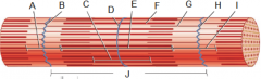
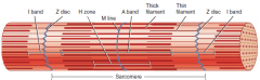
Briefly describe the function of 5 structural muscle proteins |
Titin; - large protein whichconnects the Z-disk to the M-line - has elastic propertieswhich accounts for the elasticity and flexibility of myofibrils Alphaactinin - main structural proteinof Z-discs which bind actin molecules of the thin filaments Myomesin - mainprotein of the M-line (M for myomesin).This protein also binds totitin and binds adjacent thick filaments. Nebulin - non-elastic proteinwraps around the thin filaments like a sheath. It helps to anchorthem to the Z-disk and regulates their length during development. Dystrophin - links the thinfilaments to proteins within the sarcolemma. They are thought toprovide structure to the sarcolemma and help transmit tension to thetendons |
|
|
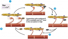
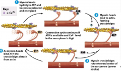
Starting with the nerve impulse, describe the process of muscle contraction |
- a nerve impulse triggers the opening of voltage-gated channels atsynaptic end bulbs. The concentration of Ca2+is higher outside of the synaptic bulb, so Ca2+begins to flow in. -the influx of Ca2+to the synaptic bulb stimulates acetylcholine-containing vesicles toundergo exocytosis – releasing acetylcholine into the synapticcleft - free acetylcholinemolecules bind to receptors in the motor end plates (2 per receptor),which opens ion channels. This allows small cations – mostcrucially Na2+ - to flow into the muscle fibres - the influx of Na+into the muscle fibres make them more positively charged – changingthe membrane potential -the change in the membrane potential triggers an action potential,which propagates along the sarcolemma into the T tubules. This, inturn, causes the sarcoplasmic reticulum to release Ca2+into the sarcoplasm. -the Ca2+ ions then bind to troponin which pushestropomyosin away from the mysoin-binding sites on the actin thinfilaments -Myosin heads posses an ATP-binding site, and a ATPase – i.e. canhydrolyse ATP to ADP + a phosphate. -the myosin heads are able to gain energy by hydrolysing ATP. Afterthis, the products – ADP + phosphate – remain bound. - with the myosin headsenergised and the troponin binding site free, the myosin heads areable to bind to the binding site on actin – forming a cross-bridge.The phosphate group is released during this binding. - binding causes the sitewhich binds ADP to open up. This causes the cross-bridges to rotate,releasing the ADP. The rotating of the cross-bridge towards thecentre of the sarcomere generates force, causing the thin filamentsto slide towards the M-line, and overlap with the thick filaments. - at the end of thestroke, the mysoin heads bind to another molecule of ATP. Thisreleases tropomyosin from troponin, releasing the cross-bridges. ThisATP molecule can again be hydrolysed to mark the beginning of thenext cycle. - this cycle can continueas long as there is Ca2+ in the sarcoplasm - with each stroke, thecross-bridges rotate, pulling the thin filaments progressively closerto the M-line (they can do this around 600 times a second. - the overall outcome ofthese successive contraction is that the Z-plates mover closer andcloser to one another – shortening the sarcomere. This in turn canpull on other sarcomeres, shortening the muscle fibre. - as the muscles fibresare connected to connective tissues, which in turn are connected tobone, this contraction allows these parts of the body to move. |
|
|
|
|
|
How is the activity of acetycholine stopped? |
- acetylcholinesterase isa membrane which exists in the extracellular matrix of the synapticcleft (space). - acetylcholinesterasebreaks down acetycholine into acetyl and choline - this ensures that theactivity of acetycholine is short lived in the synaptic cleft |
|
|
What are the three types of muscle fiber? |
Slowoxidative fibres – type 1 Fastoxidative glycolytic fibres (fast fatigue resistant) – typicallytype 2a Fastglycolytic fibres (fast fatigueable) – 2b and 2x |
|
|
Describe slow oxidative muscle fibers |
-smallest in diameter and weakest of all muscle fibres -contain large amount of myoglobin and blood capillaries – makingthe muscle bright red -contain many large mitochondria and thus produce ATP through aerobicrespiration -they are called slow in relation to the activity level ATPase in themyosin i.e. the ATPase hydrolyses ATP slowly. Because of thecontraction cycle is slower compared to other types of muscle fiber. -while slow oxidative fibres are generally weaker, they are moreresistant to fatigue, and thus able to make sustained contractionsfor many hours. -these types of muscles are maintaining posture, and activities suchas marathon running |
|
|
Describe fast oxidative glycolytic fibers |
-some major similarities to slow oxidative fibres -fast oxidative glycolytic fibres are intermediate in diameter -like slow oxidative fibres, fast oxidative glycolytic fibres havelarge amount of myoglobin and blood capillaries, and therefore alsohave a similar bright red appearance. -they also have large amount of mitchondria and therefore also produceATP through oxidative respiration -they also have large quantities of glycogen which allows them to alsogenerate ATP anaerobically through glycolysis -fast oxidative glycolytic fibres are called fast because of theirATPase: it hydrolyses ATP fast and thus allows the contraction cycleto repeat more rapidly. This allows Z-plates to reach peak tensionmore quickly than slow oxidative fibres, though they also becomefatigued quicker. -fast oxidative glycolytic fibres are used for activities such aswalking and running |
|
|
Describe fast glycolytic fibres |
-largest in diameter and most densely packed with myofibrils -have large amounts of glycogen, though low concentrations ofmyoglobin, few capillaries and few mitochondria. Because of the lowamount of myoglobin, they appear white. -because of their high glycogen content they mainly produce ATPaerobically, though in lower levels that the other muscle fibre types -high activity level of ATPase - allows the contraction cycle torepeat more rapidly. -these muscle fibres are used for intense anaerobic activities –such as weightlifting or throwing – though they also fatiguequickly |
|
|
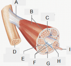
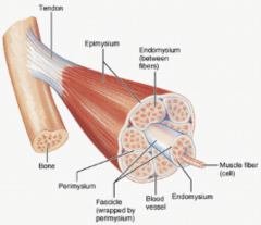
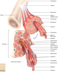
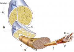
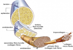
Describe the structure of tendons |
-made from multiple layer of colagenous connective tissue which uniteto form a ropelike structure - tendon -contain few cells; composed mainly of parallel collagen fibres andproteoglycan ground substance. Also contains small amount of elastin. -main type of collagen is type I, though there is also small amount oftype III and type IV (5%) -the main cell type is fibroblasts which lay is parallel spacesbetween collagen fibrils -the fibroblasts synthesise precursors for the connective tissuematrix e.g. collagen, elastin and proteoglycans -fibrils are bound together mainly by proteoglycans, glycoproteins andglycosaminoglycans, forming fascicles -fascicles are bound together by connective tissue called theendotenon, which carries blood vessels, lymphatics and nerves. Theendostenon is continuous with the periosteum (vascular connectivetissue surrounding bone) -the endostenon is surrounded by a membrane called the epitenon -in some tendons, the epitenon is surrounded by a areolar tissuesheath called the paratenon (vascular), which allows free gliding ofthe tendon against surrounding tissue. In other tendons there may betendon sheaths (avascular) or bursae -collectively the epitenon and the paratenon are known as theperitenon |
|
|
|
|
|
Give an example of 5 types of muscle injury |
-strains (1-3): causedwhen intense and rapid contraction is demanded of a muscle. Suchcontractions can be too strong, and muscle fibres may be unable toresist – causing a tear -contusion (bruise): causedby impact to a muscle. Muscle fibres compressed in this manner canbecome irritated and even torn. A bruise develops as blood pools atthe impact site. -cramp: a muscle cramp occurs when the muscle forciblycontracts and will not relax. Cramps can occur in any musclecontrolled by voluntary action. -elongation: caused when a muscle is stretched beyond its capacity.Certain muscle fibres can also become torn in this manner.- rupture: separation ofmuscle tissue -necrosis: e.g. infarct |
|
|
What are the three stages of muscle regeneration? |
-inflammatory response: macrophages are recruited for phagocytosis andpromote myogenic differentiation -activation and differentiation of satellite cells -growth and remodeling of muscle tissue – in conjunction with nerveactivity |